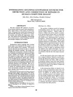
báo cáo khoa học: "Left sided inferior vena cava duplication and venous thromboembolism: case report and review of literature" ppsx
Bạn đang xem bản rút gọn của tài liệu. Xem và tải ngay bản đầy đủ của tài liệu tại đây (910.58 KB, 4 trang )
BioMed Central
Page 1 of 4
(page number not for citation purposes)
Journal of Hematology &
Open Access
Case report
Left sided inferior vena cava duplication and venous
thromboembolism: case report and review of literature
Cannon Milani*, Maria Constantinou, David Berz, James N Butera and
Gerald A Colvin
Address: Warren Alpert School of Medicine at Brown University and Department of Medicine, Rhode Island Hospital, Providence, RI, USA
Email: Cannon Milani* - ; Maria Constantinou - ; David Berz - ;
James N Butera - ; Gerald A Colvin -
* Corresponding author
Abstract
The etiology of venous thromboembolism in young patients is frequently associated with hereditary
coagulation abnormalities, immunologic diseases, and neoplasia. The advent of radiological
advances, namely Computed Tomography (CT) scans and venography has identified vena cava
malformations as a new etiologic factor worthy of consideration. In this case report, we describe
the unusual occurrence of venous thromboembolism in association with a duplicated inferior vena
cava. Duplications of the inferior vena cava (IVC) are seen with an incidence of 0.2% to 3.0% in the
general population. Embryogenesis of the IVC is a complex process involving the intricate
formation and regression of numerous anastomoses, potentially leading to various anomalies. We
present a 23-year-old Caucasian woman with IVC duplication who developed a deep venous
thrombosis and multiple pulmonary emboli. Anomaly of the IVC is a rare example of a congenital
condition that predisposes to thromboembolism, presumably by favoring venous stasis. This
diagnosis should be considered in patients under the age of 30 with spontaneous occurrence of
blood clots.
Background
Over a century ago the German physician, Rudolf Vir-
chow, was credited for elucidating the mechanism of pul-
monary thromboembolism. The factors contributing to
venous thrombosis came to be known as Virchow's triad.
The three components were 'abnormalities of blood vessel
wall', 'abnormalities of blood constituents', and 'abnor-
malities of blood flow'. [1]
The incidence of deep venous thrombosis (DVT) in west-
ern populations is estimated at 1 in 1,000 individuals per
annum. [2] This figure varies with age, and in adults aged
between 20 and 40 years, the incidence is 10 times lower.
[3]
The objective of this case report is to describe IVC malfor-
mation in a young patient as a risk factor in the develop-
ment of venous thromboembolism and the associated
clinical manifestations. This case provides the opportu-
nity to review the dysregulated embryogenesis of this phe-
nomenon, and the intricate management questions that
arise.
In duplication of the IVC, there is a normal inferior vena
cava along the right side of the spine. In addition, a left-
Published: 2 December 2008
Journal of Hematology & Oncology 2008, 1:24 doi:10.1186/1756-8722-1-24
Received: 25 September 2008
Accepted: 2 December 2008
This article is available from: />© 2008 Milani et al; licensee BioMed Central Ltd.
This is an Open Access article distributed under the terms of the Creative Commons Attribution License ( />),
which permits unrestricted use, distribution, and reproduction in any medium, provided the original work is properly cited.
Journal of Hematology & Oncology 2008, 1:24 />Page 2 of 4
(page number not for citation purposes)
sided IVC ascends to the level of the renal veins to join the
right-sided IVC through a vascular structure that may pass
either anterior or posterior to the aorta at the level of the
renal veins. The characteristic CT appearance (Figure 1) is
a single right-sided inferior vena cava at levels above the
renal veins, a vascular structure crossing either anterior or
posterior to the aorta at the level of the renal veins, and a
vascular structure to the left (i.e. the duplicated IVC) and
right of the aorta below the level of the renal veins. [4]
Case Presentation
Our 23-year-old female patient with no significant past
medical history presented to the hospital complaining of
acute shortness of breath, abdominal pain, and severe left
lower extremity pain. The patient was in her usual state of
health until the evening of her symptoms, when she sud-
denly turned in her bed and began to experience numb-
ness, tingling, and purplish discoloration of her left lower
extremity. On arrival to the emergency department, the
patient was in respiratory distress and had bilateral swell-
ing of her lower extremities, the left side larger than the
right. A bilateral lower extremity duplex ultrasound
revealed a deep vein thrombosis (DVT) in the left lower
extremity, the popliteal, and the femoral veins. In addi-
tion a CT of the chest utilizing our pulmonary embolism
protocol delineated two pulmonary emboli in the first
order pulmonary arteries and multiple segmental emboli
located in the right upper and right lower lobes. She was
subsequently started on a heparin drip.
The patient denied any tobacco use, recent immobiliza-
tion, or travel. She reported the use of an estrogen vaginal
delivery device (ethinyl estradiol vaginal ring) for the pre-
vious 6 months. Her hypercoagulable workup was reveal-
ing for heterozygosity at C677T for factor V Leiden. It
should be noted that heterozygous carriers of FVL have
been shown to have an overall 3- to 7-fold increased risk
of venous thrombosis, while homozygotes to C677T have
a 50- to 100-fold increased risk. [5]
Due to the acute presentation of her venous thromboem-
bolism, a left pelvic and lower extremity venous
thrombectomy was performed. This was followed by
stenting of a popliteal venous occlusion, and subsequent
thrombolysis with tissue plasminogen activator. Surpris-
ingly, a left lower extremity venogram revealed bilateral
inferior vena cavas.
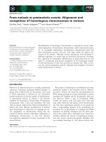
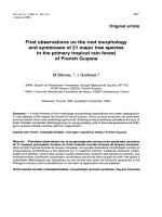
In our patient, the left-sided duplicated IVC and iliac veins
were found to be thrombosed. This was marked by the
stippled appearance of the duplicated left IVC vessel seen
in Figure 2a. The decision was made to stent the dupli-
cated IVC given the anatomic irregularity and increased
risk of repeat thrombosis. Two wall stents (Figure 2b)
were deployed at the proximal end at the level of the left
sided (duplicated) IVC. Next a balloon was used to angi-
oplasty the entire length of the stented segment of the
duplicated IVC (Figure 2b). Figure 3 depicts the dupli-
cated left IVC with wall-stent draining into the left renal
vein with a non-occlusive thrombus within it. The ortho-
topic IVC remained patent. The patient was kept on
heparin for 24-hours and then started on Coumadin.
Discussion and Conclusion
There have been few case reports with IVC anomalies and
development of deep vein thrombosis. In our comprehen-
sive English language literature review, the earliest
A: Patient in prone position delineating the stippled appear-ance of the left duplicated IVC (black arrow) in communica-tion with the left renal vein (white arrow)Figure 2
A: Patient in prone position delineating the stippled appear-
ance of the left duplicated IVC (black arrow) in communica-
tion with the left renal vein (white arrow). 2B: Patient in
supine position with balloon angioplasty of the duplicated left
IVC (thin black arrow) and iliac stent (thick black arrow),
with improved appearance and flow through the stents.
Simultaneously the right IVC undergoing catheterization
(white arrow).
The left sided IVC (thin black arrow) is shown joining the left renal vein (thick black arrow)Figure 1
The left sided IVC (thin black arrow) is shown joining the left
renal vein (thick black arrow). At the same level the right
renal artery (white arrow) is visible
Journal of Hematology & Oncology 2008, 1:24 />Page 3 of 4
(page number not for citation purposes)
reported case of duplication of the inferior vena cava was
reported in 1912 in the journal, The Anatomical Record.
[6] Over the last 100 years, case series involving IVC dupli-
cation in association with venous thromboembolism
number less than 10.
This observation was made following an extensive
PubMed literature search encompassing the following
keywords: inferior vena cava, congenital, deep venous
thrombosis, and pulmonary embolism. From an anatom-
ical standpoint in adults, IVC has 3 segments of different
embryologic origin: pre-renal, renal, and post-renal. This
type of fusion and partial re-absorption of three pairs of
vessels is dependent upon the posterior cardinal veins in
the embryo. This complicated evolutionary process can
give rise to anatomic malformations that impede vein
drainage and favor the development of thrombosis. [7,8]
Duplication of the IVC, the prominent manifestation in
our case report, occurs because the left supracardinal vein
fails to regress early in gestation resulting in large veins on
both sides of the Aorta that usually joins anterior to the
level of the renal arteries to become the suprarenal IVC.
[9] When incidentally found, treatment options include
observation, placing filters in either systems, or coil-
embolization of the duplicated segment plus placing a fil-
ter in the right IVC.
Furthermore, radiographic duplication of the IVC can be
confused with saccular aortic aneurysms, aorto-lumbar
lymphadenopathy, left pyeloureteric dilatation, retroperi-
toneal cysts, and loops of small bowel. [10] As a result it
is imperative to consider this anomaly both in an acute
presentation of venous thromboembolism in a younger
individual, and with the above-mentioned disorders as
well. A schematic diagram of the observed abnormality is
shown in Figure 4.
As noted in our literature search, there were a few case
reports of thromboembolic disease in patients with a
duplicated IVC. In patients with DVT of the legs in this set-
ting, the treatment paradigm protocol changes, caval
interruption becomes paramount. The failure to interrupt
both the right- and left-sided IVC can result in recurrent
pulmonary embolism. [10] Physicians need to be
reminded that such anomalies of the IVC exist and that
they may influence decision-making in patients with an
acute presentation of thromboembolic disease.
In all studies, age of presentation of first thrombosis has
been less than 30 years of age and incidence is similar in
men and women. Few studies [10-12] consider double
inferior vena cava to be the cause of DVT, perhaps because
it causes retrograde stasis less often. Compensatory drain-
age thru the thoracic-lumbar, pelvic, and abdominal veins
can cause symptoms in the thorax, hypogastrium, lumbar,
and genital regions, prior to those typical of DVT of the
lower extremities. Early detection could warn of the pres-
ence of cava malformations in young patients. For
instance our patient presented complaining of chest and
abdominal pain, with associated discoloration of her left
lower limb.
Some authors believe cava malformation alone can pro-
voke DVT, [13] but the fact that lifelong asymptomatic
malformations occur, [12] the findings in the case report
Duplicated left IVC with wall-stent draining into the left renal vein (black arrow)Figure 3
Duplicated left IVC with wall-stent draining into the left renal
vein (black arrow). There is a non-occlusive thrombus within
it. The orthotopic IVC (white arrow) appears widely patent.
Diagram of the duplicated IVC of our patient based on ultra-sound showing the right and left kidneys (RK and LK), the aorta (A), the right renal vein (rrv), and the suprarenal IVCFigure 4
Diagram of the duplicated IVC of our patient based on ultra-
sound showing the right and left kidneys (RK and LK), the
aorta (A), the right renal vein (rrv), and the suprarenal IVC.
The right-sided IVC (R) is patent while the left-sided IVC (L)
with thrombus (stippled) empties into the left renal vein (lrv).
Publish with BioMed Central and every
scientist can read your work free of charge
"BioMed Central will be the most significant development for
disseminating the results of biomedical research in our lifetime."
Sir Paul Nurse, Cancer Research UK
Your research papers will be:
available free of charge to the entire biomedical community
peer reviewed and published immediately upon acceptance
cited in PubMed and archived on PubMed Central
yours — you keep the copyright
Submit your manuscript here:
/>BioMedcentral
Journal of Hematology & Oncology 2008, 1:24 />Page 4 of 4
(page number not for citation purposes)
and the status of thrombosis as a multifactorial illness,
[14] suggest the presence of associated factors, both con-
genital and acquired. The complementary entities of the
patient's heterozygosity for the Factor V Leiden mutation,
and her use of an oral contraceptive intrauterine device,
could invariably have been adjunctive triggers in her clot-
ting cascade.
Ultimately there is currently no data available to guide the
use of anticoagulation in the under 30 population who
present with the additional complication of a congenital
anomaly. Apparently, the most appropriate approach to
treatment is for more than six months' anticoagulation
while the principal factor provoking thrombosis contin-
ues. In conclusion, the possibility of recurrent thrombotic
occlusion is high in these patients when anticoagulation
treatment is withdrawn even when precipitating triggers
such as oral contraceptives are removed.
Consent
The patient has provided informed consent for the publi-
cation of this case report and accompanying images.
Competing interests
The authors declare that they have no competing interests.
Authors' contributions
The original manuscript was written by CM. All authors
participated in drafting and editing the manuscript. All
authors read and approved the final manuscript.
Authors' information
The authors provide specialized, multidisciplinary clinical
care for patients with a variety of hematologic and onco-
logic malignancies.
References
1. Chung I, Lip G: Virchow's Triad Revisited: Blood Constituents.
Pathophysiol Haemost Thromb 2003, 33:449-54.
2. Anderson FA Jr, Wheeler HB, Goldberg RJ, Hosmer DW, Patward-
han NA, Jovanovic B, Forcier A, Dalen JE: A population-based per-
spective of the hospital incidence and case-fatality rates of
deep vein thrombosis and pulmonary embolism. The
Worcester DVT Study. Arch Intern Med 1991, 151:933-938.
3. Rosendaal FR: Venous thrombosis: a multicausal disease. Lan-
cet 1999, 353:1167-1173.
4. Royal SA, Callen PW: CT evaluation of anomalies of the inferior
vena cava and left renal vein. AJR Am J Roentgenol 1979,
132:759-63.
5. Press RD, Bauer KA, Kujovich JL, Heit JA: Clinical utility of factor
V Leiden (R506Q) testing for the diagnosis and management
of thromboembolic disorders. Arch Pathol Lab Med 2002,
126:1304-1318.
6. Givens MH: Duplication of the inferior vena cava in man. The
Anatomical Record 1912, 6:475-486.
7. García-Fuster MJ, Forner MJ, Flor-Lorente B, Soler J, Campos S: Infe-
rior vena cava malformations and deep venous thrombosis.
Rev Esp Cardiol 2006, 59:171-175.
8. Chuang VP, Mena CE, Hoskins PA: Congenital anomalies of the
inferior vena cava. Review of embryogenesis and presenta-
tion of a simplified classification. Br J Radiol 1974, 47:206-213.
9. Kouroukis C, Leclerc JR: Pulmonary embolism with duplicated
inferior vena cava. Chest 1996, 109:1111-1113.
10. Senecail B, Lefevre C, Person H, Meriot P: Radiologic anatomy of
duplication of the inferior vena cava: a trap in abdominal
imaging. A report of 8 cases. Surg Radiol Anat 1987, 9:151-157.
11. Chee YL, Culligan DJ, Watson HG: Inferior vena cava malforma-
tion as a risk factor for deep venous thrombosis in the young.
Br J Haematol 2001, 114:878-880.
12. Gayer G, Luboshitz J, Hertz M, Zissin R, Thaler M, Lubetsky A, Bass
A, Korat A, Apter S:
Congenital anomalies of the inferior vena
cava revealed on CT in patients with deep vein thrombosis.
AJR Am J Roentgenol 2003, 180:729-732.
13. Obernosterer A, Aschauer M, Schnedl W, Lipp RW: Anomalies of
the inferior vena cava in patients with iliac venous thrombo-
sis. Ann Intern Med 2002, 136:37-41.
14. Huntington GS, McClure CFW: The Development of the Veins
in the Domestic Cat (Felisdomestica). Anatomical Record 1920,
20:1-30.