Journal of Hematology & Oncology BioMed Central Research Open Access Radiation produces pps
Bạn đang xem bản rút gọn của tài liệu. Xem và tải ngay bản đầy đủ của tài liệu tại đây (606.42 KB, 12 trang )
BioMed Central
Page 1 of 12
(page number not for citation purposes)
Journal of Hematology & Oncology
Open Access
Research
Radiation produces differential changes in cytokine profiles in
radiation lung fibrosis sensitive and resistant mice
Xiaoping Ao
1
, Lujun Zhao
1
, Mary A Davis
1
, David M Lubman
2
,
Theodore S Lawrence
1
and Feng-Ming Kong*
1
Address:
1
Department of Radiation Oncology, University of Michigan, Ann Arbor, MI 48109, USA and
2
Department of Surgery, University of
Michigan, Ann Arbor, MI 48109, USA
Email: Xiaoping Ao - ; Lujun Zhao - ; Mary A Davis - ;
David M Lubman - ; Theodore S Lawrence - ; Feng-Ming Kong* -
* Corresponding author
Abstract
Background: Recent research has supported that a variety of cytokines play important roles
during radiation-induced lung toxicity. The present study is designed to investigate the differences
in early cytokine induction after radiation in sensitive (C57BL/6) and resistant mice (C3H).
Results: Twenty-two cytokines in the lung tissue homogenates, bronchial lavage (BAL) fluids, and
serum from 3, 6, 12, 24 hrs to 1 week after 12 Gy whole lung irradiation were profiled using a
microsphere-based multiplexed cytokine assay. The majority of cytokines had similar baseline levels
in C57BL/6 and C3H mice, but differed significantly after radiation. Many, including granulocyte
colony-stimulating factor (G-CSF), interleukin-6 (IL-6), and keratinocyte-derived chemokine (KC)
were elevated significantly in specimens from both strains. They usually peaked at about 3–6 hrs in
C57BL/6 and 6–12 hrs in C3H. At 6 hrs in lung tissue, G-CSF, IL-6, and KC increased 6, 8, and 11
fold in C57BL/6 mice, 4, 3, and 3 fold in the C3H mice, respectively. IL-6 was 10-fold higher at 6
hrs in the C57BL/6 BAL fluid than the C3H BAL fluid. MCP-1, IP-10, and IL-1α also showed some
differences between strains in the lung tissue and/or serum. For the same cytokine and within the
same strain of mice, there were significant linear correlations between lung tissue and BAL fluid
levels (R
2
ranged 0.46–0.99) and between serum and tissue (R
2
ranged 0.56–0.98).
Conclusion: Radiation induced earlier and greater temporal changes in multiple cytokines in the
pulmonary fibrosis sensitive mice. Positive correlation between serum and tissue levels suggests
that blood may be used as a surrogate marker for tissue.
Background
Radiation-induced pulmonary injury to normal lung tis-
sue is a dose-limiting complication for cancer patients
receiving radiotherapy to the chest region [1-3]. Depend-
ing on both radiation dose and volume, lung injury is
characterized by inflammation associated pneumonitis
which may progress to permanent pulmonary fibrosis. An
improved understanding of the factors leading to pneu-
monitis and fibrosis could result in an increased ability to
predict which patients are likely to develop the disease so
that they could receive appropriate treatment.
Published: 2 February 2009
Journal of Hematology & Oncology 2009, 2:6 doi:10.1186/1756-8722-2-6
Received: 8 August 2008
Accepted: 2 February 2009
This article is available from: />© 2009 Ao et al; licensee BioMed Central Ltd.
This is an Open Access article distributed under the terms of the Creative Commons Attribution License ( />),
which permits unrestricted use, distribution, and reproduction in any medium, provided the original work is properly cited.
Journal of Hematology & Oncology 2009, 2:6 />Page 2 of 12
(page number not for citation purposes)
The response to ionizing radiation involves a number of
mediators including inflammatory cytokines produced by
macrophages, epithelial cells, and fibroblasts [4,5]. An
early activation of an inflammatory reaction can lead to
the expression and maintenance of a perpetual cytokine
cascade, resulting in increased collagen production and
ultimately fibrosis [6]. For example the cytokine, trans-
forming growth factor-beta1 (TGF-β1), is thought to be a
key mediator of lung toxicity and may predict resultant
damage to normal lung following radiation [7,8]. Since a
complex cytokine network initiates and sustains the
inflammatory and fibrogenic processes associated with
radiation-induced lung injury [9], the ability to simulta-
neously quantify multiple cytokines is critical for deci-
phering how they affect radiation-induced lung toxicity.
One such assay, a microsphere-based sandwich immu-
noassay for flow cytometry, is a highly sensitive and selec-
tive multiplexed assay platform to simultaneously
measure many cytokines in low volume samples, e.g. 25
μL sample for 22 mouse cytokines/chemokines [10]. This
assay platform, the most comprehensive one available on
the market during the time of our experiment, provides a
powerful tool for multiple cytokine profiling and a more
complete picture of the complex cytokine network.
The present study was designed to take advantage of this
platform and the known differences between the C57BL/
6 and C3H mouse strains in their response to lung radia-
tion [11-14]. C57BL/6 mice are much more sensitive to
radiation-induced pulmonary fibrosis than C3H mice
[15]. Johnston et al. have extensively studied the mRNA
expression of different cytokines in mouse lung after ion-
izing radiation [6,16-18]; these studies focused on the
remodeling phase but not the initial response. Others
noted that cytokine mRNA elevation occurred early after
radiation [19,20], and an early study on TGF-β1 showed a
rapid induction of immunoreactivity in tissue at 1 hour
post radiation [21]. While most of the previous multi-
plexed cytokine studies focused on the transcriptional
mRNAs instead of cytokine proteins, proteins, rather than
mRNA, are the actual biological effectors, making it likely
that cytokine levels will better correlate with biological
outcome than mRNA levels. Therefore, we focused our
study on the cytokines themselves. We hypothesized that
there would be significant differences in cytokine profiles
immediately after radiation in these two strains of mice
with different sensitivities to radiation. We also hypothe-
sized that serum cytokine profiles would correlate with
lung tissue levels such that a panel of serum markers could
be developed which predict for radiation-induced lung
toxicity. Therefore, in this study, we treated C57BL/6 and
C3H mice with thoracic radiation and, utilizing the mul-
tiplex immunoassay platform, measured the levels of 22
cytokines in lung tissue, broncheoalveolar lavage fluid
(BAL), and serum at times from 3 hrs to 1 week after radi-
ation.
Methods and materials
Animals and radiation treatment
Five to 6 week-old male C57BL/6 and C3H mice were pur-
chased from Charles River Breeding Labs (Wilmington,
MA). A plastic jig was used to restrain the mice without
anesthesia, and lead strips were placed to shield the head
and abdomen. A Phillips 250 orthovoltage unit was used
to deliver 12 Gy at 143.27 cGy/min to the thorax. The field
size (2 × 3 cm) was set to provide adequate coverage of the
whole lung. Dosimetry was carried out using an ioniza-
tion chamber connected to an electrometer system, which
is directly traceable to a National Institute of Standards
and Technology calibration. The use of animals was in
compliance with the regulations of the University of
Michigan and with NIH guidelines. The susceptibility of
the C57BL/6 mouse strain to radiation-induced lung dam-
age [11] has been confirmed in our laboratory by meas-
urement of lung function via plethysmography at 8 weeks
post radiation [22].
Specimen preparation
Lung tissue, bronchial lavage (BAL) fluid, and blood sam-
ples were collected from controls and at 3 hrs, 6 hrs, 12
hrs, 24 hrs, and 1 week after radiation (3 mice at each time
point for each strain). Blood was drawn from anesthetized
mice via cardiac puncture followed by portal venous per-
fusion with 20 ml PBS. The right lung was lavaged with
500 μL saline, BAL fluid was then obtained (about 300 μL
each animal). The left lung which was used for cytokine
measurement was quickly frozen in 70% ethanol contain-
ing dry ice. Blood was allowed to sit for 4 hrs at room tem-
perature to allow clotting, and the supernatant (serum)
was collected after centrifugation. Serum was used as we
were also interested in assessing level of TNF-α in this
study. All samples were stored at -80°C until assay. At the
time of analysis, 25–40 mg of frozen lung tissue was aliq-
uoted using an Ohaus analytical balance, which can meas-
ure weight accurately to 0.1 mg. The frozen tissue was
then disrupted and homogenized in 200 μL tissue lysis
buffer (CelLytic™ MT Mammalian Tissue Lysis/Extraction
Reagent from Sigma-Aldrich) using a tissue grinder
(Duall
®
All-Glass from Kimble/Kontes). After homogeni-
zation, the samples were centrifuged at 10,000 × g for 5
min, and the supernatants were used for cytokine profil-
ing.
Multiplexed cytokine analysis
The cytokine concentrations in the serum, BAL fluids, and
lung tissue lysates were assayed using a Mouse Cytokine/
Chemokine Lincoplex kit (Linco Research, St. Charles,
Missouri). The kit can simultaneously quantify 22 mouse
cytokines and chemokines: Interleukin (IL)-1α, IL-1β, IL-
2, IL-4, IL-5, IL-6, IL-7, IL-9, IL-10, IL-12p70, IL-13, IL-15,
IL-17, Interferon-γ (IFN-γ), Interferon γ-inducible Protein-
10 (IP-10), Granulocyte Colony-Stimulating factor (G-
CSF), Granulocyte Macrophage Colony-Stimulating Fac-
Journal of Hematology & Oncology 2009, 2:6 />Page 3 of 12
(page number not for citation purposes)
tor (GM-CSF), Tumor Necrosis Factor-α (TNF-α), kerati-
nocyte-derived chemokine (KC), Monocyte
Chemoattractant Protein-1 (MCP-1), Macrophage Inflam-
matory Protein-1α(MIP-1α), and Regulated upon Activa-
tion, Normal T-cell Expressed, and Secreted (RANTES).
The kit contains spectrally distinct antibody-immobilized
beads (22 bead sets specifically for the above cytokines),
cytokine standard cocktail, cytokine quality control I and
II, detection antibody cocktail, streptavidin-phycoeryth-
rin, assay buffer, wash buffer, serum matrix, and a micro-
titer filter plate.
The assay was performed according to the manufacturer's
protocol. Tissue lysis buffer, saline, and serum matrix
were used as the sample matrices for tissue lysates, BAL
fluids, and serum, respectively. After preparation, samples
were processed (50 beads per bead set in 50 μL sample
size) on a Luminex 100 instrument (Luminex Corpora-
tion, Austin, TX). All the samples were run in duplicate.
The detection limit of this kit is 3.2 pg/ml for all the
included cytokines.
Statistical analysis
Data are presented as mean ± standard error of the mean
(SEM). One way ANOVA from Origin 7.0 was used to
compare the significance between two sets of data. Values
were considered significantly different when p < 0.05.
Results
Cytokine levels in lung tissue lysates
We began by analyzing cytokine levels in the lungs of con-
trol mice. Nine cytokines out of the 22 measured in the
lung (GM-CSF, G-CSF, IL-6, IL-9, IP-10, KC, MCP-1, MIP-
1α, and RANTES) were above the detection limit of the
assay for both mouse strains. IL-10 was detected at very
low levels only in the radiation sensitive mouse strain
C57BL/6 but not the radiation resistant strain C3H. The
remaining cytokines (IFN-γ, IL-12(p70), IL-13, IL-15, IL-
17, IL-1α, IL-1β, IL-2, IL-4, IL-5, IL-7, and TNF-α) were not
detectable in the tissue lysates from either mouse strain.
There was no significant difference in cytokine levels
between these two strains in control animals except for G-
CSF, IL-6 and IP-10, which were significantly higher level
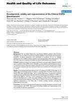
in C57BL/6 than C3H (Fig. 1).
After 12 Gy, many cytokine levels increased significantly
early after radiation. There were clear differences in time-
dependent changes between the two strains in 5 cytokines
(G-CSF, IL-6, KC, MCP1, and IP-10) with detectable eleva-
tions (Fig. 1). All of these cytokines peaked at higher levels
in C57BL/6 mice. The most striking differences occurred
in levels of IL-6 which were increased by approximately 8
fold in the C57BL/6 mice but were only slightly elevated
at 6 hours post radiation in the resistant C3H mice. In
most cases, cytokine levels peaked 3–6 hours earlier in
C57BL/6 mice.
Cytokine levels in bronchial lavage (BAL) fluid
Only three cytokines, G-CSF, IL-6 and KC, were detectable
in the BAL fluid (Fig. 2). As in lung tissue, there was no sig-
nificant difference in the levels of G-CSF and IL-6 in con-
trol C57BL/6 and C3H mice, and there were radiation-
induced peaks for both cytokines in both strains. The peak
levels were similar for G-CSF in both strains, but the peak
occurred at 6 hrs in C57BL/6 mice and at 12 hrs in C3H
mice. IL-6 increased from barely detectable (<3.2 pg/ml)
to approximately 90 pg/ml in the C57BL6J while the
increase was minimal in the C3H. Interestingly, KC levels
were significantly higher in C3H mice than in C57BL/6
mice throughout the study time course; though radiation-
induced elevation was also seen in both strains.
Cytokine levels in serum
Twelve out of 22 cytokines were above the limit of detec-
tion in the serum from both strains of mice. The detecta-
ble cytokines were G-CSF, GM-CSF, IP-10, KC, IL-6, MCP-
1, IL-1α, IL-17, IL-15, IL-13, MIP-1α, and IL-12(p70). Fig.
3 shows the dynamics of cytokines with detectable
changes after radiation. In control mice, G-CSF and IL-6
levels were not significantly different between these two
strains. The levels of KC and MCP-1 were significantly
higher, and IP-10 was lower in C57BL/6 than C3H. After
radiation, the responses of the two strains were remarka-
bly different for most of the measurable cytokines. Among
the cytokines with detectable changes, the majority of
them peaked 3–6 hrs earlier in C57BL/6 mice than in C3H
mice. There were also significant differences in the maxi-
mum extent of elevations. MCP-1 and KC levels peaked at
greater levels in C57BL/6 mice. The radiation-induced ele-
vations were slightly greater and lasted longer for IL-6 and
G-CSF in C3H mice. Radiation induced a similar level and
pattern of changes in IL-1α in the two mouse strains.
Thus, there were some differences in serum cytokine levels
prior to radiation, and there were more significant differ-
ences in time dependent responses after radiation
between these two strains.
Relationships among cytokine levels in lung tissue, BAL
fluid, and serum
There were remarkable similarities among lung tissue,
BAL fluid and serum in their changing patterns of
cytokine levels after radiation. A majority of the changes
were characterized by a peak of elevation. The peak times
of these cytokines are listed for all three types of specimen
(Table 1). Of note, in C57 mice, KC peaked about 3 hours
earlier in lung tissue than serum and BAL fluid. In C3H
mice, G-CSF peaked about 6 hrs earlier in serum than in
tissue and BAL fluid. MCP-1 and IP-10 peaked 3 hours
earlier than all other detectable cytokines in both serum
and tissue in both C57 mice and C3H mice.
Among the three cytokines detectable in all three speci-
mens, there were significant correlations of absolute levels
Journal of Hematology & Oncology 2009, 2:6 />Page 4 of 12
(page number not for citation purposes)
between BAL fluid and tissue (Fig. 4), and serum and tis-
sue (Fig. 5), though there were differences in the peak
times, which caused differential changes in G-CSF and IL-
6 in lung tissue/serum between C57BL/6 and C3H mice.
The best correlations between serum and lung tissue levels
were seen for KC, which had similar peak time in the two
compartments.
Discussion
Using a multiplex screen for 22 cytokines/chemokines at
various time-points, we demonstrated significant differ-
ences after thoracic radiation in both the extent of eleva-
tion and temporal patterns in G-CSF, IL-6, and KC levels
in the lung tissue, BAL fluid, and serum between two
mouse strains with different sensitivity to radiation lung
fibrosis. Our study is unique with respect to its measure-
Mouse lung tissue cytokine levels in C57BL/6 (C57) and C3H miceFigure 1
Mouse lung tissue cytokine levels in C57BL/6 (C57) and C3H mice. Mice were untreated or received a single dose of
12 Gy to the lung. Cytokine levels were normalized based on lung tissue mass. Data are expressed as the mean ± SEM of dupli-
cate determinations from three different mice for each time point of each strain.
0
100
200
300
400
500
600
700
800
Control 3h 6h 12h 24h 1wk
G-CSF (pg/g tissue)
C57
C3H
0
100
200
300
400
500
600
700
800
900
1000
Control 3h 6h 12h 24h 1wk
IL-6 (pg/g tissue)
C57
C3H
0
5000
10000
15000
20000
25000
Control 3h 6h 12h 24h 1wk
KC (pg/g tissue)
C57
C3H
0
2000
4000
6000
8000
10000
12000
Control 3h 6h 12h 24h 1wk
MCP-1 (pg/g tissue)
C57
C3H
0
5000
10000
15000
20000
25000
Control 3h 6h 12h 24h 1wk
IP-10 (pg/g tissue)
C57
C3H
Journal of Hematology & Oncology 2009, 2:6 />Page 5 of 12
(page number not for citation purposes)
BAL cytokine levels in C57BL/6 and C3H miceFigure 2
BAL cytokine levels in C57BL/6 and C3H mice. Mice were treated as described in Figure 1. Only three cytokines were
detectable in the BAL fluid: G-CSF, IL-6, and KC. Data are expressed as the mean ± SEM of duplicate determinations from
three different mice for each time point of each strain.
0
5
10
15
20
25
30
Control 3 h 6 h 12 h 24 h 1 wk
G-CSF (pg/mL)
C57
C3H
0
10
20
30
40
50
60
70
80
90
100
Control 3 h 6 h 12 h 24 h 1 wk
IL-6 (pg/mL)
C57
C3H
0
10
20
30
40
50
60
70
80
90
Control 3 h 6 h 12 h 24 h 1 wk
KC (pg/mL)
C57
C3H
Journal of Hematology & Oncology 2009, 2:6 />Page 6 of 12
(page number not for citation purposes)
ment of early changes in multiple cytokines as well as the
comparison of cytokines from primary lung tissue to BAL
fluid, and serum.
The cytokines which we found to be differentially
expressed in lung tissue are known to be important in ini-
tiation and maintenance of inflammatory processes.
While it is impossible to discuss all the cytokines, Table 2
summarizes the specific function of each one tested here,
and whether there is prior evidence of an effect of RT on
its expression. For example, G-CSF increases neutrophil
migration to the lung after irradiation and stimulates neu-
trophils to produce reactive oxygen species (ROS) and
proteases, thus increasing the risk of toxicity of neutrophil
products for endothelial and even epithelial cells previ-
ously injured [23,24]. G-CSF has also been reported to
Mouse serum cytokine levels after a single 12 Gy dose of thoracic irradiationFigure 3
Mouse serum cytokine levels after a single 12 Gy dose of thoracic irradiation. Data are expressed as the mean ±
SEM of duplicate determinations from three different mice for each time point of each strain.
0
100
200
300
400
500
600
700
800
900
1000
Control 3h 6h 12h 24h 1wk
G-CSF (pg/mL)
C57
C3H
0
10
20
30
40
50
60
Control 3h 6h 12h 24h 1wk
IL-6 (pg/mL)
C57
C3H
0
20
40
60
80
100
120
140
160
180
Control 3h 6h 12h 24h 1wk
KC (pg/mL)
C57
C3H
0
20
40
60
80
100
120
140
160
180
200
Control 3h 6h 12h 24h 1wk
MCP-1 (pg/mL)
C57
C3H
0
50
100
150
200
250
300
350
400
Control 3h 6h 12h 24h 1wk
IP-10 (pg/m L)
C57
C3H
0
10
20
30
40
50
60
70
Control 3h 6h 12h 24h 1wk
IL-1a (pg/mL)
C57
C3H
Journal of Hematology & Oncology 2009, 2:6 />Page 7 of 12
(page number not for citation purposes)
induce an increased synthesis of insulin-like growth fac-
tor-1 molecules by cells recruited in the lung, with possi-
ble enhancement of the fibrogenic mechanisms [25]. In
our study, G-CSF peaked significantly higher in lung tis-
sue of C57 mice, but higher in serum in C3H mice. G-CSF
local levels in the lung may contribute to the radiation-
induced lung damage in the C57BL/6 mouse. It is possible
that G-CSF produced by local lung tissue following irradi-
ation accumulates in the lung in the radiation sensitive
mouse while local G-CSF in the lung is removed to circu-
lating blood, which reduces the toxic effects on the lung
locally in the C3H mouse. G-CSF may be an important
mediator for the pathogenesis of radiation pneumonitis
[26] and deserves further study in this context.
Likewise, IL-6 was up-regulated and peaked at 6 hrs after
radiation in lung tissue, BAL fluid and blood in C57BL/6
mice (Fig. 1, Table 1), which is somewhat consistent with
previous reports [4,5,22-27]. IL-6, a major mediator of the
acute-phase inflammatory response, can be synthesized
by a variety of cells in the lung parenchyma such as fibrob-
lasts and alveolar macrophages and has been found to be
upregulated within hours following ionizing radiation
[28]. High levels of IL-6 in the C57BL/6 mouse lung (8-
fold increase compared with 2.8-fold in C3H mouse 6 hrs
post-irradiation) may exacerbate the inflammatory
response in the lung (overreacting), which ultimately
causes IL-6 leakage to bronchoalveolae and further lung
damage. Thus, IL-6 removal from local lung tissue to cir-
culating blood might help reduce the IL-6 overreacting
inflammatory response and play a protective role in the
C3H mouse lung. Additionally, the tight correlation (R
2
=
0.97) between tissue and serum levels suggests that blood
IL-6 could be a good predictor for radiation pneumonitis
[4,29,30].
KC is a neutrophil and monocyte chemoattractant and the
murine functional homolog of human IL-8, and blood IL-
8 level has been reported to have predictive value for
symptomatic radiation-induced lung injury in patients
receiving thoracic radiation [31]. Our study demonstrated
significant elevations in KC level after radiation, and we
found a significant correlation between blood and tissue
levels. During acute lung inflammation, KC produced pri-
marily by pulmonary fibroblasts acts in chemotaxis and
activation of neutrophils. Also, IL-8 has been implicated
as a significant angiogenic factor in idiopathic pulmonary
fibrosis [32]. Our data further confirm that KC is most
probably produced locally from the lung, as it peaked
approximately 3–6 hours earlier in tissue than in blood of
both C57BL/6 and C3H mice. The higher level of KC
working together with other inflammatory cytokines such
as IL-6 and G-CSF may attract more inflammatory cells
such as neutrophils, monocytes, macrophages to the
injured local lung in the C57BL/6 mouse, which ulti-
mately causes serious damage to the lung and leads to
chronic fibrosis [14].
While our study focused on cytokine protein levels, previ-
ous studies have documented radiation-induced changes
in cytokine mRNA expression in these two mouse strains
and have shown a biphasic expression in the lung: an ini-
tial transitory cytokine response and a second more per-
sistent cytokine mRNA elevation [33]. In other work,
Chiang et al. reported that both BAL and whole lung tissue
showed biphasic cytokine mRNA responses with striking
temporal differences between the two compartments and
changes in the lung tissue correlating better than BAL with
the onset of fibrosis in the C57BL/6 mouse strain during
the latent period [34]. Also, Hong et al. reported early dif-
ferences between these two mouse strains [20] in mRNA
Table 1: Cytokine peak time following a single dose 12 Gy whole lung irradiation for C57BL/6 and C3H mouse strains.
C57 (hr) C3H (hr) Note
Tissue
G-CSF 6 12
IL-6 6 6* *Or between 6 and12
KC 6^ 6 ^6 or less
MCP-1 3 6* *Or between 6 and12
IP-10 3 18* *Or between 12 and 24
Serum
G-CSF 6 6* *Or between 6 and 12, C3H higher peak
IL-6 6 6* *Or between 6 and 12, C3H Higher peak
KC 6 12
MCP-1 3 6* *Or between 6 and 12
IP-10 3 6* *Or between 6 and 12. Higher in C3H all time points
IL-1α 6 6* *Or between 6 and 12
BAL
G-CSF 6 12 C3H with higher peak
IL-6 6 6* *Or between 6 and 12
KC 6 6# # Higher in C3H all time points, peak at 6 hr to 1 wk
Journal of Hematology & Oncology 2009, 2:6 />Page 8 of 12
(page number not for citation purposes)
Correlation s of cytokine levels between tissue and bronchial lavage (BAL)Figure 4
Correlation s of cytokine levels between tissue and bronchial lavage (BAL). C57BL/6 (n = 18) and C3H (n = 18)
mice. Error bars denote the standard errors (n = 3).
y = 0.0432x + 3.3997
R
2
= 0.9262
y = 0.0235x + 1.6412
R
2
= 0.7963
0.0
10.0
20.0
30.0
0 200 400 600 800
G-CSF in Tissue (pg/g)
G-CSF in BAL (pg/mL)
C3H
C57
Linear (C3H)
Linear (C57)
y = 0.0246x + 1.2011
R
2
= 0.7153
y = 0.1095x - 8.828
R
2
= 0.9886
0
20
40
60
80
100
0 200 400 600 800 1000
IL-6 in Tissue (pg/mL)
IL-6 in BAL (pg/mL)
C3H
C57
Linear (C3H)
Linear (C57)
y = 0.0094x + 26.192
R
2
= 0.4645
y = 0.0017x + 7.3405
R
2
= 0.6223
0
20
40
60
80
100
0 5000 10000 15000 20000
KC in Tissue (pg/g)
KC in BAL (pg/mL)
C3H
C57
Linear (C3H)
Linear (C57)
Journal of Hematology & Oncology 2009, 2:6 />Page 9 of 12
(page number not for citation purposes)
Correlation s of cytokine levels between tissue and serumFigure 5
Correlation s of cytokine levels between tissue and serum. C57BL/6 (n = 18) and C3H (n = 18) mice. Error bars
denote the standard errors (n = 3).
y = 1.4085x + 73.342
R
2
= 0.5576
y = 0.5224x - 7.8752
R
2
= 0.8148
0
200
400
600
800
1000
0 200 400 600 800
G-CSF in Tissue (pg/g)
G-CSF in Serum (pg/mL)
C3H
C57
Linear (C3H)
Linear (C57)
y = 0.2479x - 13.162
R
2
= 0.885
y = 0.0366x + 0.8851
R
2
= 0.9738
0
10
20
30
40
50
60
0 200 400 600 800 1000
IL-6 in Tissue (pg/g)
IL-6 in Serum (pg/mL)
C3H
C57
Linear (C3H)
Linear (C57)
y = 0.1321x - 21.849
R
2
= 0.9843
y = 0.2123x - 222.23
R
2
= 0.9491
0
500
1000
1500
2000
2500
0 5000 10000 15000 20000
KC in Tissue (pg/g)
KC in Serum (pg/mL)
C3H
C57
Linear (C57)
Linear (C3H)
Journal of Hematology & Oncology 2009, 2:6 />Page 10 of 12
(page number not for citation purposes)
Table 2: Biological functions of the studied cytokines and some evidence on their expression related to radiation lung treatment.
Cytokine Function Prior evidence related to RT
G-CSF Induces the survival, proliferation, and differentiation of neutrophilic
granulocyte precursor cells and functionally activates mature blood neutrophils
Pulmonary toxicity
26
GM-CSF Stimulates the production of neutrophilic granulocytes, macrophages, and
mixed granulocyte-macrophage colonies from bone marrow cells and
stimulates the formation of eosinophil colonies from fetal liver progenitor cells
Elevation induced by radiation
24
IFN-γ Coordinates a diverse array of cellular programs through transcriptional
regulation of immunologically relevant genes, antiviral and antineoplastic
activity
N/A
IL-1α Plays a role in various immune responses, inflammatory processes, and
hematopoiesis.
Potential marker
4,5
; causes radiation lung toxicity
6.16,28
IL-1β Plays a role in immune defense against infection; induces fever, controls
lymphocytes, increases the number of bone marrow cells and causes
degeneration of bone joints
Uncertain correlation with RT toxicity
6
IL-2 Causes activation and differentiation of other T lymphocytes independently of
antigen
N/A
IL-4 Promotes antibody production by causing proliferation and differentiation of B-
cells
N/A
IL-5 Promotes eosinophil differentiation and activation in haematopoiesis and
triggering activated B-cells for a terminal differentiation into Ig-secreting cells
N/A
IL-6 Stimulates the growth and differentiation of B-cells and T-cells Potential marker
4,5,29,30
Cause radiation lung toxicity
28
IL-7 Promotes growth of B-cell precursors and activation of mature T-cell N/A
IL-9 Stimulates the proliferation of erythroid precursor cells N/A
IL-10 Co-regulates mast cell growth; inhibits synthesis of pro-inflammatory
cytokines; suppresses the antigen presentation capacity of antigen presenting
cells; stimulatory towards certain T cells, mast cells and B cells
Potential marker for lung toxicity
27
IL-12p70 Involved in the differentiation of naive T cells into Th1 cells, which is important
in resistance against pathogens
N/A
IL-13 Plays a role in regulating inflammatory and immune responses and has anti-
inflammatory activity
Maybe related to RT lung damage, no evidence yet
IL-15 Stimulates the proliferation of T-lymphocytes; induces B-lymphocyte
proliferation and differentiation.
N/A
IL-17 Induces and mediates pro-inflammatory responses; induces the production of
many other cytokines, chemokines and prostaglandins from many cell types
Maybe related to RT lung damage, no evidence yet
IP-10 Selectively chemoattracts Th1 lymphocytes and monocytes and inhibits
cytokine stimulated hematopoietic progenitor cell proliferation
Fibrosis related
14,32,18
KC Activates neutrophils and attracts neutrophils and T-lymphocytes Fibrosis related
28
, possible marker
31
MCP-1 Causes cellular activation of specific functions related to host defense No correlation to RT
4
, fibrosis related
14,18
MIP-1α Attracts macrophages and monocytes; stimulates macrophages, and may play a
role in regulating haematopoiesis
No significant correlation
18
Journal of Hematology & Oncology 2009, 2:6 />Page 11 of 12
(page number not for citation purposes)
of IL-6 and TNFα following lung irradiation. The lack of
agreement between our and Hong et al's data might be
due to the poor correlation between mRNA levels derived
from gene expression and protein expression levels, which
can vary up to 20-fold [35,36].
The remarkable dynamic changes in cytokine levels sug-
gest that the timing of changes in cytokine levels may be
particularly important. In most cases, in lung tissue and
BAL fluid, cytokine levels increased earlier in the more
sensitive strain than in the more resistant strain. As these
changes were relatively transient, the meaning of the ear-
lier increase in C57BL/6 is unknown. However, these data
do suggest that the time after radiation when measure-
ments are taken should be considered in the development
of a predictive assay. Furthermore, both the correlation in
levels between tissue and more easily accessible sites such
as BAL or serum and the predictive value must be consid-
ered. For example, both G-CSF and IL-6 had greater and
earlier peaks in lung tissue and BAL fluid in the more sen-
sitive C57/BL6 mice, but in serum the peak levels were
greater for these molecules in the more resistant C3H
mice. On the other hand, tissue KC and serum KC are
more positively correlated than are tissue and BAL KC.
Further study is needed to further investigate the potential
mechanisms and the values of these molecules in predict-
ing long term toxicity.
This study has some limitations. Although it provides a
high throughput and reproducible measurements, this
multiplex cytokine assessment is not optimized for meas-
urement of all cytokines. Of note, only 9, 3, and 12
cytokines were measurable in lung tissue, BAL fluid and
serum, respectively. The inability to detect other cytokines
may be due to the detection limits of the assay in addition
to un-optimized assay conditions. For instance, TNF-α
may be involved in the generation of radiation-induced
lung damage [37] but was at the borderline for its detec-
tion. Also, we chose serum as we were initially interested
in TNF-α level; however, the use of serum instead of
plasma may have resulted in measurements of cytokines
that were released from platelets during coagulation thus
making the results more difficult to interpret. In addition,
TGF-β1, known to play a major role in the lung's response
to radiation by other studies, was not measured in our
study due to the limited blood sample and the absence of
plasma samples.
In summary, this study demonstrates that thoracic radia-
tion induced significant strain-dependent early expres-
sions of G-CSF, IL-6, and KC in the lung tissue, BAL fluid,
and serum in C3H/HeN and C57/BL6 mice. Correlations
between levels in tissue and blood suggest the possibility
of using blood as a surrogate marker to estimate or predict
tissue changes and thus late radiation toxicity. Further
study is needed to elucidate the underlying mechanism of
such differences and determine which of the earlier
changes may be predictive of pneumonitis or late fibrosis.
Competing interests
The authors warrant that there is no conflict of interests,
including conflicts of a financial nature involved with any
pharmaceutical company.
Authors' contributions
XA designed and performed experiments, analyzed the
results and wrote the manuscript; LZ performed the ani-
mal experiments; MAD helped with experiments, data
interpretation and manuscript preparation; DML pro-
vided materials and helped manuscript preparation; TSL
provided funding, oversaw all steps of experiments,
helped with data analysis and interpretation, and
involved in the manuscript preparation; FMK as senior
author involved in the experimental design, data interpre-
tation, manuscript preparation, and approved the final
document.
Acknowledgements
We appreciate the help of Mark Warnock at the Mouse Coagulation Lab-
oratory, University of Michigan in running the Luminex 100 instrument.
References
1. Emami B, Lyman J, Brown A, Coia L, Goitein M, Munzenrider JE, Shank
B, Solin LJ, Wesson M: Tolerance of normal tissues to therapeu-
tic irradiation. Int J Radiat Oncol Biol Phys 1991, 21:109-122.
2. MacKay RI, Niemierko A, Goitein M, Hendry JH: Potential clinical
impact of normal-tissue intrinsic radiosensitivity testing.
Radiother Oncol 1998, 46:215-219.
3. Severin E, Greve B, Pascher E, Wedemeyer N, Hacker-Klom U, Silling
G, Kienast J, Willich N, Gohde W: Evidence for predictive valid-
RANTES Attract eosinophils, monocytes, and lymphocytes Fibrosis sensitivity related
14,18
TNF-α Regulates immune cells; causes apoptotic cell death, cellular proliferation,
differentiation, inflammation, tumorigenesis, and viral replication; induces
necrosis (death) of tumor cells and possesses a wide range of proinflammatory
actions
Causes radiation-induced lung toxicity
22,28,37
MIP-1β Attracts macrophages and monocytes; stimulates macrophages and acute lung
inflammation
RT lung injury
38
Table 2: Biological functions of the studied cytokines and some evidence on their expression related to radiation lung treatment.
Journal of Hematology & Oncology 2009, 2:6 />Page 12 of 12
(page number not for citation purposes)
ity of blood assays to evaluate individual radiosensitivity. Int
J Radiat Oncol Biol Phys 2006, 64:242-250.
4. Chen Y, Williams J, Ding I, Hernady E, Liu W, Smudzin T, Finkelstein
JN, Rubin P, Okunieff P: Radiation pneumonitis and early circu-
latory cytokine markers. Semin Radiat Oncol 2002, 12(Suppl
1):26-33.
5. Chen Y, Hyrien O, Williams J, Okunieff P, Smudzin T, Rubin P: Inter-
leukin (IL)-1A and IL-6: Applications to the predictive diag-
nostic testing of radiation pneumonitis. Int J Radiat Oncol Biol
Phys 2005, 62:260-266.
6. Rubin P, Johnston CJ, Williams JP, McDonald S, Finkelstein JN: A per-
petual cascade of cytokines post-irradiation leads to pulmo-
nary fibrosis. Int J Radiat Oncol Biol Phys 1995, 33:99-109.
7. Anscher MS, Kong FM, Marks LB, Bentel GC, Jirtle RL: Changes in
plasma transforming growth factor beta during radiother-
apy and the risk of symptomatic radiation-induced pneumo-
nitis. Int J Radiat Oncol Biol Phys 1997, 37:253-258.
8. Anscher MS, Marks LB, Shafman TD, Clough R, Huang H, Tisch A,
Munley M, Herndon JE 2nd, Garst J, Crawford J, Jirtle RL: Using
plasma transforming growth factor beta-1 during radiother-
apy to select patients for dose escalation. J Clin Oncol 2001,
19:3758-3765.
9. Mehta V: Radiation pneumonitis and pulmonary fibrosis in
non-small-cell lung cancer: pulmonary function, prediction,
and prevention. Int J Radiat Oncol Biol Phys 2005, 63:5-24.
10. Kellar KL, Iannone MA: Multiplexed microsphere-based flow
cytometric assays. Exp Hematol 2002, 30:1227-1237.
11. Franko AJ, Sharplin J, Ward WF, Hinz JM: The genetic basis of
strain-dependent differences in the early phase of radiation
injury in mouse lung. Radiat Res 1991, 126:349-356.
12. Ward WF, Sharplin J, Franko AJ, Hinz JM: Radiation-induced pul-
monary endothelial dysfunction and hydroxyproline accu-
mulation in four strains of mice. Radiat Res 1989, 120:113-120.
13. Sharplin J, Franko AJ: A quantitative histological study of strain-
dependent differences in the effects of irradiation on mouse
lung during the early phase. Radiat Res 1989, 119:1-14.
14. Johnston CJ, Williams JP, Okunieff P, Finkelstein JN: Radiation-
induced pulmonary fibrosis: Examination of chemokine and
chemokine receptor families. Radiat Res 2002, 157:256-265.
15. Dileto CL, Travis EL: Fibroblast radiosensitivity in vitro and
lung fibrosis in vivo: Comparison between a fibrosis-prone
and fibrosis-resistant mouse strain. Radiat Res 1996, 146:61-67.
16. Johnston CJ, Piedboeuf B, Rubin P, Williams JP, Baggs R, Finkelstein
JN: Early and persistant alterations in the expression of inter-
leukin-1α, interleukin-1β and tumor necrosis factor α mRNA
levels in fibrosis-resistant and sensitive mice after thoracic
irradiation. Radiat Res 1996, 145:762-767.
17. Finkelstein JN, Johnston CJ, Baggs R, Rubin P: Early alterations in
extracellular matrix and transforming growth factor β gene
expression in mouse lung indicative of late radiation fibrosis.
Int J Radiat Oncol Biol Phys 1994, 28:621-631.
18. Johnston CJ, Wright TW, Rubin P, Finkelstein JN: Alterations in the
expression of chemokine mRNA levels in fibrosis-resistant
and -sensitive mice after thoracic irradiataion. Exp Lung Res
1998, 24:321-337.
19. Rübe CE, Uthe D, Schmid KW, Richter KD, Wessel J, Schuck A,
Willich N, Rube C: Dose-dependent induction of transforming
growth factor β (TGF-β) in the lung tissue of fibrosis-prone
mice after thoracic irradiation. Int J Radiat Oncol Biol Phys 2000,
47:1033-1042.
20. Hong JH, Chiang CS, Tsao CY, Lin PY, McBride WH, Wu CJ: Rapid
induction of cytokine gene expression in the lung after single
and fractionated doses of radiation. Int J Radiat Biol 1999,
75:1421-1427.
21. Barcellos-Hoff MH: Radiation-induced transforming growth
factor beta and subsequent extracellular matrix reorganiza-
tion in murine mammary gland.
Cancer Res 1993, 53:3880-3886.
22. Zhang M, Qian J, Kong F-M, Zhao L, Chen M, Lawrence TS: Inhibi-
tion of the TNF-alpha pathway is radioprotective for the
lung. Clin Cancer Res 2008, 14:1868-1876.
23. Tate RM, Repine JE: Neutrophils and the adult respiratory dis-
tress syndrome. Am Rev Respir Dis 1983, 128:552-559.
24. Fedorocko P, Egyed A, Vacek A: Irradiation induces increased
production of haemopoietic and proinflammatory cytokines
in the mouse lung. Int J Radiat Biol 2002, 78:305-313.
25. Capoluongo E, Vento G, Ameglio F, Lulli P, Matassa PG, Carrozza C,
Santini SA, Antenucci M, Castagnola M, Giardina B, Romagnoli C,
Zuppi C: ncreased levels of IGF-1 and beta2-microglobulin in
epithelial lining fluid of preterm newborns developing
chronic lung disease. effects of rhG-CSF. Int J Immunopathol
Pharmacol 2006, 19:I57-66.
26. Azoulay E, Attalah H, Harf A, Schlemmer B, Delclaux C: Granulo-
cyte colony-stimulating factor or neutrophil-induced pulmo-
nary toxicity: myth or reality? Chest 2001, 120:1695-1701.
27. Rübe CE, Uthe D, Wilfert F, Ludwig D, Yang K, Konig J, Palm J, Schuck
A, Willich N, Remberger K, Rube C: The bronchiolar epithelium
as a prominent source of pro-inflammatory cytokines after
lung irradiation. Int J Radiat Oncol Biol Phys 2005, 61:1482-1492.
28. Rübe CE, Wilfert F, Uthe D, Konig J, Liu L, Schuck A, Willich N, Rem-
berger K, Rube C: Increased expression of pro-inflammatory
cytokines as a cause of lung toxicity after combined treat-
ment with gemcitabine and thoracic irradiation. Radiother
Oncol 2004, 72:231-241.
29. Arpin D, Perol D, Blay J-Y, Falchero L, Claude L, Vuillermoz-Blas S,
Martel-Lafay I, Ginestet C, Alberti L, Nosov D, Etienne-Mastroianni B,
Cottin V, Perol M, Guerin JC, Cordier JF, Carrie C: Early variations
of circulating interleukin-6 and interleukin-10 levels during
thoracic radiotherapy are predictive for radiation pneumo-
nitis. J Clin Oncol 2005, 23:8748-8756.
30. Chen Y, Rubin P, Williams J, Hernady E, Smudzin T, Okunieff P:
Cir-
culating IL-6 as a predictor of radiation pneumonitis. Int J
Radiat Oncol Biol Phys 2001, 49:641-648.
31. Hart J, Broadwater G, Rabbani Z, Moeller BJ, Clough R, Huang D,
Sempowski GA, Dewhirst M, Pizzo SV, Vujaskovic Z, Anscher MS:
Cytokine profiling for prediction of symptomatic radiation-
induced lung injury. Int J Radiat Oncol Biol Phys 2005, 63:1448-1454.
32. Keane MP, Arenberg DA, Lynch JP III, Whyte RI, Iannettoni MD,
Burdick MD, Wilke CA, Morris SB, Glass MC, DiGiovine B, Kunkel
SL, Strieter RM: The CXC chemokines, IL-8 and IP-10, regu-
late angiogenic activity in idiopathic pulmonary fibrosis. J
Immunol 1997, 159:1437-1443.
33. Rübe CE, Wilfert F, Palm J, Konig J, Burdak-Rothkamm S, Liu L,
Schuck A, Willich N, Rube C: Irradiation induces a biphasic
expression of pro-inflammatory cytokines in the lung. Strahl-
enther Onkol 2004, 180:442-448.
34. Chiang CS, Liu WC, Jung SM, Chen FH, Wu CR, McBride WH, Lee
CC, Hong JH: Compartment responses after thoracic irradia-
tion of mice: Strain differences. Int J Radiat Oncol Biol Phys 2005,
62:862-871.
35. Gygi SP, Rochon Y, Franza BR, Aebersold R: Correlation between
protein and mRNA abundance in yeast. Mol Cell Biol 1999,
19:1720-1730.
36. Szkanderová S, Port M, Stulík J, Hernychova L, Kasalova I, Van Beun-
ingen D, Abend M: Comparison of the abundance of 10 radia-
tion-induced proteins with their differential gene expression
in L929 cells. Int J Radiat Biol 2003, 79:623-633.
37. Rübe CE, Wilfert F, Uthe D, Schmid KW, Knoop R, Willich N, Schuck
A, Rube C: Modulation of radiation-induced tumor necrosis
factor α (TNF-α) expression in the lung tissue by pentoxifyl-
line. Radiother Oncol 2002, 64:177-187.
38. Bless NM, Huber-Lang M, Guo RF, Warner RL, Schmal H, Czermak
BJ, Shanley TP, Crouch LD, Lentsch AB, Sarma V, Mulligan MS, Friedl
HP, Ward PA: Role of CC chemokines (macrophage inflam-
matory protein-1 beta, monocyte chemoattractant protein-
1, RANTES) in acute lung injury in rats. J Immunol 2000,
164(5):2650-2659.