Biomimetics Learning from nature Part 11 pps
Bạn đang xem bản rút gọn của tài liệu. Xem và tải ngay bản đầy đủ của tài liệu tại đây (6.07 MB, 30 trang )
Podophyllotoxinandantitumorsyntheticaryltetralines.Towardabiomimeticpreparation 323
Our computational investigation of the 8-8 oxidative coupling of quinomethide radical (67)
shows that the R,R, S,S and R,S, S,S isomers of bisquinomethide (68) should be formed in
larger amounts with respect to the S,S, S,S isomer. The former, after aromatization preserves
only one R centre that gives ring closure to the trans 1S,2R absolute configuration, while the
latter after aromatization can preserve both an R or S centre, giving ring closure to both the
trans 1S,2R, and 1R,2S absolute configurations. Hence, the configuration of thomasidioic
acid amide (69) from this enantioselective synthesis is predicted to be 1S,2R.
7. References
Advani R., Horning S.J., J. Natl. Compr. Canc. Netw., 2006, 4 (3), 241-7.
Andrews, R. C., Teague, S. J., Meyers, A. I., J. Am. Chem. Soc., 1988, 110, 7854-7858.
Ayers, D., C.; Loike, J. D.; Lignans. Chemical biological and clinical properties; Cambridge
University Press, Cambridge 1990, pp 278-373.
Bernards, M. A.; Lopez, M. L.; Zajicek, J.; Lewis, N. G.; J. Biol. Chem., 1995, 270, 7382-7387.
Bett, W. R., Practitioner 1951, 166, 77.
Beutner, K. R.; Ferenczy, A. Am. J. Med. 1997, 102, 28-37.
Bolzacchini, E.; Brunow, G.; Meinardi, S.; Orlandi, M.; Rindone, B.; Rummakko, P.; Setälä,
H.; Tetrahedron Lett., 1998, 39, 3291-3294.
Bogucki; D. E., Charlton, J. L., J. Org. Chem., 1995, 60, 588-593.
Bogucki, D. E., Charlton, J. L., Can. J. Chem., 1997, 75, 1783-1794.
Bookman, M. A., McMeekin, D. S., Fracasso, P. M., Gynecologic Oncology , 2006, 103(2), 473-
478.
Botta B., Delle Monache G., Misiti D., Vitali A., Zappia G., Current medicinal Chemistry, 2001,
8 (11), 1363-81, 2001.
Bruschi. M.; Orlandi. M.; Rindone, B.; Rummakko, P.; Zoia, L.; J. Phys. Org. Chem., 2006, 19,
592-596.
Bush, E. J., Jones, D. W., J. Chem. Soc. Perkin Trans. 1, 1996, 151–155.
Capriati, V. Florio, S., Luisi, R., Perna, F. M., Salomone, A., Gasparrini, F., Org. Lett. 2005, 7,
4895–4898.
Charlton, J. L., Plourde, G. L., Koh, K., Secco, S., Can. J. Chem., 1990, 68, 2022–2027
Charlton, J. L., Koh, K., J. Org. Chem. 1992, 57, 1514–1516
Collier K., Schink C., Young A.M., How K., Seckl M., Savage P., J. Oncol. Pharm. Pract. 2008,
14 (1), 51-5.
Cragg, G., Suffness, M., Pharmacol. Ther. 1988, 37, 425–461
Creaven, P. J., Cancer Chemother. Pharmacol. 1982, 7, 133–140.
Damayanthi Y., Lown J.W., Curr Med Chem. 1998, Jun 5 (3), 205-52.
Daquino, C., Spatafora, C., Tringali, C., unpublished results.
Dow, L. W., Sinkule, J. A., Look, A. T., Horvath, A. Evans, W. E., Cancer Res. 1983, 43,
5699–5706
Engelhardt, U., Sarkar, A., Linker, T., Angew. Chem. Int. Ed.2003, 42, 2487–2489.
Feldman D.R., Bosl G.J., Scheinfeld J., Motzer R.J., JAMA, 2008, 299 (6): 672-84.
Forsey, S. P., Rajapaksa, D., Taylor, N. J., Rodrigo, R., J. Org.Chem. 1989, 54, 4280–4290
Gensler, W. J., Gatsonis, C. D., J. Org. Chem. 1966, 31, 3224–3227.
Gonzalez, A. G., Perez, J. P., Trujillo, J. M., Tetrahedron 1978, 34, 1011–1013
Gordaliza M., Castro M.A., del Corral J.M., Feliciano A.S., Curr Pharm Des., 2000, 6 (18),
1811-39.
Hande K.R., Wedlund P.J., Noone R.N. et al., Cancer Res, 1984, 44: 379-82.
Hande K.R, Eur. J. Cancer, 1998, 34 (10), 1514-21.
Hartwell, J. L., Johnson, J. M., Fitzgerald, D. B., Belkin, M., J.Am. Chem. Soc. 1953, 75, 235–
236.
Higuchi, T.; Biosynthesis of Lignin, In Biosynthesis and Biodegradation of Wood Component
Higuchi, T. Ed.; Academic Press Inc., New York 1985, pp 141-148.
Hussain S.A., Ma Y.T., Cullen M.H., Expert Rev. Anticancer Ther, 2008, 8 (5): 771-84.
Kell J., Rev. Recent Clin. Trials 2006, 1 (2), 103-11.
Keller-Juslen, C., Kuhn, M., Stahelin, H., von Wartburg, A., J.Med. Chem. 1971, 14, 936–940.
Kende, A. S., King, M. L., Curran, D. P., J. Org. Chem. 1981, 46, 2826–2828
Kennedy-Smith, J. J., Young, L. A., Toste, F. D. Org. Lett. 2004, 6, 1325–1327.
Kluin-Nelemans H.C., Zagonel V., Anastasopolou A., Bron D., Roodendaal K.J., Noordijk
E.M., Musson H., Teodorovic I., Maes B., Carbone A., Carde P., Thomas J., J. Natl.
Cancer Inst., 2001, 93 (1), 22-30.
Kuo Hsiung-Lee, Antitumor Agents 188, 2000.
Kuroda, T., Takahashi, M., Kondo, K., Iwasaki, T., J. Org. Chem.1996, 61, 9560–9563.
Ionkova, I. Pharmacognosy Reviews 2007, 1(1), 57-68.
Jardine, I., Anticancer Agents based on Natural Products, Academic Press, NewYork, 1980.
Jones, D. W., Thompson, A. M., J. Chem. Soc. Chem. Commun.1987, 1797–1798
Lajide, L., Escoubas, P., Mizutani, J. Phytochemistry, 1995, 40, 1105-1112.
Lewis, N, G.; Davin, L. B. Lignans: biosynthesis and function. In Comprehensive Natural Products
Chemistry, vol 1.; Barton, Sir D. H. R.; Nakanishi, K.; Meth-Cohn, O., (Eds.); Elsevier:
Oxford, UK, 1999; pp. 639-712.
Liu Y.Q., Yang L.M., Tian X., Curr. Bioactive Compounds, 2007, 3 (1), 37-66.
Maddaford, S. P.; Charlton, J. L; J. Org. Chem., 1993, 58, 4132-4138.
Maeda, S., Masuda, H., Tokoroyama, T., Chem. Pharm. Bull., 1994, 42, 2506-2513.
Maeda, S., Masuda, H., Tokoroyama, T., Chem. Pharm. Bull., 1995, 43, 35-40.
Martindale, R. G., The Complete Drug Reference, 35
th
edition, 2007.
Meresse, P.; Dechaux, E.; Monneret, C.; Bertounesque, E. Curr. Med. Chem. 2004, 11, 2443-
2466.
Pelter, A., Ward, R. S., Pritchard, M. C., Kay, I. T. J. Chem. Soc.Perkin Trans. 1, 1988, 1603–
1613
Pelter, A., Ward, R. S., Jones, D. M., Maddocks, P., Tetrahedron:Asymmetry 1990, 1, 855–856
Quoix E., Breton J.L., Daniel C., Jacoulet P., Debieuvre D., Paillot N., Kessler R., Moreau L.,
Liu Y.Q., Yang L.M., Tian X., Coetmeur D., Lemarié E., Milleron B., Ann. Oncol.,
2001, 12 (7), 957-62
Ragan, M. A.; Phytochemistry, 1984, 23, 2029-2032.
Rajapaksa, D., Rodrigo, R., J. Am. Chem. Soc. 1981, 103, 6208–6209
Reif S., Kingreen D., Kloft C., Grimm J., Siegert W., Schunack W., Jaehende U., Cancer
Chermother. Pharmacol, 2001, 48 (2), 134-40.
Rindone, B. Unpublished results
Rodrigo, R., J. Org. Chem. 1980, 45, 4538–4540
Rummalko, P.; Brunow, G.; Orlandi, M.; Rindone, B.; Synlett, 1999, 333-335.
Sarkanen, K. V.; Wallis, A. F. A. J. Chem. Soc. Perkin I 1973, 1869-1878.
Biomimetics,LearningfromNature324
Sellars J.D., Steel P.G., European J. of Organic Chemistry 2007, 23, 3815-28.
Van Speybroeck, R., Guo, H., Van der Eycken, J., Vandewalle, M., Tetrahedron 1991, 47, 4675–
4682.
Ward, R. S., Chem. Soc. Rev. 1982, 11, 75–125.
Ward, R. S., Tetrahedron 1990, 46, 5029–5041.
Ward, R. S., Synthesis 1992, 719–730.
Ward, R. S. Lignans, neolignans and related compounds. Nat. Prod. Rep. 1997, 14, 43-74.
Ward, R. S., Pelter, A., Brizzi, A., Sega, A., Paoli, P., J. Chem. Res, 1998, 226-227.
Ward, R. S., Phytochem. Rev. 2004, 2, 391–400
Weiss, S. G., Tin-Wa, M., Perdue, R. E., Farnsworth, N. R., J.Pharm. Sci. 1975, 64, 95–98
Xiao, Z., Vance, J. R., Bastow, K. F., Brossi, A., Wang, H. K., Lee K. H., , Bioorg. Med. Chem.
2004, 12, 3363–3369.
Yee D., Danielson B., Roa W., Rev. Recent Clin. Trials, 2008, 3 (2), 150-5.
Zoia, L., Bruschi, M. Orlandi, M., Tollpa, E. L., Rindone, B., Molecules, 2008, 13, 129-148.
Superhydrophobicity,LearnfromtheLotusLeaf 325
Superhydrophobicity,LearnfromtheLotusLeaf
MengnanQu,JinmeiHeandJunyanZhang
X
Superhydrophobicity, Learn from the Lotus Leaf
Mengnan Qu
a
, Jinmei He
a
and Junyan Zhang
b
a
College of Chemistry and Chemical Engineering,
Xi’an University of Science and Technology
Xi’an 710054, P.R. China
b
State Key Laboratory of Solid Lubrication,
Lanzhou Institute of Chemical Physics,
Chinese Academy of Sciences,
Lanzhou 730000, P.R. China
1. Introduction
As early as the eleventh century, the Song dynasty of China, one scholar named Zhou Dunyi
(1017–1073), had planted the lotus all over the poll in his home and wrote an article named
Ode to A Lotus Flower. From then on, in the East Asian countries and regions, especially the
ancient China, the lotus flower and its leaves are frequently compared to one’s noble spirit
and purity because of “live in the silt but not sullied”. Zhou Dunyi was thus memorized by
this ode and the sentence “live in the silt but not sullied” was also came down to people
today from that time.
This sentence displays an interesting phenomenon to us: the lotus’ flowers and leaves
unfold and stayed immaculacy by the pollution even when emerging from mud and muddy
waters. Furthermore, in a pond after a rainfall, spherical water droplets on the lotus leaves,
carrying effortlessly the contaminations attached on the leaves when the surface is slightly
tilted, showing a self-cleaning function (Fig. 1a). The lotus, furthermore, is not the only type
of plant in nature that the spherical water droplets can float on the leaves. Rice, for example,
the main source of food for over half of the world population, is cultivated over a
geographical range from 53
°N to 40°S and to elevations of more than 2500 m (
a
Guo & Liu,
2007). According to soil and water habitat, rice is generally classified into four broad
categories: irrigated or paddy-grown rice, lowland rainfed rice, upland rice, and deep-water
rice. Whatever the kind of rice is, we can easily find the interesting phenomenon that the
rice leaf is very similar to the lotus leaves: their surfaces have the ability to resist water, and
water droplets cannot wet on the leave surfaces.
In addition to the leaves of plants, a number of insects, their wings also have the ability to
resists water to spread on their surfaces. The most representative example is the water
strider (Gerris remigis). The water striders are famous for their nonwetting legs that enable
them to stand on water effortlessly (Fig. 2a). The maximal supporting force of a single leg is
152 dyn (1 dyn = 1 × 10
–5
N), which is about 15 times the weight of the insect (Gao & Jiang,
16
Biomimetics,LearningfromNature326
2004). Furthermore, butterflies and cicadas, the evolution bestowed them the self-cleaning
ability which can keep them uncontaminated by removing dust particles, dew or water
droplets easily from their wings, and bestowed them water-repellent ability which can keep
their wings not be wetting in the rain. Many poultry, such as the duck and the swan, have
also the ability that their feathers can resist the water to spread out on the whole body
surfaces when they are floating on the water.
On the surface of the lotus leaves, the almost spherical water droplets will not come to rest
and simply roll off if the surface is tilted even slightly, which is now usually referred to as
the “Lotus Effect”. This effect belongs to the subfield of the wettability of solid surface and
is also named as the “Superhydrophobicity”. The wetting behaviour of solid surfaces by a
liquid is a very important aspect of surface chemistry, which may have a variety of practical
applications. When a liquid droplet contacts a solid substrate, it will either remain as a
droplet or spread out on the surface to form a thin liquid film, a property which is normally
characterized by means of the contact angle measurements. For a solid substrate, when the
contact angle of water or oil on it is larger than 150°, it is called superhydrophobic or
superoleophobic, respectively. On the other hand, when the contact angel of water or oil on
a surface is almost 0°, it is called superhydrophilic or superoleophilic, respectively. Among
the four kinds of surfaces, the superhydrophobic surfaces are referred to as self-cleaning
surfaces and the contamination on them is easily removed by rolling droplets and as such
this type of surface has obviously great potential uses, as water will not “stick” to it.
Fig. 1. (a) An almost ballshaped water droplet on a non-wettable plant leaf (Blossey, 2003).
(b) Low- and (c) high-magnification scanning electron microscope images of the surface
structures on the lotus leaf. Every epidermal cell forms a micrometer-scale papilla and has a
dense layer of epicuticular waxes superimposed on it. Each of the papillae consists of
branchlike nanostructures (Zhai et al., 2002). (Reproduced with permission from the Nature
Publishing Group, Copyright 2003, and from the Chinese Physical Society, Copyright 2002.)
People have noticed these interesting nature phenomena quite a long time, while it is
impossible to find out the essence under the science conditions at ancient time. The
developments of analytical instruments are always promoting the level of human cognition.
In the past two scores years, by means of scanning electron microscope, the studies of
biological surfaces have revealed an incredible microstructural diversity of the outer
surfaces of plants. Not until W. Barthlott and C. Neinhuis, Boon University, Germany, have
research the lotus leaves systematically did people completely realized the mechanism of
the lotus leaves to resist water. Barthlott and coworkers investigated the micro-structure of
the lotus leaves with a scanning electron microscope and hold that the surface roughness in
micro-meter scale papillae and the wax layer of the surface were synergistic bestowed the
superhydrophobicity to the surface of lotus leaves (Barthlott & Neinhuis, 1997). Further,
detailed scanning electron microscopy images of lotus leaves indicated that their surfaces
are composed of micro- and nanometer-scale hierarchical structures, that is, fine-branched
nanostructures (ca. 120 nm) on top of micropapillae (5–9 μm) (Fig. 1b and 1c). The
cooperation of these special double-scale surface structures and hydrophobic cuticular
waxes is believed to be the reason for the superhydrophobicity (
a
Feng et al., 2002; Zhai et al.,
2002). Jiang and coworkers investigated the water strider’s legs by the means of scanning
electron microscope and revealed that the leg is composed of numerous needle-shaped setae
with diameters on the microscale and that each microseta is composed of many elaborate
nanoscale grooves (Fig. 2b and 2c). Such a hierarchical surface structure together with the
hydrophobic, secreted wax is considered to be the origin of the superhydrophobicity of the
water strider’s legs (Gao & Jiang, 2004).
Fig. 2. The non-wetting leg of a water strider. (a) Typical sideview of a maximal-depth
dimple (4.38±0.02 mm) just before the leg pierces the water surface. Inset, water droplet on
a leg; this makes a contact angle of 167.6±4.4°. (b), (c) Scanning electron microscope images
of a leg showing numerous oriented spindly microsetae (b) and the fine nanoscale grooved
structures on a seta (c). Scale bars: (b), 20 μm; (c), 200 nm. (Gao & Jiang, 2004). (Reproduced
with permission from the Nature Publishing Group, Copyright 2004.)
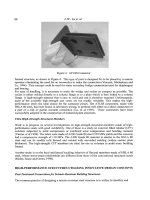
2. The Related Fundamental Theories
The shape of a liquid droplets on solid surface, may be flat, hemisphere or spherical, and is
governed by the surface tensions. Figure 3 showed the two typical states of the liquid
droplet on a solid surface. The surface tensions γ
s-l
and γ
v-l
attempt to make the droplet to
shrink, while the tension γ
s-v
attempts to make the droplet to spread out on the surface.
When the droplets on surface reached equilibrium, the angle between the solid/liquid
interface and the liquid/vapour interface was named as contact angle (θ). The value of the
contact angle describes the degree of the liquid wetting the solid surface. The relationship
between these parameters is commonly given by the famous Young’s equation:
cosθ = (γ
s-v
− γ
s-l
) / γ
v-l
Superhydrophobicity,LearnfromtheLotusLeaf 327
2004). Furthermore, butterflies and cicadas, the evolution bestowed them the self-cleaning
ability which can keep them uncontaminated by removing dust particles, dew or water
droplets easily from their wings, and bestowed them water-repellent ability which can keep
their wings not be wetting in the rain. Many poultry, such as the duck and the swan, have
also the ability that their feathers can resist the water to spread out on the whole body
surfaces when they are floating on the water.
On the surface of the lotus leaves, the almost spherical water droplets will not come to rest
and simply roll off if the surface is tilted even slightly, which is now usually referred to as
the “Lotus Effect”. This effect belongs to the subfield of the wettability of solid surface and
is also named as the “Superhydrophobicity”. The wetting behaviour of solid surfaces by a
liquid is a very important aspect of surface chemistry, which may have a variety of practical
applications. When a liquid droplet contacts a solid substrate, it will either remain as a
droplet or spread out on the surface to form a thin liquid film, a property which is normally
characterized by means of the contact angle measurements. For a solid substrate, when the
contact angle of water or oil on it is larger than 150°, it is called superhydrophobic or
superoleophobic, respectively. On the other hand, when the contact angel of water or oil on
a surface is almost 0°, it is called superhydrophilic or superoleophilic, respectively. Among
the four kinds of surfaces, the superhydrophobic surfaces are referred to as self-cleaning
surfaces and the contamination on them is easily removed by rolling droplets and as such
this type of surface has obviously great potential uses, as water will not “stick” to it.
Fig. 1. (a) An almost ballshaped water droplet on a non-wettable plant leaf (Blossey, 2003).
(b) Low- and (c) high-magnification scanning electron microscope images of the surface
structures on the lotus leaf. Every epidermal cell forms a micrometer-scale papilla and has a
dense layer of epicuticular waxes superimposed on it. Each of the papillae consists of
branchlike nanostructures (Zhai et al., 2002). (Reproduced with permission from the Nature
Publishing Group, Copyright 2003, and from the Chinese Physical Society, Copyright 2002.)
People have noticed these interesting nature phenomena quite a long time, while it is
impossible to find out the essence under the science conditions at ancient time. The
developments of analytical instruments are always promoting the level of human cognition.
In the past two scores years, by means of scanning electron microscope, the studies of
biological surfaces have revealed an incredible microstructural diversity of the outer
surfaces of plants. Not until W. Barthlott and C. Neinhuis, Boon University, Germany, have
research the lotus leaves systematically did people completely realized the mechanism of
the lotus leaves to resist water. Barthlott and coworkers investigated the micro-structure of
the lotus leaves with a scanning electron microscope and hold that the surface roughness in
micro-meter scale papillae and the wax layer of the surface were synergistic bestowed the
superhydrophobicity to the surface of lotus leaves (Barthlott & Neinhuis, 1997). Further,
detailed scanning electron microscopy images of lotus leaves indicated that their surfaces
are composed of micro- and nanometer-scale hierarchical structures, that is, fine-branched
nanostructures (ca. 120 nm) on top of micropapillae (5–9 μm) (Fig. 1b and 1c). The
cooperation of these special double-scale surface structures and hydrophobic cuticular
waxes is believed to be the reason for the superhydrophobicity (
a
Feng et al., 2002; Zhai et al.,
2002). Jiang and coworkers investigated the water strider’s legs by the means of scanning
electron microscope and revealed that the leg is composed of numerous needle-shaped setae
with diameters on the microscale and that each microseta is composed of many elaborate
nanoscale grooves (Fig. 2b and 2c). Such a hierarchical surface structure together with the
hydrophobic, secreted wax is considered to be the origin of the superhydrophobicity of the
water strider’s legs (Gao & Jiang, 2004).
Fig. 2. The non-wetting leg of a water strider. (a) Typical sideview of a maximal-depth
dimple (4.38±0.02 mm) just before the leg pierces the water surface. Inset, water droplet on
a leg; this makes a contact angle of 167.6±4.4°. (b), (c) Scanning electron microscope images
of a leg showing numerous oriented spindly microsetae (b) and the fine nanoscale grooved
structures on a seta (c). Scale bars: (b), 20 μm; (c), 200 nm. (Gao & Jiang, 2004). (Reproduced
with permission from the Nature Publishing Group, Copyright 2004.)
2. The Related Fundamental Theories
The shape of a liquid droplets on solid surface, may be flat, hemisphere or spherical, and is
governed by the surface tensions. Figure 3 showed the two typical states of the liquid
droplet on a solid surface. The surface tensions γ
s-l
and γ
v-l
attempt to make the droplet to
shrink, while the tension γ
s-v
attempts to make the droplet to spread out on the surface.
When the droplets on surface reached equilibrium, the angle between the solid/liquid
interface and the liquid/vapour interface was named as contact angle (θ). The value of the
contact angle describes the degree of the liquid wetting the solid surface. The relationship
between these parameters is commonly given by the famous Young’s equation:
cosθ = (γ
s-v
− γ
s-l
) / γ
v-l
Biomimetics,LearningfromNature328
Fig. 3. The two typical states of the liquid droplets on a solid surface.
The Young’s equation can be only applied for the chemical homogeneous and ideal flat
surfaces. In actuality, few solid surfaces are truly flat, therefore, the surface roughness factor
must be considered during the evaluation of the surface wettability. Wenzel and Cassie have
developed Young’s equation and worked out the Wenzel’s equation and Cassie’s equation,
respectively. The two equations are commonly used to correlate the surface roughness with
the contact angle of a liquid droplet on a solid surface. This improvement has made their
application scope more wide than the Young’s equation.
In 1936, Wenzel found that the surface roughness must be considered during the evaluation
of the surface wettability (Wenzel, 1936). He hold that the liquid completely fills the grooves
of the rough surface where they contact (Fig. 4a). The situation is described by equation:
cosθ
W
= r (γ
s-v
− γ
s-l
) / γ
v-l
= r cosθ
where
θ
W
is the contact angle in the Wenzel mode and r is the surface roughness factor.
From this equation, it can be found that if the contact angle of a liquid on a smooth surface is
less than 90°, the contact angle on a rough surface will be smaller, while the contact angle of
a liquid on a smooth surface is more than 90°, the angle on a rough surface will be larger.
These two situations can be described as: for
θ < 90°, θ
W
< θ; for θ > 90°, θ
W
> θ.
In 1944, based on Wenzel’s model, Cassie further developed and revised the Young’s
equation. He presented that the solids rough surface should be regarded as a solid-vapour
composite interface and the vapour pockets were assumed to be trapped underneath the
liquid (Fig. 4b). In this case, the solid-liquid-vapour three phase contact area can be
represented by the f
s
and f
v
, which are the area fractions of the solid and vapour on the
composite surface. Defining the contact angle in the Cassie mode as
θ
C
, θ
C
can be correlated
to the chemical heterogeneity of a rough surface by equation:
cosθ
C
= f
s
cosθ
s
+ f
v
cosθ
v
Since f
s
+ f
v
= 1, θ
s
=θ, θ
v
= 180°, the above equation can be written as equation:
cosθ
C
= f
s
(cosθ + 1) – 1
From the above equation it can be easily found that for a true contact angle more than 90°,
the surface roughness will increase the apparent angle. This is unlike the Wenzel case,
because even when the intrinsic contact angle of a liquid on a smooth surface is less than 90°,
the contact angle can still be enhanced as a result of the as trapped superhydrophobic
vapour pockets.
Fig. 4. (a) Wetted contact between the liquid and the rough substrate (Wenzel’s model). (b)
Non-wetted contact between the liquid and the rough substrate (Cassie’s model).
The achievements of the Wenzel’s and Cassie’s models are that they have expressed the
contact state between the liquid and the rough solid surface more realistically and exactly.
Heretofore, Wenzel’s and Cassie’s models and equations are numerously applied for
illustrating the mechanism of the superhydrophobic surfaces which were prepared by the
material researchers in their articles.
With the emergence of the nanometer materials in 1960’s, it promoted greatly the progress
of the science and technology. Preparation and studies on the surface properties of the
nanomaterials are the foundation of the nanoscience research. The emergence of the
nanometer materials provides a good platform for the biomimetic materials research.
Inspired by the microstructure of the natural water-resister, and based on the rapidly
developed nanoscience and technology, material researchers have strong motivation to
mimic the structure and the chemical component of the lotus leave surface for the
biomimetic preparation of the superhydrophobic materials.
Heretofore, a variety of methods have been reported for constructing superhydrophobic
surfaces by mimicking the surface of lotus leaves. These artificial superhydrophobic surfaces
have been fabricated mostly by controlling the roughness and topography of hydrophobic
surfaces and using techniques such as anodic oxidation, electrodeposition and chemical
etching, plasma etching, laser treating, electrospinning, chemical vapour deposition, sol–gel
processing, phase separation and so on. The materials that were used to fabricate the surface
morphology ranged from carbon nanotubes, nanoparticles and nanofibers, mental oxide
Superhydrophobicity,LearnfromtheLotusLeaf 329
Fig. 3. The two typical states of the liquid droplets on a solid surface.
The Young’s equation can be only applied for the chemical homogeneous and ideal flat
surfaces. In actuality, few solid surfaces are truly flat, therefore, the surface roughness factor
must be considered during the evaluation of the surface wettability. Wenzel and Cassie have
developed Young’s equation and worked out the Wenzel’s equation and Cassie’s equation,
respectively. The two equations are commonly used to correlate the surface roughness with
the contact angle of a liquid droplet on a solid surface. This improvement has made their
application scope more wide than the Young’s equation.
In 1936, Wenzel found that the surface roughness must be considered during the evaluation
of the surface wettability (Wenzel, 1936). He hold that the liquid completely fills the grooves
of the rough surface where they contact (Fig. 4a). The situation is described by equation:
cosθ
W
= r (γ
s-v
− γ
s-l
) / γ
v-l
= r cosθ
where
θ
W
is the contact angle in the Wenzel mode and r is the surface roughness factor.
From this equation, it can be found that if the contact angle of a liquid on a smooth surface is
less than 90°, the contact angle on a rough surface will be smaller, while the contact angle of
a liquid on a smooth surface is more than 90°, the angle on a rough surface will be larger.
These two situations can be described as: for
θ < 90°, θ
W
< θ; for θ > 90°, θ
W
> θ.
In 1944, based on Wenzel’s model, Cassie further developed and revised the Young’s
equation. He presented that the solids rough surface should be regarded as a solid-vapour
composite interface and the vapour pockets were assumed to be trapped underneath the
liquid (Fig. 4b). In this case, the solid-liquid-vapour three phase contact area can be
represented by the f
s
and f
v
, which are the area fractions of the solid and vapour on the
composite surface. Defining the contact angle in the Cassie mode as
θ
C
, θ
C
can be correlated
to the chemical heterogeneity of a rough surface by equation:
cosθ
C
= f
s
cosθ
s
+ f
v
cosθ
v
Since f
s
+ f
v
= 1, θ
s
=θ, θ
v
= 180°, the above equation can be written as equation:
cosθ
C
= f
s
(cosθ + 1) – 1
From the above equation it can be easily found that for a true contact angle more than 90°,
the surface roughness will increase the apparent angle. This is unlike the Wenzel case,
because even when the intrinsic contact angle of a liquid on a smooth surface is less than 90°,
the contact angle can still be enhanced as a result of the as trapped superhydrophobic
vapour pockets.
Fig. 4. (a) Wetted contact between the liquid and the rough substrate (Wenzel’s model). (b)
Non-wetted contact between the liquid and the rough substrate (Cassie’s model).
The achievements of the Wenzel’s and Cassie’s models are that they have expressed the
contact state between the liquid and the rough solid surface more realistically and exactly.
Heretofore, Wenzel’s and Cassie’s models and equations are numerously applied for
illustrating the mechanism of the superhydrophobic surfaces which were prepared by the
material researchers in their articles.
With the emergence of the nanometer materials in 1960’s, it promoted greatly the progress
of the science and technology. Preparation and studies on the surface properties of the
nanomaterials are the foundation of the nanoscience research. The emergence of the
nanometer materials provides a good platform for the biomimetic materials research.
Inspired by the microstructure of the natural water-resister, and based on the rapidly
developed nanoscience and technology, material researchers have strong motivation to
mimic the structure and the chemical component of the lotus leave surface for the
biomimetic preparation of the superhydrophobic materials.
Heretofore, a variety of methods have been reported for constructing superhydrophobic
surfaces by mimicking the surface of lotus leaves. These artificial superhydrophobic surfaces
have been fabricated mostly by controlling the roughness and topography of hydrophobic
surfaces and using techniques such as anodic oxidation, electrodeposition and chemical
etching, plasma etching, laser treating, electrospinning, chemical vapour deposition, sol–gel
processing, phase separation and so on. The materials that were used to fabricate the surface
morphology ranged from carbon nanotubes, nanoparticles and nanofibers, mental oxide
Biomimetics,LearningfromNature330
nanorods, polymers to engineering alloys materials. In the following text, some most
common and important preparation methods and the categories of the artificial
superhydrophobic surfaces are introduced.
3. Methods for the Preparation of the Superhydrophobic Surfaces
3.1 Layer-by-Layer and colloidal assembly
The Layer-by-Layer assembly technique, which was developed by Decher’s group, has been
proved to be a simple and inexpensive way to build controllable chemical composition and
micro- and nanometer scale (Decher & Hong, 1991). The greatest strength of the Lay-by-
Layer technique is to control the thickness and the chemical properties of the thin film in
molecular level by virtue of the electrostatic interaction and the hydrogen bond interaction
between the molecules. Cohen, Rubner and coworkers prepared a surface structure that
mimics the water harvesting wing surface of the Namib Desert beetle by means of Lay-by-
Layer technique. The Stenocara beetle, which lived in the areas of limited water, uses their
hydrophilic/superhydrophobic patterned surface of its wings to collect drinking water from
fog-laden wind. In a foggy dawn, the Stenocara beetle tilts its body forward into the wind to
capture small water droplets in the fog. After these small water droplets coalesce into bigger
droplets, they roll down into the beetle’s mouth, providing the beetle with a fresh morning
drink. Cohen, Rubner and coworkers created the hydrophilic patterns on superhydrophobic
surfaces by selectively delivering polyelectrolytes to the surface in a mixed water/2-
propanol solvent to produce surfaces with extreme hydrophobic contrast (Zhai et al., 2006).
Potential applications of such surfaces include water harvesting surfaces, controlled drug
release coatings, open-air microchannel devices, and lab-on-chip devices. Sun and
coworkers reported a facile method for preparing a superhydrophobic surface was
developed by layer-by-layer deposition of poly(diallyldimethylammonium
chloride)/sodium silicate multilayer films on a silica-sphere-coated substrate followed with
a fluorination treatment. The superhydrophobic surface has a water contact angle of 157.1°
and sliding angle of 3.1° (Zhang et al., 2007). The easy availability of the materials and
simplicity of this method might make the superhydrophobic surface potentially useful in a
variety of applications.
3.2 Electrochemical reaction and deposition
The electrochemical reaction and the electrochemical deposition are widely used for the
preparation of the superhydrophobic materials. Zhang and coworkers reported a surface
covered with dendritic gold clusters, which is formed by electrochemical deposition onto an
indium tin oxide electrode modified with a polyelectrolyte multilayer, shows
superhydrophobic properties after further chemisorption of a self-assembled monolayer of
n-dodecanethiol (Zhang et al., 2004). When the deposition time exceeds 1000s, the contact
angle reaches a constant value as high as 156°. Yan, Tusjii and coworkers reported a
poly(alkylpyrrole) conductive films with a water contact angle larger than 150° (Fig. 5). The
films were obtained by electrochemical polymerization of alkylpyrrole and are stable to
temperature, organic solvents and oils. The surface of the film is a fractal and consists of an
array of perpendicular needle-like structures (Yan et al., 2005).
Fig. 5. Scanning electron microscopic image of the super water-repellent poly(alkylpyrrole)
film (scale bar: 15 μm). Left inset: scanning electron microscopic image of the cross section of
the film (bar: 15 μm). Right inset: image of a water droplet on the film (bar: 500 μm) (Yan et
al., 2005). (Reproduced with permission from Wiley-VCH Verlag GmbH & Co. KGaA,
Copyright 2005.)
Our group reported a Pt nanowire array superhydrophobic surface on a Ti/Si substrate by
utilizing electrodeposition of Pt into the pores of anodic aluminium oxide templates and
surface fluorination. The method can be extended to other metals to which the recently
developed chemical etching method is not applicable (
a
Qu et al., 2008). Zhou and coworkers
reported a fabrication of superhydrophobic materials with a water contact angle of 178°
using a perpendicular brucite-type cobalt hydroxide nanopin film fabricated with a bottom-
up process (Fig. 6) (Hosono et al., 2005).
Fig. 6. (a,b) Field-emission scanning electron microscopic images of the brucite-type cobalt
hydroxide films observed from the top and side, respectively. (c) Transmission electron
microscope images of the films. (d) A simple model of the film with the fractal structure.
Inset: image of a water droplet on the film with a contact angle of 178° (Hosono et al., 2005).
(Reproduced with permission from the American Chemical Society, Copyright 2005.)
3.3 Sol-Gel Processing
For many materials, the sol-gel processing can also bestow the surface superhydrophobicty.
Many research results showed that the surfaces can be made superhydrophobic while it
Superhydrophobicity,LearnfromtheLotusLeaf 331
nanorods, polymers to engineering alloys materials. In the following text, some most
common and important preparation methods and the categories of the artificial
superhydrophobic surfaces are introduced.
3. Methods for the Preparation of the Superhydrophobic Surfaces
3.1 Layer-by-Layer and colloidal assembly
The Layer-by-Layer assembly technique, which was developed by Decher’s group, has been
proved to be a simple and inexpensive way to build controllable chemical composition and
micro- and nanometer scale (Decher & Hong, 1991). The greatest strength of the Lay-by-
Layer technique is to control the thickness and the chemical properties of the thin film in
molecular level by virtue of the electrostatic interaction and the hydrogen bond interaction
between the molecules. Cohen, Rubner and coworkers prepared a surface structure that
mimics the water harvesting wing surface of the Namib Desert beetle by means of Lay-by-
Layer technique. The Stenocara beetle, which lived in the areas of limited water, uses their
hydrophilic/superhydrophobic patterned surface of its wings to collect drinking water from
fog-laden wind. In a foggy dawn, the Stenocara beetle tilts its body forward into the wind to
capture small water droplets in the fog. After these small water droplets coalesce into bigger
droplets, they roll down into the beetle’s mouth, providing the beetle with a fresh morning
drink. Cohen, Rubner and coworkers created the hydrophilic patterns on superhydrophobic
surfaces by selectively delivering polyelectrolytes to the surface in a mixed water/2-
propanol solvent to produce surfaces with extreme hydrophobic contrast (Zhai et al., 2006).
Potential applications of such surfaces include water harvesting surfaces, controlled drug
release coatings, open-air microchannel devices, and lab-on-chip devices. Sun and
coworkers reported a facile method for preparing a superhydrophobic surface was
developed by layer-by-layer deposition of poly(diallyldimethylammonium
chloride)/sodium silicate multilayer films on a silica-sphere-coated substrate followed with
a fluorination treatment. The superhydrophobic surface has a water contact angle of 157.1°
and sliding angle of 3.1° (Zhang et al., 2007). The easy availability of the materials and
simplicity of this method might make the superhydrophobic surface potentially useful in a
variety of applications.
3.2 Electrochemical reaction and deposition
The electrochemical reaction and the electrochemical deposition are widely used for the
preparation of the superhydrophobic materials. Zhang and coworkers reported a surface
covered with dendritic gold clusters, which is formed by electrochemical deposition onto an
indium tin oxide electrode modified with a polyelectrolyte multilayer, shows
superhydrophobic properties after further chemisorption of a self-assembled monolayer of
n-dodecanethiol (Zhang et al., 2004). When the deposition time exceeds 1000s, the contact
angle reaches a constant value as high as 156°. Yan, Tusjii and coworkers reported a
poly(alkylpyrrole) conductive films with a water contact angle larger than 150° (Fig. 5). The
films were obtained by electrochemical polymerization of alkylpyrrole and are stable to
temperature, organic solvents and oils. The surface of the film is a fractal and consists of an
array of perpendicular needle-like structures (Yan et al., 2005).
Fig. 5. Scanning electron microscopic image of the super water-repellent poly(alkylpyrrole)
film (scale bar: 15 μm). Left inset: scanning electron microscopic image of the cross section of
the film (bar: 15 μm). Right inset: image of a water droplet on the film (bar: 500 μm) (Yan et
al., 2005). (Reproduced with permission from Wiley-VCH Verlag GmbH & Co. KGaA,
Copyright 2005.)
Our group reported a Pt nanowire array superhydrophobic surface on a Ti/Si substrate by
utilizing electrodeposition of Pt into the pores of anodic aluminium oxide templates and
surface fluorination. The method can be extended to other metals to which the recently
developed chemical etching method is not applicable (
a
Qu et al., 2008). Zhou and coworkers
reported a fabrication of superhydrophobic materials with a water contact angle of 178°
using a perpendicular brucite-type cobalt hydroxide nanopin film fabricated with a bottom-
up process (Fig. 6) (Hosono et al., 2005).
Fig. 6. (a,b) Field-emission scanning electron microscopic images of the brucite-type cobalt
hydroxide films observed from the top and side, respectively. (c) Transmission electron
microscope images of the films. (d) A simple model of the film with the fractal structure.
Inset: image of a water droplet on the film with a contact angle of 178° (Hosono et al., 2005).
(Reproduced with permission from the American Chemical Society, Copyright 2005.)
3.3 Sol-Gel Processing
For many materials, the sol-gel processing can also bestow the surface superhydrophobicty.
Many research results showed that the surfaces can be made superhydrophobic while it
Biomimetics,LearningfromNature332
needs not the surface hydrophobic process after the sol-gel processing because that the low
surface energy materials already exist in the sol-gel process. Shirtcliffe and coworkers
reported superhydrophobic foams with contact angles greater than 150° which were
prepared using a sol-gel phase-separation process. A rapid hydrophobic to hydrophilic
transition was presented in the surface at around 400 °C, generating a material that
absorbed water rapidly (Shirtcliffe et al., 2003). Cho and coworkers reported a fabrication of
superhydrophobic surface from a supramolecular organosilane with quadruple hydrogen
bonding by a simple sol-gel processing at room temperature. Compared with other template
syntheses, this approach to fabricating a phase-separated continuous material is a very
simple way of producing a superhydrophobic coating and is made possible by the
supramolecular characteristics of the novel organosilane (Han et al., 2004). Wu and
coworkers prepared the ZnO surface with micro- and nanostructure via a wet chemical
route. The surface showed superhydrophobic after the surface chemical modification with
the moderate-length alkanoic acids (Wu et al., 2005).
3.4 Etching and Lithography
Etching is the most efficient way for the construction of rough surface. The detailed methods
are plasma etching, laser etching, chemical etching et al. These methods have been greatly
applied for the biomimic fabrication of the superhydrophobic surface. Teshima and
coworkers formed a ultra water-repellent polymer sheets on a poly(ethylene terephthalate)
substrate. Its nanotexture was formed on a poly(ethylene terephthalate) substrate surface via
selective oxygen plasma etching and subsequent hydrophobic coating by means of low
temperature chemical vapor deposition or plasma-enhanced chemical vapour deposition
(Teshima et al., 2005). The as-prepared polymer sheets are transparent and ultra water-
repellent, showing a water contact angle greater than 150°. Shen and coworkers reported
fabrication of superhydrophobic surfaces by a dislocation-selective chemical etching on
aluminium, copper, and zinc substrates (Qian & Shen, 2005). Our group developed a
solution-immersion process to fabricate of superhydrophobic surfaces on engineering
materials, such as steel, copper alloy and titanium alloy by wet chemical etching and surface
coating with fluoroalkylsilane (Qu et al., 2007). The synergistic effect of the two-lengthscale
surface microstructures and the low surface energy of the fluorinated surface are considered
to be responsible for this superhydrophobicity. Compared with the other methods, it is
convenient, time-saving, and inexpensive. The as-fabricated superhydrophobic surfaces
show long-term stability and are able to withstand salt solutions in a wide range of
concentrations.
For the fabrication of large proportion and periodic micro- and nanopatterns, lithography,
such as the electronic beam lithography, light lithography, X-ray lithography and
nanospheres lithography, are fairly good methods. Riehle and coworkers fabricated ordered
arrays of nanopits and nanopillars by an electronic beam writer with the desired pattern and
investigated their dynamic wettability before and after chemical hydrophobization
(Martines et al., 2007). These ordered patterns showed superhydrophobic after the surfaces
were coated with octadecyltricholorosilane. Tatsuma and coworkers reported
superhydrophobic and superhydrophilic gold surfaces which were prepared by modifying
microstructured gold surfaces with thiols (Notsu et al., 2005). The patterns required by the
superhydrophobic surface were obtained by photocatalytic lithography using a TiO
2
-coated
photomask. The perfluorodecanethiol modified rough gold surface can be converted from
superhydrophobic to superhydrophilic by photocatalytic remote oxidation using the TiO
2
film. On the basis of this technique, enzymes and algal cells can be patterned on the gold
surfaces to fabricate biochips.
3.5 Chemical Vapor Deposition and Physical Vapor Deposition
The chemical and physical vapour depositions have been also widely used for the
nanostructure fabrication and the chemical modification in the surface chemistry. Lau and
coworkers deposited vertically aligned carbon nanotube forest with a plasma enhanced
chemical vapor deposition technique, which is a fairly good technique that produces
perfectly aligned, untangled (i.e., individually standing) carbon nanotubes whose height
and diameter can be conveniently controlled (Lau et al., 2003). While after the depositing a
thin hydrophobic poly(tetrafluoroethylene) coating on the surface of the nanotubes through
a hot filament chemical vapor deposition process, the surface showed stable
superhydrophobicty with advancing and receding contact angles are 170° and 160°,
respectively. Furthermore, Lau and coworkers also reported a formation of a stable
superhydrophobic surface via aligned carbon nanotubes coated with a zinc oxide thin film.
The carbon nanotubes template was synthesized by chemical vapor deposition on a Fe−N
catalyst layer. The ZnO film, with a low surface energy, was deposited on the carbon
nanotubes template by the filtered cathodic vacuum arc technique. The ZnO-coated carbon
nanotubes surface shows no sign of water seepage even after a prolonged period of time.
The wettability of the surface can be reversibly changed from superhydrophobicity to
hydrophilicity by alternation of ultraviolet irradiation and dark storage. Contact angle
measurement reveals that the surface of the ZnO-coated carbon nanotubes is
superhydrophobic with water contact angle of 159° (Huang et al., 2005). Jiang and
coworkers demonstrated a honeycomb-like aligned carbon nanotube films which were
grown by pyrolysis of iron phthalocyanine in the Ar/H
2
atmosphere by the physical vapour
deposition (Li et al., 2002). Wettability studies revealed the film surface showed a
superhydrophobic property with much higher contact angle (163.4 ± 1.4°) and lower sliding
angle (less than 5°).
3.6 Electrospinning
Electrospinning is a very good method for the fabrication of the ultra-thin fibers. Heretofore,
many groups have applied this technique to the preparation of the superhydrophobic
surfaces. The merit of electrospinning is that the superhydrophobic surface can be obtained
within one step. Rutledge and coworkers produced a block copolymer poly(styrene-b-
dimethylsiloxane) fibers via electrospinning from solution in tetrahydrofuran and
dimethylformamide (Ma et al., 2005). The submicrometer diameters of the fibers were in the
range 150–400 nm and the contact angle measurements indicate that the nonwoven fibrous
mats are superhydrophobic, with a contact angle of 163°. Jiang and coworkers reported a
polyaniline/polystyrene composite film which was prepared via the simple electrospinning
method (Zhu et al., 2006). The as-prepared superhydrophobic surface showed stable
superhydrophobicity and conductivity, even in many corrosive solutions, such as acidic or
basic solutions over a wide pH range, and also in oxidizing solutions.
Superhydrophobicity,LearnfromtheLotusLeaf 333
needs not the surface hydrophobic process after the sol-gel processing because that the low
surface energy materials already exist in the sol-gel process. Shirtcliffe and coworkers
reported superhydrophobic foams with contact angles greater than 150° which were
prepared using a sol-gel phase-separation process. A rapid hydrophobic to hydrophilic
transition was presented in the surface at around 400 °C, generating a material that
absorbed water rapidly (Shirtcliffe et al., 2003). Cho and coworkers reported a fabrication of
superhydrophobic surface from a supramolecular organosilane with quadruple hydrogen
bonding by a simple sol-gel processing at room temperature. Compared with other template
syntheses, this approach to fabricating a phase-separated continuous material is a very
simple way of producing a superhydrophobic coating and is made possible by the
supramolecular characteristics of the novel organosilane (Han et al., 2004). Wu and
coworkers prepared the ZnO surface with micro- and nanostructure via a wet chemical
route. The surface showed superhydrophobic after the surface chemical modification with
the moderate-length alkanoic acids (Wu et al., 2005).
3.4 Etching and Lithography
Etching is the most efficient way for the construction of rough surface. The detailed methods
are plasma etching, laser etching, chemical etching et al. These methods have been greatly
applied for the biomimic fabrication of the superhydrophobic surface. Teshima and
coworkers formed a ultra water-repellent polymer sheets on a poly(ethylene terephthalate)
substrate. Its nanotexture was formed on a poly(ethylene terephthalate) substrate surface via
selective oxygen plasma etching and subsequent hydrophobic coating by means of low
temperature chemical vapor deposition or plasma-enhanced chemical vapour deposition
(Teshima et al., 2005). The as-prepared polymer sheets are transparent and ultra water-
repellent, showing a water contact angle greater than 150°. Shen and coworkers reported
fabrication of superhydrophobic surfaces by a dislocation-selective chemical etching on
aluminium, copper, and zinc substrates (Qian & Shen, 2005). Our group developed a
solution-immersion process to fabricate of superhydrophobic surfaces on engineering
materials, such as steel, copper alloy and titanium alloy by wet chemical etching and surface
coating with fluoroalkylsilane (Qu et al., 2007). The synergistic effect of the two-lengthscale
surface microstructures and the low surface energy of the fluorinated surface are considered
to be responsible for this superhydrophobicity. Compared with the other methods, it is
convenient, time-saving, and inexpensive. The as-fabricated superhydrophobic surfaces
show long-term stability and are able to withstand salt solutions in a wide range of
concentrations.
For the fabrication of large proportion and periodic micro- and nanopatterns, lithography,
such as the electronic beam lithography, light lithography, X-ray lithography and
nanospheres lithography, are fairly good methods. Riehle and coworkers fabricated ordered
arrays of nanopits and nanopillars by an electronic beam writer with the desired pattern and
investigated their dynamic wettability before and after chemical hydrophobization
(Martines et al., 2007). These ordered patterns showed superhydrophobic after the surfaces
were coated with octadecyltricholorosilane. Tatsuma and coworkers reported
superhydrophobic and superhydrophilic gold surfaces which were prepared by modifying
microstructured gold surfaces with thiols (Notsu et al., 2005). The patterns required by the
superhydrophobic surface were obtained by photocatalytic lithography using a TiO
2
-coated
photomask. The perfluorodecanethiol modified rough gold surface can be converted from
superhydrophobic to superhydrophilic by photocatalytic remote oxidation using the TiO
2
film. On the basis of this technique, enzymes and algal cells can be patterned on the gold
surfaces to fabricate biochips.
3.5 Chemical Vapor Deposition and Physical Vapor Deposition
The chemical and physical vapour depositions have been also widely used for the
nanostructure fabrication and the chemical modification in the surface chemistry. Lau and
coworkers deposited vertically aligned carbon nanotube forest with a plasma enhanced
chemical vapor deposition technique, which is a fairly good technique that produces
perfectly aligned, untangled (i.e., individually standing) carbon nanotubes whose height
and diameter can be conveniently controlled (Lau et al., 2003). While after the depositing a
thin hydrophobic poly(tetrafluoroethylene) coating on the surface of the nanotubes through
a hot filament chemical vapor deposition process, the surface showed stable
superhydrophobicty with advancing and receding contact angles are 170° and 160°,
respectively. Furthermore, Lau and coworkers also reported a formation of a stable
superhydrophobic surface via aligned carbon nanotubes coated with a zinc oxide thin film.
The carbon nanotubes template was synthesized by chemical vapor deposition on a Fe−N
catalyst layer. The ZnO film, with a low surface energy, was deposited on the carbon
nanotubes template by the filtered cathodic vacuum arc technique. The ZnO-coated carbon
nanotubes surface shows no sign of water seepage even after a prolonged period of time.
The wettability of the surface can be reversibly changed from superhydrophobicity to
hydrophilicity by alternation of ultraviolet irradiation and dark storage. Contact angle
measurement reveals that the surface of the ZnO-coated carbon nanotubes is
superhydrophobic with water contact angle of 159° (Huang et al., 2005). Jiang and
coworkers demonstrated a honeycomb-like aligned carbon nanotube films which were
grown by pyrolysis of iron phthalocyanine in the Ar/H
2
atmosphere by the physical vapour
deposition (Li et al., 2002). Wettability studies revealed the film surface showed a
superhydrophobic property with much higher contact angle (163.4 ± 1.4°) and lower sliding
angle (less than 5°).
3.6 Electrospinning
Electrospinning is a very good method for the fabrication of the ultra-thin fibers. Heretofore,
many groups have applied this technique to the preparation of the superhydrophobic
surfaces. The merit of electrospinning is that the superhydrophobic surface can be obtained
within one step. Rutledge and coworkers produced a block copolymer poly(styrene-b-
dimethylsiloxane) fibers via electrospinning from solution in tetrahydrofuran and
dimethylformamide (Ma et al., 2005). The submicrometer diameters of the fibers were in the
range 150–400 nm and the contact angle measurements indicate that the nonwoven fibrous
mats are superhydrophobic, with a contact angle of 163°. Jiang and coworkers reported a
polyaniline/polystyrene composite film which was prepared via the simple electrospinning
method (Zhu et al., 2006). The as-prepared superhydrophobic surface showed stable
superhydrophobicity and conductivity, even in many corrosive solutions, such as acidic or
basic solutions over a wide pH range, and also in oxidizing solutions.
Biomimetics,LearningfromNature334
4. The Category of the Artificial Superhydrophobic Materials
4.1 Carbon nanotubes
Carbon nanotubes are new type of carbon structures which was discovered in 1991. Due to
their excellent electrical and mechanical properties, the carbon nanotubes are widely used in
both fundamental and applied research. Jiang and coworkers prepared an aligned carbon
nanotubes films with micro- and nanometer structure. The aligned carbon nanotube films
showed superamphiphobic properties after the surface modification with a fluoroalkylsilane
coating. The surface showed high contact angles for both water and rapeseed oil on the film
and the values of the contact angles were 171° and 161°, respectively (Li et al., 2001). Lau
and coworkers demonstrated a creation of a stable, superhydrophobic surface using the
nanoscale roughness inherent in a vertically aligned carbon nanotube forest together with a
thin, conformal hydrophobic poly(tetrafluoroethylene) coating on the surface of the
nanotubes (Lau et al., 2003).
4.2 Metallic compounds nanorods and nanoparticles
Fig. 7. A metallic model “pond skater” (body length 28 mm) standing on a water surface.
Note the deformation of the water surface around the legs (Larmour et al., 2007).
(Reproduced with permission from Wiley-VCH Verlag GmbH & Co. KGaA, Copyright 2007.)
With the development of the research on inorganic materials, the superhydrophobic
inorganic materials were also reported numerously. For example, ZnO is a novel II - IV
semiconductor material with a direct bandgap of 3.2 eV, excellent lattice, photovoltaic,
pizeoelectric and dielectric properties, and it is non-toxic and low cost from cheap and
abundant raw materials. Jiang and coworkers reported a controllable wettability of aligned
ZnO nanorod films. This inorganic oxide films show superhydrophobicity and
superhydrophilicity at different conditions, and the wettability can be reversibly switched
by alternation of ultraviolet irradiation and dark storage (Feng et al., 2003). This effect is
believed to be due to the cooperation of the surface photosensitivity and the aligned
nanostructure of the films. Such special wettability will greatly extend the applications of
ZnO films to many other important fields. Futherore, Jiang and coworkers deposited similar
TiO
2
nanorod films and aligned SnO
2
nanorod films on glass substrates for the preparation
of the superhydrophobic surface. The two kinds of superhydrophobic surfaces can all be
switched between superhydrophobicity and superhydrophilicity by the alternation of
ultraviolet irradiation and dark storage (Feng et al., 2005; Zhu et al., 2006). Bell and
coworkers reported a remarkably straightforward method for treating metals uses
electroless galvanic deposition to coat a metal substrate with a textured layer of a second
metal to fabricate superhydrophobic surfaces on metal surface (Larmour et al., 2007). The
process is carried out under ambient conditions using readily available starting materials
and laboratory equipment. The as-prepared superhydrophobic surfaces show
approximately 180° contact angle. It is very striking and interesting that they have applied
this preparation method to the four legs of a metallic model “pond skater” (Gerridae) and
made this metallic model with the capacity of floating on the water (Fig. 7).
4.3 Engineering Alloy Materials
Fig. 8. Image of water droplets with different sizes on the superhydrophobic surface of steel
having a contact angle of 161 ± 1° and on the superhydrophobic surface of copper alloy with
a contact angle of 158 ± 1° respectively (Qu et al., 2007). (Reproduced with permission from
Wiley-VCH Verlag GmbH & Co. KGaA, Copyright 2007.)
Engineering materials, such as steel, aluminium and its alloy, copper alloy and titanium
alloy, have diverse technological applications in the marine, auto, aviation, and space
industries. Superhydrophobicity will greatly extend their applications as engineering
materials. Liu and coworkers reported a simple and inexpensive method to produce super-
hydrophobic surfaces on aluminium and its alloy by oxidation and chemical modification
(Guo et al., 2005). The superhydrophobic surfaces show long-term stability overall wide pH
range. Our group reported a novel mixed-solution system for the fabrication of
superhydrophobic surfaces on steel, copper alloy and titanium alloy by a chemical etching
method (Fig. 8). The superhydrophobic surfaces are able to withstand salt solutions in a
wide range of concentrations, which may open a new avenue in applications especially for
the marine engineering materials where salt resistance is required. We expect that this
technique will accelerate the large-scale production of superhydrophobic engineering
materials with new industrial applications (Qu et al., 2007).
Superhydrophobicity,LearnfromtheLotusLeaf 335
4. The Category of the Artificial Superhydrophobic Materials
4.1 Carbon nanotubes
Carbon nanotubes are new type of carbon structures which was discovered in 1991. Due to
their excellent electrical and mechanical properties, the carbon nanotubes are widely used in
both fundamental and applied research. Jiang and coworkers prepared an aligned carbon
nanotubes films with micro- and nanometer structure. The aligned carbon nanotube films
showed superamphiphobic properties after the surface modification with a fluoroalkylsilane
coating. The surface showed high contact angles for both water and rapeseed oil on the film
and the values of the contact angles were 171° and 161°, respectively (Li et al., 2001). Lau
and coworkers demonstrated a creation of a stable, superhydrophobic surface using the
nanoscale roughness inherent in a vertically aligned carbon nanotube forest together with a
thin, conformal hydrophobic poly(tetrafluoroethylene) coating on the surface of the
nanotubes (Lau et al., 2003).
4.2 Metallic compounds nanorods and nanoparticles
Fig. 7. A metallic model “pond skater” (body length 28 mm) standing on a water surface.
Note the deformation of the water surface around the legs (Larmour et al., 2007).
(Reproduced with permission from Wiley-VCH Verlag GmbH & Co. KGaA, Copyright 2007.)
With the development of the research on inorganic materials, the superhydrophobic
inorganic materials were also reported numerously. For example, ZnO is a novel II - IV
semiconductor material with a direct bandgap of 3.2 eV, excellent lattice, photovoltaic,
pizeoelectric and dielectric properties, and it is non-toxic and low cost from cheap and
abundant raw materials. Jiang and coworkers reported a controllable wettability of aligned
ZnO nanorod films. This inorganic oxide films show superhydrophobicity and
superhydrophilicity at different conditions, and the wettability can be reversibly switched
by alternation of ultraviolet irradiation and dark storage (Feng et al., 2003). This effect is
believed to be due to the cooperation of the surface photosensitivity and the aligned
nanostructure of the films. Such special wettability will greatly extend the applications of
ZnO films to many other important fields. Futherore, Jiang and coworkers deposited similar
TiO
2
nanorod films and aligned SnO
2
nanorod films on glass substrates for the preparation
of the superhydrophobic surface. The two kinds of superhydrophobic surfaces can all be
switched between superhydrophobicity and superhydrophilicity by the alternation of
ultraviolet irradiation and dark storage (Feng et al., 2005; Zhu et al., 2006). Bell and
coworkers reported a remarkably straightforward method for treating metals uses
electroless galvanic deposition to coat a metal substrate with a textured layer of a second
metal to fabricate superhydrophobic surfaces on metal surface (Larmour et al., 2007). The
process is carried out under ambient conditions using readily available starting materials
and laboratory equipment. The as-prepared superhydrophobic surfaces show
approximately 180° contact angle. It is very striking and interesting that they have applied
this preparation method to the four legs of a metallic model “pond skater” (Gerridae) and
made this metallic model with the capacity of floating on the water (Fig. 7).
4.3 Engineering Alloy Materials
Fig. 8. Image of water droplets with different sizes on the superhydrophobic surface of steel
having a contact angle of 161 ± 1° and on the superhydrophobic surface of copper alloy with
a contact angle of 158 ± 1° respectively (Qu et al., 2007). (Reproduced with permission from
Wiley-VCH Verlag GmbH & Co. KGaA, Copyright 2007.)
Engineering materials, such as steel, aluminium and its alloy, copper alloy and titanium
alloy, have diverse technological applications in the marine, auto, aviation, and space
industries. Superhydrophobicity will greatly extend their applications as engineering
materials. Liu and coworkers reported a simple and inexpensive method to produce super-
hydrophobic surfaces on aluminium and its alloy by oxidation and chemical modification
(Guo et al., 2005). The superhydrophobic surfaces show long-term stability overall wide pH
range. Our group reported a novel mixed-solution system for the fabrication of
superhydrophobic surfaces on steel, copper alloy and titanium alloy by a chemical etching
method (Fig. 8). The superhydrophobic surfaces are able to withstand salt solutions in a
wide range of concentrations, which may open a new avenue in applications especially for
the marine engineering materials where salt resistance is required. We expect that this
technique will accelerate the large-scale production of superhydrophobic engineering
materials with new industrial applications (Qu et al., 2007).
Biomimetics,LearningfromNature336
4.4 Polymer Materials
Jiang and coworkers synthesized superhydrophobic needle-like polyacrylonitrile nanofibers
via extrusion of the polyacrylonitrile precursor solution into the solidifying solution under
pressure. The aligned nanofibers with different diameters and densities can be easily
obtained by using anodic aluminium oxide membrane with different pore diameters, and
the alignment process can be applied to different polymer precursors such as poly(vinyl
alcohol), polystyrene, polyesters, and polyamides (
b
Feng et al., 2002). The
superhydrophobicity is believed that not only the nanostructure of the nanofibers but also
their lower density contributes to the very large fraction of air in the surface. McCarthy and
coworkers fabricated superhydrophobic polypropylene surfaces by the simultaneous
etching of polypropylene and etching/sputtering of poly(tetrafluoroethylene) using
inductively coupled radio frequency argon plasma. The as-prepared surfaces showed
superhydrophobicity with a water contact angle of 172° (Youngblood & McCarthy, 1999).
Shimomura and coworkers fabricated a honeycomb patterned fluorinated polymer films by
casting of the polymer solution under humid conditions. Such honeycomb patterned films
have application as transparent and superhydrophobic polymer films and it films can be
formed from a large variety of materials and on a wide variety of substrates (Yabu &
Shimomura, 2005). Our group prepared a polymer superhydrophobic surface on Ti/Si
substrates via the fabrication of conductive polyaniline nanowire film. The polyaniline
nanowire film was synthesized by electrodeposition of aniline into the pores of an anodic
aluminum oxide template on Ti/Si substrate followed by the removal of the template (
b
Qu
et al., 2008). The surface showed conductivity and superhydrophobicity, even in many
corrosive solutions, such as acidic or basic solutions over a wide pH range. Compared with
the electrospining method, the method in this paper is cheap and time-saving and avoided
high-voltage power, and the method can be easily applied to other conducting polymers.
5. The Superhydrophobic Surfaces Related Properties and Application
With more and more in-depth study on the preparation of the superhydrophobic surfaces,
the materials researchers are not only satisfy with the preparation and the contact model of
the superhydrophobic surface, but the application and the related properties of the
superhydrophobic surfaces. With the increase of the surface roughness, however, the
surface will lost some important properties, such as the optical transparence and the
mechanics property. These unfavorable factors will limit the widespread application of
superhydrophobic surface greatly. Thus more and more groups have devoted to the
preparation of the multi-functional superhydrophobic surfaces.
5.1 The Superhydrophobic Surfaces with the Anticorrosive Property
The pure water (pH value is 7) was commonly used for the contact angle measurements.
Recently, the measurements for contact angel in whole pH range have aroused considerable
interest from many researchers because of the wide application environments of this kind of
superhydrophobic materials. For the engineering materials, undoubtedly, the resistance to
the water or corrosive liquid will greatly enhance their anticorrosive ability, broaden its
application environment and extend their service life. The superhydrophobic surfaces are
able to withstand salt solutions in a wide range of concentrations, which may open a new
avenue in applications especially for the marine engineering materials where salt resistance
is required. Liu’s group and our group reported the superhydrophobic engineering
materials such as the, steel, copper, alloy aluminium and its alloy et al (Guo et al., 2005; Qu
et al., 2007). These superhydrophobic engineering materials showed superhydrophobicity in
nearly the entire pH range, so they can be used in strongly corrosive environments.
Furthermore, graphite carbon has intrinsic thermal and chemical resistance. Jiang and
coworkers reported a nanostructured carbon films by pyrolyzing nanostructured
polyacrylontrile films (Feng et al., 2003). The films also showed superhydrophobicity in
nearly the entire pH range.
5.2 The Superhydrophobic surfaces with the Optical Property
Fig. 9. Image of a glass slide coated with a transparent, superhydrophobic multilayer with
antireflection properties (Bravo et al. 2007). (Reproduced with permission from the
American Chemical Society, Copyright 2007.)
For many devices, such as the car windscreen and the glasses, the optical transparency is a
very special and important property. Preparing the transparent superhydrophobic surface
has aroused considerable interest for many materials researchers. Hydrophobicity and
transparency, however, are two contradictory properties of the surface. Increasing the
surface roughness is beneficial for the hydrophobicity, while the transparency decreases due
to the light-scattering losses. Therefore, controlling of surface roughness to an appropriate
position is to meet the requirements for both the two key factor. Watanabe and coworkers
reported a sol–gel method for producing transparent boehmite films on glass substrates. The
surface roughness could be precisely controlled in the range between 20 and 50 nm
(Nakajima et al. 1999). This method, however, requires as high as 500 °C heating process
(500 °C), which is incompatible with many optical devices. To solve this problem, a
microwave plasma-enhanced chemical vapour deposition process was adapted to prepare
transparent superhydrophobic films at temperatures as low as 100 °C (Hozumi & Takai,
1998; Wu et al. 2002). Jiang and coworkers prepared multifunctional ZnO nanorod films
with visible-light transparency and superhydrophobic properties through controlling the
diameter and length of nanorods using a low-temperature solution approach. The diameter
and the spacing between the nanorods are both less than 100 nm. Such surface
nanostructures are small enough not to give rise to visible light scattering. Cohen, Rubner
and coworkers demonstrate a Layer-by-Layer processing scheme that can be utilized to
Superhydrophobicity,LearnfromtheLotusLeaf 337
4.4 Polymer Materials
Jiang and coworkers synthesized superhydrophobic needle-like polyacrylonitrile nanofibers
via extrusion of the polyacrylonitrile precursor solution into the solidifying solution under
pressure. The aligned nanofibers with different diameters and densities can be easily
obtained by using anodic aluminium oxide membrane with different pore diameters, and
the alignment process can be applied to different polymer precursors such as poly(vinyl
alcohol), polystyrene, polyesters, and polyamides (
b
Feng et al., 2002). The
superhydrophobicity is believed that not only the nanostructure of the nanofibers but also
their lower density contributes to the very large fraction of air in the surface. McCarthy and
coworkers fabricated superhydrophobic polypropylene surfaces by the simultaneous
etching of polypropylene and etching/sputtering of poly(tetrafluoroethylene) using
inductively coupled radio frequency argon plasma. The as-prepared surfaces showed
superhydrophobicity with a water contact angle of 172° (Youngblood & McCarthy, 1999).
Shimomura and coworkers fabricated a honeycomb patterned fluorinated polymer films by
casting of the polymer solution under humid conditions. Such honeycomb patterned films
have application as transparent and superhydrophobic polymer films and it films can be
formed from a large variety of materials and on a wide variety of substrates (Yabu &
Shimomura, 2005). Our group prepared a polymer superhydrophobic surface on Ti/Si
substrates via the fabrication of conductive polyaniline nanowire film. The polyaniline
nanowire film was synthesized by electrodeposition of aniline into the pores of an anodic
aluminum oxide template on Ti/Si substrate followed by the removal of the template (
b
Qu
et al., 2008). The surface showed conductivity and superhydrophobicity, even in many
corrosive solutions, such as acidic or basic solutions over a wide pH range. Compared with
the electrospining method, the method in this paper is cheap and time-saving and avoided
high-voltage power, and the method can be easily applied to other conducting polymers.
5. The Superhydrophobic Surfaces Related Properties and Application
With more and more in-depth study on the preparation of the superhydrophobic surfaces,
the materials researchers are not only satisfy with the preparation and the contact model of
the superhydrophobic surface, but the application and the related properties of the
superhydrophobic surfaces. With the increase of the surface roughness, however, the
surface will lost some important properties, such as the optical transparence and the
mechanics property. These unfavorable factors will limit the widespread application of
superhydrophobic surface greatly. Thus more and more groups have devoted to the
preparation of the multi-functional superhydrophobic surfaces.
5.1 The Superhydrophobic Surfaces with the Anticorrosive Property
The pure water (pH value is 7) was commonly used for the contact angle measurements.
Recently, the measurements for contact angel in whole pH range have aroused considerable
interest from many researchers because of the wide application environments of this kind of
superhydrophobic materials. For the engineering materials, undoubtedly, the resistance to
the water or corrosive liquid will greatly enhance their anticorrosive ability, broaden its
application environment and extend their service life. The superhydrophobic surfaces are
able to withstand salt solutions in a wide range of concentrations, which may open a new
avenue in applications especially for the marine engineering materials where salt resistance
is required. Liu’s group and our group reported the superhydrophobic engineering
materials such as the, steel, copper, alloy aluminium and its alloy et al (Guo et al., 2005; Qu
et al., 2007). These superhydrophobic engineering materials showed superhydrophobicity in
nearly the entire pH range, so they can be used in strongly corrosive environments.
Furthermore, graphite carbon has intrinsic thermal and chemical resistance. Jiang and
coworkers reported a nanostructured carbon films by pyrolyzing nanostructured
polyacrylontrile films (Feng et al., 2003). The films also showed superhydrophobicity in
nearly the entire pH range.
5.2 The Superhydrophobic surfaces with the Optical Property
Fig. 9. Image of a glass slide coated with a transparent, superhydrophobic multilayer with
antireflection properties (Bravo et al. 2007). (Reproduced with permission from the
American Chemical Society, Copyright 2007.)
For many devices, such as the car windscreen and the glasses, the optical transparency is a
very special and important property. Preparing the transparent superhydrophobic surface
has aroused considerable interest for many materials researchers. Hydrophobicity and
transparency, however, are two contradictory properties of the surface. Increasing the
surface roughness is beneficial for the hydrophobicity, while the transparency decreases due
to the light-scattering losses. Therefore, controlling of surface roughness to an appropriate
position is to meet the requirements for both the two key factor. Watanabe and coworkers
reported a sol–gel method for producing transparent boehmite films on glass substrates. The
surface roughness could be precisely controlled in the range between 20 and 50 nm
(Nakajima et al. 1999). This method, however, requires as high as 500 °C heating process
(500 °C), which is incompatible with many optical devices. To solve this problem, a
microwave plasma-enhanced chemical vapour deposition process was adapted to prepare
transparent superhydrophobic films at temperatures as low as 100 °C (Hozumi & Takai,
1998; Wu et al. 2002). Jiang and coworkers prepared multifunctional ZnO nanorod films
with visible-light transparency and superhydrophobic properties through controlling the
diameter and length of nanorods using a low-temperature solution approach. The diameter
and the spacing between the nanorods are both less than 100 nm. Such surface
nanostructures are small enough not to give rise to visible light scattering. Cohen, Rubner
and coworkers demonstrate a Layer-by-Layer processing scheme that can be utilized to
Biomimetics,LearningfromNature338
create transparent superhydrophobic films from SiO
2
nanoparticles of various sizes (Fig. 9).
By controlling the placement and level of aggregation of differently sized nanoparticles
within the resultant multilayer thin film, it is possible to optimize the level of surface
roughness to achieve superhydrophobic behaviour with limited light scattering (Bravo et al.
2007).
5.3 The Superhydrophobic Surfaces with Highly Adhesive Forces
It is easy and acknowledged to image that a surface with a high water contact angle and
small contact area should be associated with a low adhesive force. However, some research
results on superhydrophobic surfaces indicate that it is not true in any situation. Feng, Jiang
and coworkers reported a superhydrophobic aligned polystyrene nanotube layer via a
simple template-wetting method. The surface shows superhydrophobicity, while it can hold
a spherical water droplet even when it is turned upside down (Jin et al., 2005). They hold
that the large contact area (the as-prepared polystyrene films are composed of about 6.76 ×
10
6
nanotubes mm
–2
) induces a strong interacting force between the water droplet and the
polystyrene nanotube films. The mechanism described here is similar to the one geckos use
in nature while the difference is that the latter interactions are between two solids. Liu and
coworkers also reported sticky superhydrophobic surface which were fabricated on
aluminium alloy by suitable aqueous solution to control the surface roughness (
b
Guo & Liu,
2007). These superhydrophobic surfaces with high adhesive force to liquid are expected to
be used as a “mechanical hand” to transfer mini liquid droplets for microsample analyses in
the future.
5.4 The Superhydrophobic Surfaces with High Electrical Conductivity
Electrical conductivity is a very important property required for many kinds of
microelectrical devices, such as field-effect transistors, light-emitting diodes, and thin-film
transistors. In some applications, such as biotechnology, corrosion protection, antistatics,
conductive textiles and antifouling coatings, the superhydrophobic surfaces prepared with
conducting material would be very useful and vital. Our group prepared conductive
superhydrophobic surfaces with polyaniline by means of ananodic deposition technique on
Ti/Si substrate. The method was also general to other conductive polymers, such as
polythiophenes and polypyrroles. The as-prepared surface showed conductivity and
superhydrophobicity, even in many corrosive solutions, such as acidic or basic solutions
over a wide pH range (
b
Qu et al., 2008). Jiang and coworkers prepared conductive
hydrophobic zinc oxide thin films by means of electrochemical deposition (Li et al., 2003).
They expected that the superhydrophobic conductive thin films materials have potential use,
such as microfluidic devices, in the future. In fact, many reported superhydrophobic
surfaces based on the metal and metallic nanomaterials are conductive naturally, while their
intrinsic nature and the potential uses were ignored at the time.
6. Outlook and Summary
In this chapter, we have presented the origin model of the superhydrophobicty in nature,
the lotus leaf. In the following text, the mechanism of the surface resisting water, the recent
studies on the biomimetic preparation and the properties of the superhydrophobic surface
were elaborated.
The superhydrophobic surface, because of the novel aspects of surface physics and
important applications ranging from selfcleaning materials, marine coatings, antiadhesive
coatings, and nanobattery to microfluidic devices, has aroused considerable interest and
resulted in a growing number of reports in the recent years. In addition to the
superhydrophobic research, many and many researcher focused on the other wetting
properties of solid surface, that is, superhydrophilicity, superoleophobicity, and
superoleophilicity. For example, when superhydrophobicity/superoleophobicity or
superhydrophilicity/superoleophobicity coexist, separation of water from oil (or oil from
water) can be realized. Moreover, the smart surfaces whose wettability can be modulated
reversibly between superhydrophobicity and superhydrophilicity or superoleophobicity
and superoleophilicity are attracting more and more researchers devoted to them. Although
it is very difficult that to achieve the superoleophobic surface which resists the oil, such as
the chloroform or hexane, just like the superhydrophobic surfaces resists water, the
preparation of the superoleophobic materials must be a research focus in the near future.
With millions of years of evolution, creatures in nature possess amazing and mysterious
properties that we do not yet know. Therefore, further exploration and explanation of
surfaces with special wetting behavior in nature is also necessary. Learning from nature will
give us inspiration to develop simple and cheap methods to construct biomimetic multi-
functional surfaces and materials.
Acknowledgment
This work was financially supported by the Natural Science Foundation of Shannxi Province,
China (Grant No. 2009JQ2010), the Natural Science Research Project of Education
Department of Shannxi Province, China (Grant No. 09JK580) and the Peiyu Fund of Xi’an
University of Science and Technology (Grant No. 200818).
7. References
Barthlott, W. & Neinhuis, C. (1997). Purity of the Sacred Lotus, or Escape from
Contamination in Biological Surfaces. Planta, 202, 1-8.
Blossey, R. (2003). Self-cleaning surfaces — virtual realities. Nature Materials, 2, 301-306.
Bravo, J.; Zhai, L.; Wu, Z.; Cohen, R. E. & Rubner, M. F. (2007). Transparent
Superhydrophobic Films Based on Silica Nanoparticles. Langmuir, 23, 7293-7298.
Cassie, A. B. D. & Baxter, S. (1944). Wettability of Porous Surfaces. Trans. Faraday Soc., 40,
546-551.
Decher, G. & Hong, J. (1991). Buildup of Ultrathin Multilayer Films by a Self-Assembly
Process. I. Consecutive Adsorption of Anionic and Cationic Bipolar Amphiphiles
on Charged Surfaces. Makromol. Chem. Macromol. Symp., 46, 321-327.
a
Feng, L.; Li, S.; Li, Y.; Li, H.; Zhang, L.; Zhai, J.; Song, Y.; Liu, B.; Jiang, L. & Zhu, D. (2002).
Super-Hydrophobic Surfaces, From Natural to Artificial. Advanced Materials, 14,
1857-1860.
Superhydrophobicity,LearnfromtheLotusLeaf 339
create transparent superhydrophobic films from SiO
2
nanoparticles of various sizes (Fig. 9).
By controlling the placement and level of aggregation of differently sized nanoparticles
within the resultant multilayer thin film, it is possible to optimize the level of surface
roughness to achieve superhydrophobic behaviour with limited light scattering (Bravo et al.
2007).
5.3 The Superhydrophobic Surfaces with Highly Adhesive Forces
It is easy and acknowledged to image that a surface with a high water contact angle and
small contact area should be associated with a low adhesive force. However, some research
results on superhydrophobic surfaces indicate that it is not true in any situation. Feng, Jiang
and coworkers reported a superhydrophobic aligned polystyrene nanotube layer via a
simple template-wetting method. The surface shows superhydrophobicity, while it can hold
a spherical water droplet even when it is turned upside down (Jin et al., 2005). They hold
that the large contact area (the as-prepared polystyrene films are composed of about 6.76 ×
10
6
nanotubes mm
–2
) induces a strong interacting force between the water droplet and the
polystyrene nanotube films. The mechanism described here is similar to the one geckos use
in nature while the difference is that the latter interactions are between two solids. Liu and
coworkers also reported sticky superhydrophobic surface which were fabricated on
aluminium alloy by suitable aqueous solution to control the surface roughness (
b
Guo & Liu,
2007). These superhydrophobic surfaces with high adhesive force to liquid are expected to
be used as a “mechanical hand” to transfer mini liquid droplets for microsample analyses in
the future.
5.4 The Superhydrophobic Surfaces with High Electrical Conductivity
Electrical conductivity is a very important property required for many kinds of
microelectrical devices, such as field-effect transistors, light-emitting diodes, and thin-film
transistors. In some applications, such as biotechnology, corrosion protection, antistatics,
conductive textiles and antifouling coatings, the superhydrophobic surfaces prepared with
conducting material would be very useful and vital. Our group prepared conductive
superhydrophobic surfaces with polyaniline by means of ananodic deposition technique on
Ti/Si substrate. The method was also general to other conductive polymers, such as
polythiophenes and polypyrroles. The as-prepared surface showed conductivity and
superhydrophobicity, even in many corrosive solutions, such as acidic or basic solutions
over a wide pH range (
b
Qu et al., 2008). Jiang and coworkers prepared conductive
hydrophobic zinc oxide thin films by means of electrochemical deposition (Li et al., 2003).
They expected that the superhydrophobic conductive thin films materials have potential use,
such as microfluidic devices, in the future. In fact, many reported superhydrophobic
surfaces based on the metal and metallic nanomaterials are conductive naturally, while their
intrinsic nature and the potential uses were ignored at the time.
6. Outlook and Summary
In this chapter, we have presented the origin model of the superhydrophobicty in nature,
the lotus leaf. In the following text, the mechanism of the surface resisting water, the recent
studies on the biomimetic preparation and the properties of the superhydrophobic surface
were elaborated.
The superhydrophobic surface, because of the novel aspects of surface physics and
important applications ranging from selfcleaning materials, marine coatings, antiadhesive
coatings, and nanobattery to microfluidic devices, has aroused considerable interest and
resulted in a growing number of reports in the recent years. In addition to the
superhydrophobic research, many and many researcher focused on the other wetting
properties of solid surface, that is, superhydrophilicity, superoleophobicity, and
superoleophilicity. For example, when superhydrophobicity/superoleophobicity or
superhydrophilicity/superoleophobicity coexist, separation of water from oil (or oil from
water) can be realized. Moreover, the smart surfaces whose wettability can be modulated
reversibly between superhydrophobicity and superhydrophilicity or superoleophobicity
and superoleophilicity are attracting more and more researchers devoted to them. Although
it is very difficult that to achieve the superoleophobic surface which resists the oil, such as
the chloroform or hexane, just like the superhydrophobic surfaces resists water, the
preparation of the superoleophobic materials must be a research focus in the near future.
With millions of years of evolution, creatures in nature possess amazing and mysterious
properties that we do not yet know. Therefore, further exploration and explanation of
surfaces with special wetting behavior in nature is also necessary. Learning from nature will
give us inspiration to develop simple and cheap methods to construct biomimetic multi-
functional surfaces and materials.
Acknowledgment
This work was financially supported by the Natural Science Foundation of Shannxi Province,
China (Grant No. 2009JQ2010), the Natural Science Research Project of Education
Department of Shannxi Province, China (Grant No. 09JK580) and the Peiyu Fund of Xi’an
University of Science and Technology (Grant No. 200818).
7. References
Barthlott, W. & Neinhuis, C. (1997). Purity of the Sacred Lotus, or Escape from
Contamination in Biological Surfaces. Planta, 202, 1-8.
Blossey, R. (2003). Self-cleaning surfaces — virtual realities. Nature Materials, 2, 301-306.
Bravo, J.; Zhai, L.; Wu, Z.; Cohen, R. E. & Rubner, M. F. (2007). Transparent
Superhydrophobic Films Based on Silica Nanoparticles. Langmuir, 23, 7293-7298.
Cassie, A. B. D. & Baxter, S. (1944). Wettability of Porous Surfaces. Trans. Faraday Soc., 40,
546-551.
Decher, G. & Hong, J. (1991). Buildup of Ultrathin Multilayer Films by a Self-Assembly
Process. I. Consecutive Adsorption of Anionic and Cationic Bipolar Amphiphiles
on Charged Surfaces. Makromol. Chem. Macromol. Symp., 46, 321-327.
a
Feng, L.; Li, S.; Li, Y.; Li, H.; Zhang, L.; Zhai, J.; Song, Y.; Liu, B.; Jiang, L. & Zhu, D. (2002).
Super-Hydrophobic Surfaces, From Natural to Artificial. Advanced Materials, 14,
1857-1860.
Biomimetics,LearningfromNature340
b
Feng, L.; Li, S.; Li, H.; Zhai, J.; Song, Y.; Jiang, L. & Zhu, D. (2002). Super-Hydrophobic
Surface of Aligned Polyacrylonitrile Nanofibers. Angewandte Chemie International
Edition, 41, 1221-1223.
Feng, L.; Song, Y.; Zhai, J.; Liu, B.; Xu, J.; Jiang, L. & Zhu, D. (2003). Creation of a
Superhydrophobic Surface from an Amphiphilic Polymer. Angewandte Chemie
International Edition, 42, 800-802.
Feng, X.; Feng, L.; Jin, M.; Zhai, J.; Jiang, L. & Zhu, D. (2004). Reversible Super-
hydrophobicity to Super-hydrophilicity Transition of Aligned ZnO Nanorod Films.
Journal of the American Chemical Society, 126, 62-63.
Feng, X.; Zhai, J. & Jiang, L. (2005). The Fabrication and Switchable Superhydrophobicity of
TiO
2
Nanorod Films. Angewandte Chemie International Edition, 44, 5115-5118.
Gao, X. & Jiang, L. (2004). Water-Repellent Legs of Water Striders. Nature, 432, 36.
Guo, Z.; Zhou, F.; Hao, J. & Liu, W. (2005). Stable Biomimetic Super-Hydrophobic
Engineering Materials. Journal of the American Chemical Society, 127, 15670-15671.
a
Guo, Z. & Liu, W. (2007). Biomimic from the Superhydrophobic Pplant Leaves in Nature:
Binary Structure and Unitary Structure. Plant Science, 172, 1103–1112.
b
Guo, Z. & Liu, W. (2007). Sticky Superhydrophobic Surface. Applied Physics Letters, 90,
223111.
Han, J. T.; Lee, D. H.; Ryu, C. Y. & Cho, K. (2004). Fabrication of Superhydrophobic Surface
from a Supramolecular Organosilane with Quadruple Hydrogen Bonding. Journal
of the American Chemical Society, 2004, 126, 4796-4797.
Hosono, E.; Fujihara, S.; Honma, I. & Zhou, H. (2005) Superhydrophobic Perpendicular
Nanopin Film by the Bottom-Up Process. Journal of the American Chemical Society,
127, 13458-13459.
Hozumi, A. & Takai, O. (1998). Preparation of Silicon Oxide Films having a Water-Repellent
Layer by Multiple-Step Microwave Plasma-Enhanced Chemical Vapor Deposition.
Thin Solid Films, 334, 54-59.
Huang, L.; Lau, S. P.; Yang, H. Y.; Leong, E. S. P.; Yu, S. F. & Prawer, S. (2005). Stable
Superhydrophobic Surface via Carbon Nanotubes Coated with a ZnO Thin Film.
Journal of Physical Chemistry B, 109, 7746-7748.
Jin, M.; Feng, X.; Feng, L.; Sun, T.; Zhai, J.; Li, T. & Jiang, L. (2005). Superhydrophobic
Aligned Polystyrene Nanotube Films with High Adhesive Force. Advanced
Materials, 17, 1977.
Larmour, I. A.; Bell, S. E. J. & Saunders, G. C. (2007). Remarkably Simple Fabrication of
Superhydrophobic Surfaces Using Electroless Galvanic Deposition. Angewandte
Chemie International Edition, 46, 1710-1712.
Lau, K. K. S.; Bico, J.; Teo, K. B. K.; Chhowalla, M.; Amaratunga, G. A. J.; Milne, W. I.;
McKinley, G. H. & Gleason, K. K. (2003). Superhydrophobic Carbon Nanotube
Forests. Nano Letters, 3, 1701-1705.
Li, H.; Wang, X.; Song, Y.; Liu,Y.; Li, Q.; Jiang,L. & Zhu, D. (2001). Super-“Amphiphobic”
Aligned Carbon Nanotube Films. Angewandte Chemie International Edition, 40, 1743-
1746.
Li, M.; Zhai, J.; Liu, H.; Song, Y.; Jiang, L. & Zhu, D. (2003). Electrochemical Deposition of
Conductive Superhydrophobic Zinc Oxide Thin Films. Journal of Physical Chemistry
B, 107, 9954-9957.
Li, S.; Li, H.; Wang, X.; Song, Y.; Liu, Y.; Jiang, L. & Zhu, D. (2002). Super-Hydrophobicity of
Large-Area Honeycomb-Like Aligned Carbon Nanotubes. Journal of Physical
Chemistry B, 106, 9274-9276.
Ma, M.; Hill, R. M.; Lowery, J. L.; Fridrikh, S. V. & Rutledge, G. C. (2005). Electrospun
Poly(Styrene-block-dimethylsiloxane) Block Copolymer Fibers Exhibiting
Superhydrophobicity. Langmuir, 21, 5549-5554.
Martines, E.; Seunarine, K.; Morgan, H.; Gadegaard, N.; Wilkinson, C. D. W. & Riehle, M. O.
(2005). Superhydrophobicity and Superhydrophilicity of Regular Nanopatterns.
Nano Letters, 5, 2097-2103.
Nakajima, A.; Fujishima, A.; Hashimoto, K. & Watanabe, T. (1999). Preparation of
Transparent Superhydrophobic Boehmite and Silica Films by Sublimation of
Aluminum Acetylacetonate. Advanced Materials, 11, 1365-1368.
Notsu, H.; Kubo, W.; Shitanda I. & Tatsuma T. (2005) Super-Hydrophobic/Super-
Hydrophilic Patterning of Gold Surfaces by Photocatalytic Lithography. Journal of
Materials Chemistry, 15, 1523-1527.
Qian, B. & Shen, Z. (2005). Fabrication of Superhydrophobic Surfaces by Dislocation-
Selective Chemical Etching on Aluminum, Copper, and Zinc Substrates. Langmuir,
21, 9007-9009.
Qu, M.; Zhang, B.; Song, S.; Chen, L.; Zhang, J. & Cao, X. (2007). Fabrication of
Superhydrophobic Surfaces on Engineering Materials by a Solution-Immersion
Process. Advanced Functional Materials, 17, 593-596.
a
Qu, M.; Zhao, G.; Wang, Q.; Cao, X. & Zhang, J. (2008). Fabrication of Superhydrophobic
Surfaces by a Pt nanowire Array on Ti/Si Substrates. Nanotechnology, 19, 055707.
b
Qu, M.; Zhao, G.; Cao, X. & Zhang, J. (2008). Biomimetic Fabrication of Lotus-Leaf-like
Structured Polyaniline Film with Stable Superhydrophobic and Conductive
Properties. Langmuir, 24, 4185-4189.
Shirtcliffe, N. J.; McHale, G.; Newton, M. I. & Perry, C. C. (2003). Intrinsically
Superhydrophobic Organosilica Sol-Gel Foams. Langmuir, 19, 5626-5631.
Teshimaa, K.; Sugimurab, H.; Inouec, Y.; Takaic, O. & Takano, A. (2005). Transparent Ultra
Water-Repellent Poly(ethylene terephthalate) Substrates Fabricated by Oxygen
Plasma Treatment and Subsequent Hydrophobic Coating. Applied Surface Science,
244, 619-622.
Wenzel, R. N. (1936). Resistance of Solid Surface to Wetting by Water. Ind. Eng. Chem., 28,
988-994.
Wu, X.; Zheng, L. & Wu, D. (2005). Fabrication of Superhydrophobic Surfaces from
Microstructured ZnO-Based Surfaces via a Wet-Chemical Route. Langmuir, 21,
2665-2667.
Wu, Y.; Sugimura, H.; Inoue, Y. & Takai, O. (2002). Thin Films with Nanotextures for
Transparent and Ultra Water-Repellent Coatings Produced from
Trimethylmethoxysilane by Microwave Plasma CVD. Chemical Vapor Deposition, 8,
47-50.
Yabu, H. & Shimomura, M. (2005). Single-Step Fabrication of Transparent
Superhydrophobic Porous Polymer Films. Chemistry of Materials, 17, 5231-5234.
Yan, H.; Kurogi, K.; Mayama, H. & Tsujii, K. (2005). Environmentally Stable Super Water-
Repellent Poly(alkylpyrrole) Films. Angewandte Chemie International Edition, 44,
3453-3456.
Superhydrophobicity,LearnfromtheLotusLeaf 341
b
Feng, L.; Li, S.; Li, H.; Zhai, J.; Song, Y.; Jiang, L. & Zhu, D. (2002). Super-Hydrophobic
Surface of Aligned Polyacrylonitrile Nanofibers. Angewandte Chemie International
Edition, 41, 1221-1223.
Feng, L.; Song, Y.; Zhai, J.; Liu, B.; Xu, J.; Jiang, L. & Zhu, D. (2003). Creation of a
Superhydrophobic Surface from an Amphiphilic Polymer. Angewandte Chemie
International Edition, 42, 800-802.
Feng, X.; Feng, L.; Jin, M.; Zhai, J.; Jiang, L. & Zhu, D. (2004). Reversible Super-
hydrophobicity to Super-hydrophilicity Transition of Aligned ZnO Nanorod Films.
Journal of the American Chemical Society, 126, 62-63.
Feng, X.; Zhai, J. & Jiang, L. (2005). The Fabrication and Switchable Superhydrophobicity of
TiO
2
Nanorod Films. Angewandte Chemie International Edition, 44, 5115-5118.
Gao, X. & Jiang, L. (2004). Water-Repellent Legs of Water Striders. Nature, 432, 36.
Guo, Z.; Zhou, F.; Hao, J. & Liu, W. (2005). Stable Biomimetic Super-Hydrophobic
Engineering Materials. Journal of the American Chemical Society, 127, 15670-15671.
a
Guo, Z. & Liu, W. (2007). Biomimic from the Superhydrophobic Pplant Leaves in Nature:
Binary Structure and Unitary Structure. Plant Science, 172, 1103–1112.
b
Guo, Z. & Liu, W. (2007). Sticky Superhydrophobic Surface. Applied Physics Letters, 90,
223111.
Han, J. T.; Lee, D. H.; Ryu, C. Y. & Cho, K. (2004). Fabrication of Superhydrophobic Surface
from a Supramolecular Organosilane with Quadruple Hydrogen Bonding. Journal
of the American Chemical Society, 2004, 126, 4796-4797.
Hosono, E.; Fujihara, S.; Honma, I. & Zhou, H. (2005) Superhydrophobic Perpendicular
Nanopin Film by the Bottom-Up Process. Journal of the American Chemical Society,
127, 13458-13459.
Hozumi, A. & Takai, O. (1998). Preparation of Silicon Oxide Films having a Water-Repellent
Layer by Multiple-Step Microwave Plasma-Enhanced Chemical Vapor Deposition.
Thin Solid Films, 334, 54-59.
Huang, L.; Lau, S. P.; Yang, H. Y.; Leong, E. S. P.; Yu, S. F. & Prawer, S. (2005). Stable
Superhydrophobic Surface via Carbon Nanotubes Coated with a ZnO Thin Film.
Journal of Physical Chemistry B, 109, 7746-7748.
Jin, M.; Feng, X.; Feng, L.; Sun, T.; Zhai, J.; Li, T. & Jiang, L. (2005). Superhydrophobic
Aligned Polystyrene Nanotube Films with High Adhesive Force. Advanced
Materials, 17, 1977.
Larmour, I. A.; Bell, S. E. J. & Saunders, G. C. (2007). Remarkably Simple Fabrication of
Superhydrophobic Surfaces Using Electroless Galvanic Deposition. Angewandte
Chemie International Edition, 46, 1710-1712.
Lau, K. K. S.; Bico, J.; Teo, K. B. K.; Chhowalla, M.; Amaratunga, G. A. J.; Milne, W. I.;
McKinley, G. H. & Gleason, K. K. (2003). Superhydrophobic Carbon Nanotube
Forests. Nano Letters, 3, 1701-1705.
Li, H.; Wang, X.; Song, Y.; Liu,Y.; Li, Q.; Jiang,L. & Zhu, D. (2001). Super-“Amphiphobic”
Aligned Carbon Nanotube Films. Angewandte Chemie International Edition, 40, 1743-
1746.
Li, M.; Zhai, J.; Liu, H.; Song, Y.; Jiang, L. & Zhu, D. (2003). Electrochemical Deposition of
Conductive Superhydrophobic Zinc Oxide Thin Films. Journal of Physical Chemistry
B, 107, 9954-9957.
Li, S.; Li, H.; Wang, X.; Song, Y.; Liu, Y.; Jiang, L. & Zhu, D. (2002). Super-Hydrophobicity of
Large-Area Honeycomb-Like Aligned Carbon Nanotubes. Journal of Physical
Chemistry B, 106, 9274-9276.
Ma, M.; Hill, R. M.; Lowery, J. L.; Fridrikh, S. V. & Rutledge, G. C. (2005). Electrospun
Poly(Styrene-block-dimethylsiloxane) Block Copolymer Fibers Exhibiting
Superhydrophobicity. Langmuir, 21, 5549-5554.
Martines, E.; Seunarine, K.; Morgan, H.; Gadegaard, N.; Wilkinson, C. D. W. & Riehle, M. O.
(2005). Superhydrophobicity and Superhydrophilicity of Regular Nanopatterns.
Nano Letters, 5, 2097-2103.
Nakajima, A.; Fujishima, A.; Hashimoto, K. & Watanabe, T. (1999). Preparation of
Transparent Superhydrophobic Boehmite and Silica Films by Sublimation of
Aluminum Acetylacetonate. Advanced Materials, 11, 1365-1368.
Notsu, H.; Kubo, W.; Shitanda I. & Tatsuma T. (2005) Super-Hydrophobic/Super-
Hydrophilic Patterning of Gold Surfaces by Photocatalytic Lithography. Journal of
Materials Chemistry, 15, 1523-1527.
Qian, B. & Shen, Z. (2005). Fabrication of Superhydrophobic Surfaces by Dislocation-
Selective Chemical Etching on Aluminum, Copper, and Zinc Substrates. Langmuir,
21, 9007-9009.
Qu, M.; Zhang, B.; Song, S.; Chen, L.; Zhang, J. & Cao, X. (2007). Fabrication of
Superhydrophobic Surfaces on Engineering Materials by a Solution-Immersion
Process. Advanced Functional Materials, 17, 593-596.
a
Qu, M.; Zhao, G.; Wang, Q.; Cao, X. & Zhang, J. (2008). Fabrication of Superhydrophobic
Surfaces by a Pt nanowire Array on Ti/Si Substrates. Nanotechnology, 19, 055707.
b
Qu, M.; Zhao, G.; Cao, X. & Zhang, J. (2008). Biomimetic Fabrication of Lotus-Leaf-like
Structured Polyaniline Film with Stable Superhydrophobic and Conductive
Properties. Langmuir, 24, 4185-4189.
Shirtcliffe, N. J.; McHale, G.; Newton, M. I. & Perry, C. C. (2003). Intrinsically
Superhydrophobic Organosilica Sol-Gel Foams. Langmuir, 19, 5626-5631.
Teshimaa, K.; Sugimurab, H.; Inouec, Y.; Takaic, O. & Takano, A. (2005). Transparent Ultra
Water-Repellent Poly(ethylene terephthalate) Substrates Fabricated by Oxygen
Plasma Treatment and Subsequent Hydrophobic Coating. Applied Surface Science,
244, 619-622.
Wenzel, R. N. (1936). Resistance of Solid Surface to Wetting by Water. Ind. Eng. Chem., 28,
988-994.
Wu, X.; Zheng, L. & Wu, D. (2005). Fabrication of Superhydrophobic Surfaces from
Microstructured ZnO-Based Surfaces via a Wet-Chemical Route. Langmuir, 21,
2665-2667.
Wu, Y.; Sugimura, H.; Inoue, Y. & Takai, O. (2002). Thin Films with Nanotextures for
Transparent and Ultra Water-Repellent Coatings Produced from
Trimethylmethoxysilane by Microwave Plasma CVD. Chemical Vapor Deposition, 8,
47-50.
Yabu, H. & Shimomura, M. (2005). Single-Step Fabrication of Transparent
Superhydrophobic Porous Polymer Films. Chemistry of Materials, 17, 5231-5234.
Yan, H.; Kurogi, K.; Mayama, H. & Tsujii, K. (2005). Environmentally Stable Super Water-
Repellent Poly(alkylpyrrole) Films. Angewandte Chemie International Edition, 44,
3453-3456.
Biomimetics,LearningfromNature342
Youngblood, J. P. & McCarthy, T. J. (1999). Ultrahydrophobic Polymer Surfaces Prepared by
Simultaneous Ablation of Polypropylene and Sputtering of Poly(tetrafluoro
ethylene) Using Radio Frequency Plasma. Macromolecules, 32, 6800-6806.
Zhai, L.; Berg, M. C.; Cebeci, F. Ç.; Kim, Y.; Milwid, J. M.; Rubner, M. F. & Cohen, R. E.
(2006). Patterned Superhydrophobic Surfaces: Toward a Synthetic Mimic of the
Namib Desert Beetle. Nano Letters, 6, 1213-1217.
Zhai, J.; Li, H. J.; Li, Y. S.; Li, S. H. & Jiang, L. (2002). Discovery of Super-Amphiphobic
Properties of Aligned Carbon Nanotube Films. Physics, 31, 483-486.
Zhang, L.; Chen, H.; Sun, J. & Shen, J. (2007). Layer-by-Layer Deposition of
Poly(diallyldimethyl ammonium chloride) and Sodium Silicate Multilayers on
Silica-Sphere-Coated Substrate-Facile Method to Prepare a Superhydrophobic
Surface. Chemistry of Materials, 19, 948-953.
Zhang, X.; Shi, F.; Yu, X.; Liu, H.; Fu, Y.; Wang, Z.; Jiang, L. & Li, X. (2004). Polyelectrolyte
Multilayer as Matrix for Electrochemical Deposition of Gold Clusters: Toward
Super-Hydrophobic Surface. Journal of the American Chemical Society, 126, 3064-3065.
Zhu, W.; Feng, X.; Feng, L. & Jiang, L. (2006). UV-Manipulated Wettability between
Superhydrophobicity and Superhydrophilicity on a Transparent and Conductive
SnO
2
Nanorod Film. Chemical Communications, 2753.
Zhu, Y.; Zhang, J.; Zheng, Y.; Huang, Z.; Feng, L. & Jiang, L. (2006). Stable,
Superhydrophobic, and Conductive Polyaniline/Polystyrene Films for Corrosive
Environments. Advanced Functional Materials, 2006, 16, 568-574.
MicroSwimmingRobotsBasedonSmallAquaticCreatures 343
MicroSwimmingRobotsBasedonSmallAquaticCreatures
SeiichiSudo
X
Micro Swimming Robots Based
on Small Aquatic Creatures
Seiichi Sudo
Akita Prefectural University
Japan
1. Introduction
Earth is a sphere of life. All living things are capable of movement. Animals occur almost
everywhere and make up more than half of all the living things on this planet. Most animals
move about using legs, wings, or fins. The swimming of a variety of aquatic living creatures
and the flying of insects have been a source of continuous fascination to scientists working
in many fields. The importance of the locomotive functions of animals is well recognized
with respect to a variety of robot developments. Extensive investigations on the biokinetics
of swimming and flying animals have been conducted and reported by a number of
researchers (Alexander, 1984; Azuma, 1992; Dickinson et al., 2000). Body size and shape of
living creatures are a natural result of the adaptation of the manner of movement to
environmental conditions (Azuma, 1992). In general, swimming is a far more economical
way to move to a distant point than flying. Almost all swimming creatures are considerably
longer in the swimming direction. The swimming motions of small aquatic creatures are
fascinating to behold. With increasing interest hydrodynamicists have studied the
interactions of these creatures with their surrounding fluid medium (Blake, 1972; Dresdner
et al., 1980; Daniel, 1984; Jiang et al., 2002a; 2002b; 2002c). In spite of many investigations,
however, there still remains a wide unexplored domain. Especially, in order to develop
minute micro robots and micro mechanisms, the swimming analyses of smaller living
creatures are demanded (Sudo et al., 2009). In this chapter, the swimming behaviour of
small aquatic creatures was analyzed by a digital high-speed video camera system. Various
swimming modes of small aquatic creatures were clarified experimentally. Furthermore,
based on the swimming analyses of small aquatic creatures, some wireless micro swimming
robots were made for trial purposes. Those micro swimming robots composed of a
permanent magnet were driven by the external alternating magnetic field. The swimming
characteristics of those micro robots were also examined, and frequency characteristics for
swimming velocity of micro robots were revealed.
2. Experimental Apparatus and Procedure
Experiments on small aquatic creature swimming are conducted with a high-speed video
camera system shown in authors' previous paper (Sudo et al., 2008). A schematic diagram of
17
Biomimetics,LearningfromNature344
Fig. 1. Schematic diagram of experimental apparatus for free swimming analysis of small
aquatic creatures
the experimental apparatus is shown in Fig.1. Some kinds of rectangular and cylindrical
containers with different sizes were used in the experiments. The experimental apparatus
consists of the swimming water container system, the measurement system, and the analysis
system. The containers were produced with transparent acrylic plastic for optical
observation. The sizes of the containers were changed by test aquatic creatures. The
containers were filled up to the certain depth with water or seawater according to the test
aquatic creatures. The small aquatic creatures were released in the water container. Free
swimming of the test aquatic creature was observed optically with the high-speed video
camera. A series of frames of free swimming behaviour of test aquatic creature were
analyzed by the personal computer. In the experiment of tethered swimming analyses, the
test live aquatic creature was pasted up on a human hair or a thin needle with the adhesive.
The motions of the swimming legs of test aquatic creature were recorded by the high-speed
video camera. A series of frames of the leg motions during swimming behaviour were also
analyzed by the personal computer. In the experiment for flow visualization, the powder of
chaff was scattered in the water. Movement of powder was photographed with 35mm
camera. The experiments were conducted under the room temperature in summer (water
temperature 18-22℃). For the purpose of the investigation on the morphological structure of
swimming legs of small aquatic creatures, microscopic observations were conducted with a
scanning electron microscope.
3. Swimming of Diving Beetles
3.1 Family Dytiscidae
Family Dytiscidae is one of large aquatic families with 177 species in 37 genera(Zborowski &
Storey, 1995). They are found in streams, shallow lakes and ponds, brackish pools, and
themal springs. They range from 1 to 40 mm in length, and are very smooth and mostly oval
for rapid underwater movement. Most species are black or dark brown but some have
yellow, blown, or green marking. The hind legs are enlarged and the tarsi have a border of
Fig. 2. Dorsal view of diving beetle, Cybister japonicas Sharp
Fig. 3. A typical adult diving beetle illustrating the major parts of the body
hairs acting as oars when the legs beat in unison. The hind coxae extend to the elytra on the
sides and the antennae are filiform. Highly adapted for aquatic life, they breathe by
comming to the surface backwards and exposing the tip of the abdomen which draws in air
to store under the elytra. They are fierce predators, attacking various preys, from insects to
frogs and small fish. In this experiment, test insects were collected in stream and ponds. A
diving beetle, Cybister japonicus Sharp, captured at Yurihonjo in Japan is shown in Fig.2. The
rowing hind legs with hairs are clear in Fig.2. Figure 3 shows a schematic diagram of an
MicroSwimmingRobotsBasedonSmallAquaticCreatures 345
Fig. 1. Schematic diagram of experimental apparatus for free swimming analysis of small
aquatic creatures
the experimental apparatus is shown in Fig.1. Some kinds of rectangular and cylindrical
containers with different sizes were used in the experiments. The experimental apparatus
consists of the swimming water container system, the measurement system, and the analysis
system. The containers were produced with transparent acrylic plastic for optical
observation. The sizes of the containers were changed by test aquatic creatures. The
containers were filled up to the certain depth with water or seawater according to the test
aquatic creatures. The small aquatic creatures were released in the water container. Free
swimming of the test aquatic creature was observed optically with the high-speed video
camera. A series of frames of free swimming behaviour of test aquatic creature were
analyzed by the personal computer. In the experiment of tethered swimming analyses, the
test live aquatic creature was pasted up on a human hair or a thin needle with the adhesive.
The motions of the swimming legs of test aquatic creature were recorded by the high-speed
video camera. A series of frames of the leg motions during swimming behaviour were also
analyzed by the personal computer. In the experiment for flow visualization, the powder of
chaff was scattered in the water. Movement of powder was photographed with 35mm
camera. The experiments were conducted under the room temperature in summer (water
temperature 18-22℃). For the purpose of the investigation on the morphological structure of
swimming legs of small aquatic creatures, microscopic observations were conducted with a
scanning electron microscope.
3. Swimming of Diving Beetles
3.1 Family Dytiscidae
Family Dytiscidae is one of large aquatic families with 177 species in 37 genera(Zborowski &
Storey, 1995). They are found in streams, shallow lakes and ponds, brackish pools, and
themal springs. They range from 1 to 40 mm in length, and are very smooth and mostly oval
for rapid underwater movement. Most species are black or dark brown but some have
yellow, blown, or green marking. The hind legs are enlarged and the tarsi have a border of
Fig. 2. Dorsal view of diving beetle, Cybister japonicas Sharp
Fig. 3. A typical adult diving beetle illustrating the major parts of the body
hairs acting as oars when the legs beat in unison. The hind coxae extend to the elytra on the
sides and the antennae are filiform. Highly adapted for aquatic life, they breathe by
comming to the surface backwards and exposing the tip of the abdomen which draws in air
to store under the elytra. They are fierce predators, attacking various preys, from insects to
frogs and small fish. In this experiment, test insects were collected in stream and ponds. A
diving beetle, Cybister japonicus Sharp, captured at Yurihonjo in Japan is shown in Fig.2. The
rowing hind legs with hairs are clear in Fig.2. Figure 3 shows a schematic diagram of an
Biomimetics,LearningfromNature346
adult diving beetle and the named body parts. Adult insects have a general body plan of
three main divisions; head, thorax and abdomen. In the experiment, test diving beetles were
Gaurodytes japonicas, Cybister lewisianus, Cybister japonicus, and Hydrogyphus japonicus.
3.2 Swimming Behavior of diving beetle
As was stated previously, shape of diving beetles is often distinctive; elongate-oval, convex,
streamlined. The hind legs are flattened and fringed with hairs. The hind tarsi have 1 or 2
claws. They are excellent swimmers, and usually when swimming move the hind legs in
unison. Dytiscid beetles are the most extensively studied of these insect rowers, with
excellent measurements of limb and body kinematics and drag coefficients (Nachtigall, 1980).
However, the research data on the swimming behaviour of diving beetles are insufficient,
and there still remains a wide unexplored domain. In this paragraph, the swimming
behaviour of diving beetle in water container was examined. Figure 4 shows a sequence of
photographs showing the free swimming behaviour of diving beetle. It can be seen that the
Fig. 4. A sequence of photographs showing the swimming behaviour of the diving beetle
diving beetle swims by flexing his hind legs together. During the power stroke, they are
stretched and move backward. The thrust-generating mechanism is related to the motion of
the hind legs. Four characteristic points on the diving beetle body were defined by the signs
shown in Fig.5. These points correspond to head (H0), tail (T0), and right and left hind
legtips (R3 and L3 respectively). Figure 6 shows the legtip, head, and tail orbits and the body
orbit during swimming of the diving beetle. In Fig.6, L is the body length of the diving
beetle, and δ
t
is the time interval of plotting data. Arrows indicate the direction of the
movement of the diving beetle. The orbits of right and left legtips show almost the same.
The diving beetle swims by paddling its flexible hindlegs. The articulated hindlegs with
hairs are fully extended in power stroke, but folded and narrowed in the recovery stroke.
Let us consider the thrust-generating mechanism in a diving beetle moved with a constant
velocity U. To simplify the following calculations, the leg is assumed to move backward
along a straight line with a constant velocity V
p
. The driving force T
p
generated by the fluid
Fig. 5. Definition of signs of measurement points on the diving beetle
x
0
mm
y
0
mm
H0
T0
R3
L3
Cybister japonicus Sharp
L = 33.71 mm
t
= 3.56 ms
0 25 50 75 100 125
25
50
75
100
125
Fig. 6. Swimming trajectories of hind legtips, head, and tail of the diving beetle
dynamic drag of the leg, which is proportional to the dynamic pressure of the relative speed
V
p
-U, can be expressed as follows (Azuma, 1992);
Dpppp
CSUVUVT
2
1
(1)
where S
p
is the frontal area, and C
D
is the drag coefficient. Here the inertia force has been
neglected. The power P required to drive the drag is given by Eq. (2).
MicroSwimmingRobotsBasedonSmallAquaticCreatures 347
adult diving beetle and the named body parts. Adult insects have a general body plan of
three main divisions; head, thorax and abdomen. In the experiment, test diving beetles were
Gaurodytes japonicas, Cybister lewisianus, Cybister japonicus, and Hydrogyphus japonicus.
3.2 Swimming Behavior of diving beetle
As was stated previously, shape of diving beetles is often distinctive; elongate-oval, convex,
streamlined. The hind legs are flattened and fringed with hairs. The hind tarsi have 1 or 2
claws. They are excellent swimmers, and usually when swimming move the hind legs in
unison. Dytiscid beetles are the most extensively studied of these insect rowers, with
excellent measurements of limb and body kinematics and drag coefficients (Nachtigall, 1980).
However, the research data on the swimming behaviour of diving beetles are insufficient,
and there still remains a wide unexplored domain. In this paragraph, the swimming
behaviour of diving beetle in water container was examined. Figure 4 shows a sequence of
photographs showing the free swimming behaviour of diving beetle. It can be seen that the
Fig. 4. A sequence of photographs showing the swimming behaviour of the diving beetle
diving beetle swims by flexing his hind legs together. During the power stroke, they are
stretched and move backward. The thrust-generating mechanism is related to the motion of
the hind legs. Four characteristic points on the diving beetle body were defined by the signs
shown in Fig.5. These points correspond to head (H0), tail (T0), and right and left hind
legtips (R3 and L3 respectively). Figure 6 shows the legtip, head, and tail orbits and the body
orbit during swimming of the diving beetle. In Fig.6, L is the body length of the diving
beetle, and δ
t
is the time interval of plotting data. Arrows indicate the direction of the
movement of the diving beetle. The orbits of right and left legtips show almost the same.
The diving beetle swims by paddling its flexible hindlegs. The articulated hindlegs with
hairs are fully extended in power stroke, but folded and narrowed in the recovery stroke.
Let us consider the thrust-generating mechanism in a diving beetle moved with a constant
velocity U. To simplify the following calculations, the leg is assumed to move backward
along a straight line with a constant velocity V
p
. The driving force T
p
generated by the fluid
Fig. 5. Definition of signs of measurement points on the diving beetle
x
0
mm
y
0
mm
H0
T0
R3
L3
Cybister japonicus Sharp
L = 33.71 mm
t
= 3.56 ms
0 25 50 75 100 125
25
50
75
100
125
Fig. 6. Swimming trajectories of hind legtips, head, and tail of the diving beetle
dynamic drag of the leg, which is proportional to the dynamic pressure of the relative speed
V
p
-U, can be expressed as follows (Azuma, 1992);
Dpppp
CSUVUVT
2
1
(1)
where S
p
is the frontal area, and C
D
is the drag coefficient. Here the inertia force has been
neglected. The power P required to drive the drag is given by Eq. (2).