báo cáo khoa học: "Pigmented villonodular synovitis of the hip in systemic lupus erythematosus: a case report" pptx
Bạn đang xem bản rút gọn của tài liệu. Xem và tải ngay bản đầy đủ của tài liệu tại đây (992.43 KB, 3 trang )
CAS E REP O R T Open Access
Pigmented villonodular synovitis of the hip in
systemic lupus erythematosus: a case report
Hans-Joachim Anders
Abstract
Introduction: Pigmented villonodular synovitis is a rare disease of unknown etiology mostly affecting the knee
and foot. Until now an association with autoimmune diseases has not been reported.
Case presentation: The diagnosis of systemic lupus erythematosus was made in a 15-year-old Caucasian girl
based on otherwise unexplained fatigue, arthralgia, tenosynovitis, leukopenia, low platelets and the presence of
antinuclear and deoxyribonucleic antibodies. At the age of 20 a renal biopsy revealed lupus nephritis class IV and
she went into complete remission with mycophenolate mofetil and steroids. She was kept on mycophenolate
mofetil for maintenance therapy. At the age of 24 she experienced a flare-up of lupus nephritis with nephrotic
syndrome and new onset of pain in her right hip. Magnetic resonance imaging, arthroscopy and subtotal
synovectomy identified pigmented villonodular synovitis as the underlying diagnosis. Although her systemic lupus
erythematosus went into remission with another course of steroids and higher doses of mycophenolate mofetil,
the pigmented villonodular synovitis persisted and she had to undergo open synovectomy to control her
symptoms.
Conclusion: Systemic lupus erythematosus is associated with many different musculoskeletal manifestations
including synovitis and arthritis. Pigmented villonodular synovitis has not previously been reported in association
with systemic lupus erythematosus, but as its etiology is still unknown, the present case raises the question about
a causal relationship between systemic lupus erythematosus and pigmented villonodular synovitis.
Introduction
Pigmented villonodular synovitis (PVNS) is a rare
monoarticular proliferative synovial disorder of
unknown etiology mostly affecting the knee, foot or the
hip [1]. Metastatic disease was not observed in large
cases series, therefore PVNS is considered to represent a
benign synovial tumor [2]. However, the fibrocellular
nature of PVNS tissue can cause pain, disability and
progressive destruction of cartilage and bone, especially
when the hips are affected [1-5]. The male to female
ratio of patients with PVNS is around 2:3 [1,2]. Diffuse
forms of P VNS in large joints frequently relapse even
after synovectomy [6].
Systemic lupus erythematosus (SLE) is a rare autoim-
mune disorder directed against ubiquitous nuclear auto-
antigens, immune complex disease and various forms of
organ inflammation [7]. The male to female ratio of SLE
patients is 1:9 [7]. Musculoskeletal manifestations of
SLE include arthralgia, myalgia, myositis, and rarely
synovitis, although periarticular and destructive ligamen-
tal inflammation can occur. Although both diseases are
most prevalent in adolescents a rigorous PubMed/Med-
line search did not reveal any previous report about
PVNS in SLE.
Case presentation
A previously healthy 15-year-old Caucasian girl with Ita-
lian-German parents presented with new o nset of fati-
gue, diffuse arthralgia, butterfly rash, tenosynovitis of
the wrist, lymphopenia and thrombocytopenia. In the
absence of other explanations and the presence of anti-
nuclear antibodies (ANA, 1:7680, granular pattern), anti-
double stranded deoxyribonucleic acid (dsDNA) antibo-
dies (69 U/mL) and hypoco mplementemia the diagnosis
of SLE was made. All symptoms resolved with 130 mg
prednisolone followed by dose-tapering and azathioprine
at a dose of 100 mg/d.
Correspondence:
Department of Nephrology, Medizinische Poliklinik, University of Munich,
Munich, Germany
Anders Journal of Medical Case Reports 2011, 5:443
/>JOURNAL OF MEDICAL
CASE REPORTS
© 2011 Anders ; licensee BioMed Centra l Ltd. This is an Open Access article distributed under the terms of the Creative Commons
Attribution License ( which permits unrestricted use, distribution, and rep roduction in
any medium, provided the original work is properly cited.
At the age of 20 acute appendicitis (treated by open
surgery) was followed by persistent high fever and
rashes, fatigue, diffuse arthralgia, leucopenia and hypo-
complementemia. A lupus flare-up was suspected.
ANAs and anti-dsDNA were 1:7680 and 2713 U/mL,
respectively. The prednisolone maintenance dose of 5
mg/ d was increased to 1 mg/kg body weight along with
100 mg azathioprine. However, because of 2.5 g protei-
nuria over 24 hours and dysmorphic erythrocyturia
(serum creatinine 1.1 mg/dL), a renal biopsy was per-
formed and displayed diffuse proliferative lupus nephri-
tis (class IV). Our patient received six 500 mg pulses of
cyclophosphamide according to the Euro-Lupus proto-
col [8] and was subsequently treated with 2 g/d myco-
phenolate mofetil as a maintenance therapy. Complete
remission of proteinuria was reached 18 months after
initiation of this regimen so the low dose prednisolone
was stopped.
Three years later after stepwise reduction of the myco-
phenolate mofetil down to 1 g/d our patient developed
pain in her right hip , lymphopenia, hypocomplemente-
mia, erythrocyturia and massive proteinuria of 10 g/d. A
flare-up of SLE and lupus nephritis was suspected. She
was put on prednisolone 1 mg/kg body weight and the
dose of mycophenolate mofetil was in creased to 2 g/d,
and later to 3 g/d. She was also put on chloroquine but
stopped it shortly after because of new-onset of alopecia.
Partial improvement of her proteinuria was reached one
year later (200 mg/d). Since then her SLE-rel ated symp-
toms and l aboratory parameters have remained stable
suggesting sustained remission. Only her hip pain per-
sisted and had not at all responded to the high doses of
prednisolone. Magnetic resonance imaging (MRI) of her
right hip suggested the diagnosis of villonodular synovi-
tis and subsequent arthroscopy of her right hip and sub-
total synovectomy confirmed the diagnosis of PVNS.
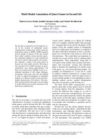
Her hip pain resolved but reoccurred two years later
when another MRI indicated remittent PVNS without
evidence of osteoarthritis, arthritis, or osteonecrosis
(Figure 1). Plain X-rays were normal. Our patient under-
went rigorous synovectomy by open surgery, which sub-
sequently controlled all PVNS-related symptoms.
Discussion
A rigorous PubMed/Medline research did not reveal any
previous reports about an association between PVNS
and t he key words “ lupus”, autoimmunity”, “kidney” or
“proteinuria” , rendering a causal relationship between
the underlying SLE or lupus nephritis and PVNS to be
unlikely. It is of note that the reports on larger series of
PVNS mostly lack a detailed description of comorbid-
ities. However, in our patient the symptoms of PVNS
clearly developed in a temporal association with a flare-
up of SLE and lupus nephritis. We considered that
A
B
C
Figure 1 MRI of her right hip joint. (A) Th e T1-we ighted coronal
image illustrates the synovial fluid effusion (white) in the dorsal
recessus of the joint around the femoral head. (B) Gadolinium
contrast of sagittal images shows diffuse enhancement in synovial
tissue along the zona orbicularis to the posterior joint cavity
surrounding a contrast-free corpus librum of 4 mm diameter. (C)
The synovial proliferation appears in dark grey in the T2-weighted
image at the same location. Bone or cartilage did not display
erosions or thinning, respectively.
Anders Journal of Medical Case Reports 2011, 5:443
/>Page 2 of 3
pigmented synovitis could be secondary to chloroquine
treatment which often causes hyperpigmentation of the
skin and mucus membranes. However, our patient had
not been exposed to antimalarial drugs before the PVNS
diagnosis was made and an association be tween PVNS
and chloroquine treatment has also not been reported.
PVNS is almost equally prevalent in ma les and females
while SLE has a 1:9 male to female ratio, which also
argues against a shared pathogenesis. This includes a
potential role of estrogens which clearly contribute to
onset and disease activity of SLE while an association of
estrogens and PVNS remains speculative [9]. Further-
more, SLE remains a recurrent disease with flares of
synovitis while open synovectomy can result in persis-
tent cure of PVNS [2-5]. As the precise cause of PVNS
to date remains unknown it might still be worthwhile to
consider that either the pathomechanisms that drive
SLE disease activity or its consequences on tissue home-
ostasis h ave an impact on the factors that d rive PVNS.
For example, a study that compared histopathological
characteristics of synovitis in rheumatoid arthritis and
diffuse PVNS found an overlapping pattern of proliferat-
ing macrophages and fibroblasts [10]. CD68/CD163+
synoviocytes were preferentially located in the vicinity of
the synovial lining layer of rheumatoid arthritis patients
while they were randomly distributed in PVNS [10]. In
addition, 20% of synoviocyt es were aneuploid in diffuse
PVNS while all samples of focal PVNS or rheumatoid
arthritis were diploid [10]. It will depend on future
reports to see whether PVNS and SLE represent an acci-
dental coincidence in our case or whether there is an
association between these two disorders that has not
been previously recognized. Monoarticular arthralgia not
responding to immunosuppressive therapy in lupus
patients should raise suspicion of alternative diagnoses
such as PVNS.
Conclusion
This is the first reported association between PVNS and
SLE which might simply represent an accidental coinci-
dence of two rare diseases or indicate that they share
triggers for synovial overgrowth.
Consent
Written informed consent was obtained from the patient
at adult age for publication of this case report and any
accompanying images. A copy of the written consent is
available for review by the Editor-in-Chief of this
journal.
Acknowledgements
The author thanks Dr G Luttke, OMC Radiology Clinic, Munich, for
performing and assessing the MRI images.
Competing interests
The authors declare that they have no competing interests.
Received: 18 April 2011 Accepted: 7 September 2011
Published: 7 September 2011
References
1. Ottaviani S, Ayral X, Dougados M, Gossec L: Pigmented villonodular
synovitis: a retrospective single-center study of 122 cases and review of
the literature. Semin Arthritis Rheum 2010, 40:539-546.
2. Mankin H, Trahan C, Hornicek F: Pigmented villonodular synovitis of
joints. J Surg Oncol 2011, 103(5):386-9.
3. Cotten A, Flipo RM, Chastanet P, Desvigne-Noulet MC, Duquesnoy B,
Delcambre B: Pigmented villonodular synovitis of the hip: review of
radiographic features in 58 patients. Skeletal Radiol 1995, 24:1-6.
4. Gonzalez Della Valle A, Piccaluga F, Potter HG, Salvati EA, Pusso R:
Pigmented villonodular synovitis of the hip: 2- to 23-year followup
study. Clin Orthop Relat Res 2001, 388:187-199.
5. Vastel L, Lambert P, De Pinieux G, Charrois O, Kerboull M, Courpied JP:
Surgical treatment of pigmented villonodular synovitis of the hip. J Bone
Joint Surg Am 2005, 87:1019-1024.
6. Murphey MD, Rhee JH, Lewis RB, Fanburg-Smith JC, Flemming DJ,
Walker EA: Pigmented villonodular synovitis: radiologic-pathologic
correlation. Radiographics 2008, 28:1493-1518.
7. Rahman A, Isenberg DA: Systemic lupus erythematosus. N Engl J Med
2008, 358:929-939.
8. Houssiau FA, Vasconcelos C, D’Cruz D, Sebastiani GD, Garrido Ed Ede R,
Danieli MG, Abramovicz D, Blockmans D, Mathieu A, Direskeneli H,
Galeazzi M, Gül A, Levy Y, Petera P, Popovic R, Petrovic R, Sinico RA,
Cattaneo R, Font J, Depresseux G, Cosyns JP, Cervera R:
Immunosuppressive therapy in lupus nephritis: the Euro-Lupus Nephritis
Trial, a randomized trial of low-dose versus high-dose intravenous
cyclophosphamide. Arthritis Rheum 2002, 46:2121-2131.
9. Walker SE: Estrogen and autoimmune disease. Clin Rev Allergy Immunol
2011, 40:60-65.
10. Berger I, Weckauf H, Helmchen B, Ehemann V, Penzel R, Fink B, Bernd L,
Autschbach F: Rheumatoid arthritis and pigmented villonodular synovitis:
comparative analysis of cell polyploidy, cell cycle phases and expression
of macrophage and fibroblast markers in proliferating synovial cells.
Histopathology 2005, 46:490-497.
doi:10.1186/1752-1947-5-443
Cite this article as: Anders: Pigmented villonodular synovitis of the hip
in systemic lupus erythematosus: a case report. Journal of Medical Case
Reports 2011 5:443.
Submit your next manuscript to BioMed Central
and take full advantage of:
• Convenient online submission
• Thorough peer review
• No space constraints or color figure charges
• Immediate publication on acceptance
• Inclusion in PubMed, CAS, Scopus and Google Scholar
• Research which is freely available for redistribution
Submit your manuscript at
www.biomedcentral.com/submit
Anders Journal of Medical Case Reports 2011, 5:443
/>Page 3 of 3