báo cáo khoa học: "Isolated adult hypoganglionosis presenting as sigmoid volvulus: a case report" docx
Bạn đang xem bản rút gọn của tài liệu. Xem và tải ngay bản đầy đủ của tài liệu tại đây (3.29 MB, 5 trang )
CASE REP O R T Open Access
Isolated adult hypoganglionosis presenting as
sigmoid volvulus: a case report
Irfan Qadir
1
, Muhammad Musa Salick
1
, Abrar Barakzai
2
and Hasnain Zafar
1*
Abstract
Introduction: Isolated hypoganglionosis is a rare cause of intestinal innervation defects. It is characterized by
sparse and small myenteric ganglia, absent or low acetylcholinesterase activity in the lamina propria and
hypertrophy of the muscularis mucosae, principally in the region of the colon and rectum. It accounts for 5% of all
intestinal neuronal malformations. To the best of our knowledge, only 92 cases of isolated hypoganglionosis were
reported from 1978 to 2009. Isolated hypoganglionosis usually manifests as enterocolitis or poor bowel function,
and is diagnosed in infancy or childhood. We report the first case of isolated hypoganglionosis presenting with
sigmoid volvulus in a 34-year-old woman.
Case presentation: A 34-year-old Asian woman had progressively increasing abdominal pain and had not passed
stool or flatus for two days. A physical examination revealed a distended abdomen with sluggish gut sounds. A
computerized tomography (CT) scan demon strated gross dilatation of the sigmoid colon (maximal diameter 14.3
cm) suggestive of sigmoid volvulus. During emerg ency laparotomy, sigmoidectomy with a side-to-side col orectal
anastomosis was performed. Histopathology of the resected specimen showed occasional ganglion cells and
hypertrophied nerve bundles in the muscle layers, suggesting hypoganglionosis. Colonoscopy was performed, and
multiple full-thickness biopsies were taken that showed hypoganglionosis of the entire large bowel. Our patient
underwent total colectomy with an ileorectal anastomosis. Subsequently our patient reported a dramatic
improvement in her bowel function.
Conclusions: Isolated hypoganglionosis is a rare cause of intestinal dysganglionosis and cannot be differentiated
from Hirschsprung’s disease based on clinical presentation. This case report describes an atypical presentation of
the disease. A definitive diagnosis requi res histopathological analysis of full-thickness intestinal biopsies. Treatment
should be tailored to the extent of hypoganglionosis.
Introduction
Hypoganglionosis is a hypogenetic variant of intestinal
dysganglionosis, characterized by sparse and small
myenteric ganglia, absent or low acetylcholinesterase
(AchE) activity in the lamina propria and hypertrophy of
the muscularis mucosae, principally in the region of the
colon and rectum [1]. Hy poganglionosis occurs in two
subtypes: the isolated form, and hy poganglion osis asso-
ciated with Hirschsprung disease [2]. Isolated disease is
rare, and accounts for 5% of all the intestinal neuronal
malformations [1]. Although such developmental
abnormalities as a cause of visceral neuropathy are
usually symptomatic and diagnosed in infancy or
childhood, we report a case of isolated hypoganglionosis
in a 34-year-old woman.
Case presentation
A 34-year-old Asian woman was admitted to our
emergency department with complaints of progres-
sively increasing abdominal pain. She had been unable
to pass stool or flatus f or two days. Our patient also
had a history of chronic constipation that was attribu-
ted to irritable bowel syndrome, and had been treated
with laxatives and prokinetics without lasting success.
She was also reported to have pseudodextrocardia due
to elevation of her left hemidiaphragm by dilated loops
of bowel. Her medical history and family history were
non-contributory.
* Correspondence:
1
General Surgery Department, Aga Khan University Hospital, Stadium Road,
Karachi 74800, Pakistan
Full list of author information is available at the end of the article
Qadir et al. Journal of Medical Case Reports 2011, 5:445
/>JOURNAL OF MEDICAL
CASE REPORTS
© 2011 Qadir et al; licensee BioMed Central Ltd. This is an Open Access article distributed under t he terms of the Cre ative Commons
Attribution License (http://creativecomm ons.org/licenses/by/2.0), which permits unrestricted use, distribution, and reproduction in
any medium, provided the or iginal work is properly cited.
A physical examination was significant for a dis-
tended abdomen with sluggi sh gut sounds. A digital
rectal examination was normal. Initial laboratory test
results were also within normal limits. Abdominal
radiography demonstrated a massively distended sig-
moid colon and rectum with dissipated faeces, giving a
ground-glass appearance. There was no evidence of
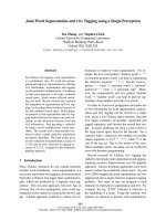
pneumoperitoneum. A computerized tomography (CT)
scan demonstrated gross dilatation of the sigmoid
colon (maximal diameter 14.3 cm) with swirling of
mesenteric vessels in the left iliac fossa suggestive of
sigmoid volvulus (Figure 1).
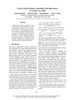
During an emergency laparotomy procedure, we
observed megacolon with concurrent sigmoid volvulus
with a twist at the mid-descending colon level. Figure 2
shows the large sigmoid volvulus peri-operatively. The
bowel was found to be viable and without contamina-
tion. After resection of the sigmoid colon, a side-to-side
colorectal anastomosis was p erformed followed by an
ileostomy 30 to 40 cm from the ileocolic junction. The
sigmoidectomy sample w as sent for histopathology
analysis.
A post-operative CT scan showed no evidence of leak-
age and our patient remained well. The histopathology
examination of sigmoid colon showed occasional gang-
lion cells and hypertrophied nerve bundles in the muscle
layers, suggesting hypoganglionosis. Our patient under-
went colonoscopy with biopsy to determine the extent
of hypoganglionosis. The colonoscopy showed a dilated
bowel with non-obstructing bezoars in the rectum and
ascending colon and an anastomotic stricture 25 cm
from the anal verge. Full-thickness biopsies were taken
from the descending, transverse and ascending colon.
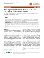
The biopsy specimens showed hypoganglionosis in all
parts of colon (Figure 3).
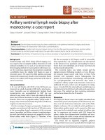
Our patient underwent total colectomy with an ileor-
ectal anastomosis followed by reversal of the ileostomy.
Figure 4 shows her resected colon with two large
bezoars.Ourpatienthasbeenfollowed-upforovera
year and has reported a dramatic improvement in her
bowel function and quality of life.
Discussion
Hypoganglionosis is a controversial condition because
(1) there is no definitive diagnostic criterion for hypo-
ganglionosis based on the location of pathology or the
clinical course; (2) hypoganglionosis occurs in two sub-
types, isolated hypoganglionosis (IH) and hypoganglio-
nosis associated with Hirschsprung disease (HD) and
intestinal neuronal dysplasia; and (3) finally, hypogan-
glionosis also normally exists immediately above the
dentate line. Regardless, isolated hypogangli onosis
should be regarded as a d istinct condition in which
there is decreased number of ganglion cells in the
myenteric plexus whereas the submuscosal plexus is
normal.However,HDaffectsbothsubmucosaland
myenteric plexuses and the transit ional segment
(between aganglionic and normal segments of intest ine)
is called hypoganglionosis [2].
Due to the rarity of the condition, the number of
reported cases in the literature is limited. To the best of
our knowledge only 92 cases were reported from 1978
Figure 1 Computerized tomography scan of the abdomen
showing sigmoid volvulus.
Figure 2 Operative findings showing large sigmoid volvulus.
Qadir et al. Journal of Medical Case Reports 2011, 5:445
/>Page 2 of 5
to 2009 (69 men and 23 women) [1]. Table 1 gives a
review of the literature on isolated hypoganglionosis
from 1978 to 2009.
The clinical and epidemiological features of patients
with IH resemble Hirsch sprun g’ s disease, although the
median age at diagnosis is significantly higher; that is,
4.85 years (range 3.3 days to 17 years ). Our patient was
diagnosed at 34 years of age.
Patients usually present with severe acute and chronic
constipation, pseudo-obstruction or enterocolitis [1]. In
our patient’s case, she presented with sigmoid volvulus.
To date there has been no report of isolated hypogan-
glionosis associated with sigmoid volvulus.
According to the literature, a diagnosis of hypogan-
glionosis can only be established by histopathological
staining of full-thickness bowel specimens. Immunohis-
tochemical staining of bowel specimens for acetylcholi-
nesterase, showing low or absent activity of AchE, is
used to confirm the diagnosis. This method has been
reported as a diagnostic modality in 10 out of 11
reports describing IH from 1978 to 2009 [1]. Morpho-
metric measurements in IH using AchE staining, per-
formed by Meier-Ruge et al., represents one of the
cornerstones of the diagnostic criteria for the disease.
Using resected bowel specimens from patients with
hypoganglionosis, they found that the number of nerve
cells was only about 40% that of a normal innervated
colon and the distance between the ganglia were
doubled (hypoganglionosis: 421 ± 98 μm; normal: 174
±60μm). The mean area of the ganglia in hypogan-
glionosis was three times smaller than the normal
innervated colon (hypoganglionosis: 8.48 ± 2.40 mm
2
;
normal: 21.88 ± 5.12 mm
2
)[3].
Other commonly used additional markers include
staining of the biopsy for lactate dehydrogenase (LDH),
succinate dehydrogenase (SDH), nicotinamide adenine
dinucleotide phosphate (NADPH)-diaphorase, c-kit,
interstitial cells of Cajal (ICC) or silver staining and S-
100 staining [1].
Figure 3 Immunohistochemical staining of biopsy showing hypertrophied nerve bundles and occasional ganglion cells. (A) Hematoxylin
and eosin stain; (B) calretinin stain; (C) S100 stain; (D) neurofilament stain.
Figure 4 Resected colon showing two large bezoars.
Qadir et al. Journal of Medical Case Reports 2011, 5:445
/>Page 3 of 5
In our patient, the presence of concurrent sigmoid
volvulus necessitated urgent treatment wit h emergency
laparotomy and sigmo idectomy, whereas diagnosis was
made later by histopathological a nalysis of resected
bowel specimens. Beside standard hematoxyl in and
eosin stain, immunohistochemical stains such as calreti-
nin, neurofilament and S100 were used to identify the
ganglion cells and nerve bundles. Immunohistochemical
evaluation of acetylcholinesterase, which i s the most fre-
quently described s taining method for IH in the litera-
ture, can only be performed on frozen sections and was
therefore not used in this case.
According to the literature, surgery is the definitive
treatment method for adult hypoganglionosis and adult
Hirschsprung’ s disease [1]. The principles of pull-
through surgery are first to remove all the hypoganglio-
nic segments and second to achieve bowel continuity
between the normally innervated bowel and the anal
canal in order to provide bowel continence in the long
term. The surgical procedures developed to treat the
disease in children have been applied to adults without
significant differences [4].
The four most commonly used procedures for pull-
through surgery are the rectosigmoidectomy developed
by Swenson and Bill, the retrorectal approach developed
by Duhamel, endorectal procedure developed by Soave
and deep anterior colorectal anastomosis developed by
Reub en. Of the 67 patie nts with reported operative pro-
cedures, 54 underwent resection/pull-through surgery.
Enterostomy was employed as treatment strategy in 11
patients and sphincter myotomy in two patients.
During a post-operative follow-up of seven months to
12 years, typical complications such as enterocolitis,
chronic constipation, overflow encopresis, and the need
for repeat pull-through surgery for residual disease have
been reported. In the 92 cases of isolated hypoganglio-
nosis reported between 1978 and 2009, the overall mor-
tality was 8%. Six of the seven patients that died were
newborns suffering from severe enterocolitis; the other
patient died owing to total parenteral nutrition-asso-
ciated complications during follow-up. Our patient has
been observed for over a year without any complication.
Conclusions
Isolated hypoganglionosis is a rare disease with cl inical
and epidemiological features similar to Hirschsprung’s
disease, although the age at diagnosis is higher. Care ful
examination of full-thicknessbiopsiesisrequiredto
make a definitive diagnosis.
Consent
Written informed consent was obtained from the patient
for publication of this case report and any accompany-
ing images. A co py of the written consent is avail able
for review by the Editor-in-Chief of this journal.
Author details
1
General Surgery Department, Aga Khan University Hospital, Stadium Road,
Karachi 74800, Pakistan.
2
Histopathology Department, Aga Khan University
Hospital, Stadium Road, Karachi 74800, Pakistan.
Authors’ contributions
IQ and MMS were involved in data collection, literature review and
manuscript preparation. AB selected and annotated appropriate images from
Table 1 Review of literature on isolated hypoganglionosis from 1978 to 2009
First author/
reference
No. of
cases
Specimen Staining Treatment Mortality
Zhang et al. [5] 17 Rectal suction and full-thickness
biopsy
AchE Resection and pull through No
Rolle et al. [6] 6 Rectal suction and full-thickness
biopsy
AchE, NADPH, ICC, c-
kit
5 Resection and pull through, 1
enterostomy
No
Kobayashi et al. [2] 3 Full-thickness biopsy AchE Enterostomy All died
Kubota et al. [7] 6 Full-thickness biopsy S-100 Enterostomy All died
Meier-Ruge et al. [3] 7 Full-thickness biopsy AchE, LDH, SDH Resection and pull through No
Schaerli et al. [8] 7 Full-thickness biopsy AchE, LDH, SDH Resection and pull through No
Ure et al. [9] 9 Rectal suction biopsy AchE, LDH, SDH 7 resection and pull through, 2 sphincter
myotomy
1 died
Yamataka et al. [10] 1 Full-thickness biopsy AchE, ICC, c-kit Enterostomy No
Munakata et al. [11] 12 Rectal suction and full-thickness
biopsy
AchE, silver
impregnation
11 resection and pull through, 1 not
reported
No
Meier-Ruge [12] 18 Full-thickness biopsy AchE, LDH, SDH Not reported No
Munakata et al. [13] 6 Full-thickness biopsy AchE, silver
impregnation
Not reported No
Adapted from Dingemann J, Puri P: Isolated hypoganglionosis: systematic review of a rare intestinal innervation defect. Pediatr Surg Int 26:1111-1115 [1]
(Permission granted).
AchE = acetylcholinesterase; ICC = interstitial cells of Cajal; LDH = lactate dehydrogenase; NADPH = nicotinamide adenine dinucleotide phosphate;
SDH = succinate dehydrogenase.
Qadir et al. Journal of Medical Case Reports 2011, 5:445
/>Page 4 of 5
histopathology slides and reviewed all available histology to ensure an
accurate diagnosis was made. HZ was involved in patient care and revised
and corrected the manuscript. All authors have read and approved the final
manuscript.
Competing interests
The authors declare that they have no competing interests.
Received: 22 March 2011 Accepted: 8 September 2011
Published: 8 September 2011
References
1. Dingemann J, Puri P: Isolated hypoganglionosis: systematic review of a
rare intestinal innervation defect. Pediatr Surg Int 26:1111-1115.
2. Kobayashi H, Yamataka A, Lane GJ, Miyano T: Pathophysiology of
hypoganglionosis. J Pediatr Gastroenterol Nutr 2002, 34:231-235.
3. Meier-Ruge WA, Brunner LA, Engert J, Heminghaus M, Holschneider AM,
Jordan P, Piket G, Posselt HG, Scharli A: A correlative morphometric and
clinical investigation of hypoganglionosis of the colon in children. Eur J
Pediatr Surg 1999, 9:67-74.
4. Miyamoto M, Egami K, Maeda S, Ohkawa K, Tanaka N, Uchida E, Tajiri T:
Hirschsprung’s disease in adults: report of a case and review of the
literature. J Nippon Med Sch 2005, 72:113-120.
5. Zhang HY, Feng JX, Huang L, Wang G, Wei MF, Weng YZ: Diagnosis and
surgical treatment of isolated hypoganglionosis. World J Pediatr 2008,
4:295-300.
6. Rolle U, Yoneda A, Solari V, Nemeth L, Puri P: Abnormalities of C-Kit-
positive cellular network in isolated hypoganglionosis. J Pediatr Surg
2002, 37:709-714.
7. Kubota A, Yamauchi K, Yonekura T, Kosumi T, Oyanagi H, Mushiake S,
Nakayama M, Imura K, Okada A: Clinicopathologic relationship of
hypoganglionosis. J Pediatr Surg 2001, 36:898-900.
8. Scharli AF, Sossai R: Hypoganglionosis. Semin Pediatr Surg 1998, 7:187-191.
9. Ure BM, Holschneider AM, Schulten D, Meier-Ruge W: Clinical impact of
intestinal neuronal malformations: a prospective study in 141 patients.
Pediatr Surg Int 1997, 12:377-382.
10. Yamataka A, Fujiwara T, Nishiye H, Sunagawa M, Miyano T: Localization of
intestinal pacemaker cells and synapses in the muscle layers of a
patient with colonic hypoganglionosis. J Pediatr Surg 1996, 31:584-587.
11. Munakata K, Okabe I, Morita K: Hypoganglionosis. Pediatr Surg Int 1992,
7:8-11.
12. Meier-Ruge W: Epidemiology of congenital innervation defects of the
distal colon. Virchows Arch A Pathol Anat Histopathol 1992, 420:171-177.
13. Munakata K, Okabe I, Morita K: Histologic studies of rectocolic
aganglionosis and allied diseases. J Pediatr Surg 1978, 13:67-75.
doi:10.1186/1752-1947-5-445
Cite this article as: Qadir et al.: Isolated adult hypoganglionosis
presenting as sigmoid volvulus: a case report. Journal of Medical Case
Reports 2011 5:445.
Submit your next manuscript to BioMed Central
and take full advantage of:
• Convenient online submission
• Thorough peer review
• No space constraints or color figure charges
• Immediate publication on acceptance
• Inclusion in PubMed, CAS, Scopus and Google Scholar
• Research which is freely available for redistribution
Submit your manuscript at
www.biomedcentral.com/submit
Qadir et al. Journal of Medical Case Reports 2011, 5:445
/>Page 5 of 5