báo cáo khoa học: "Challenges in clinical and laboratory diagnosis of androgen insensitivity syndrome: a case report" ppt
Bạn đang xem bản rút gọn của tài liệu. Xem và tải ngay bản đầy đủ của tài liệu tại đây (657.72 KB, 6 trang )
CAS E REP O R T Open Access
Challenges in clinical and laboratory diagnosis of
androgen insensitivity syndrome: a case report
Caroline OA Melo
1,2
, Daniela M Silva
1,2,4*
and Aparecido D da Cruz
1,3
Abstract
Introduction: Androgen is a generic term usually applied to describe a group of sex steroid hormones. Androgens
are responsible for male sex differentiation during embryogenesis at the sixth or seventh week of gestation,
triggering the development of the testes and penis in male fetuses, and are directed by the testicular determining
factor: the gene SRY (sex determining region on Y chromosome) located on the short arm of chromosome Y. The
differentiation of male external genitalia (penis, scrotum and penile urethra) occurs between the 9th and 13th
weeks of pregnancy and requires adequate concentration of testosterone and the conversion of this to another
more potent androgen, dihydrotestosterone, through the action of 5a-reductase in target tissues.
Case presentation: This report describes the case of a teenage girl presenting with a male karyotype, and aims to
determine the extension of the mutation that affecte d the AR gene. A Caucasian girl aged 15 was referred to our
laboratory for genetic testing due to primary amenorrhea. Physical examination, karyotype testing and molecular
analysis of the androgen receptor were critical in making the correct diagnosis of complete androgen insensitivity
syndrome.
Conclusions: Sex determination and differentiation depend on a cascade of events that begins with the
establishment of chromosomal sex at fertilization and ends with sexual maturation at puberty, subsequently
leading to fertility. Mutations affecting the AR gene may cause either complete or partial androgen insensitivity
syndrome. The case reported here is consistent with complete androgen insensitivity syndrome, misdiagnosed at
birth, and consequently our patient was raised both socially and educationally as a female. It is critical that health
care providers understand the importance of properly diagnosing a newbor n manifesting ambiguous genitalia.
Furthermore, a child with a pseudohermaphrodite phenotype should always undergo adequate endocrine and
genetic testing to reach a conclusive diagnosis before gender is assigned and surgical interventions are carried out.
Our results show that extreme care must be taken in selecting the genetic tools that are utilized for the diagnosis
for androgen insensitivity syndrome.
Introduction
Androgen i s a generic term usually applied to describe a
group of sex steroid hormones. Androgens are produced
primarily by a male’ s testes. However, some small
amounts are also produced by the ovaries in females
and b y the adrenal gland in both sexes. Androgens are
responsible for male sex differentiation during embryo-
genesis at the sixth or seventh week of gestation, trig-
gering the development of thetestesandpenisinmale
fetuses, and are directed by the testicular determining
factor: the gene SRY (sex determining region on Y chro-
mosome) located on the short arm of chromosome Y.
The differentiation of male external genitalia in the
penis, scrotum and penile urethra occurs between the
9th and 13th weeks of pre gnancy and requires adequate
concentration of testosterone and con version of this to
another more potent androgen, dihydrotestosterone
(DHT), through the action of 5a-reductase in target tis-
sues [1]. The actions of testosterone and DHT require
the presence of functional androgen receptors that, after
signal from these hormones, activate the transcription of
specific genes in target tissues. Thus, any abnormality in
the production or action of androgens in a fetus 46, XY
between the 9th and 13th weeks of pregnancy causes
* Correspondence:
1
Pontifícia Universidade Católica de Goiás, Departamento de Biologia, Núcleo
de Pesquisas Replicon, Goiânia, Goiás, Brazil
Full list of author information is available at the end of the article
Melo et al. Journal of Medical Case Reports 2011, 5:446
/>JOURNAL OF MEDICAL
CASE REPORTS
© 2011 Melo et al; licensee BioMed Central Ltd. This is an Open Access article distributed under the te rms of the Creative Commons
Attribution License ( censes/by/2.0), which permits unrestricted use, di stribution, and reproduction in
any medium, provided the original wor k is properly cited.
incomplete masculinization, resulting in male pseudo-
hermaphroditism. In men, androgens also trigger the
complex p rocess of puberty, affecting the development
of facial, body, and pubic hair, deepening of the voice,
and muscle development. Physiologically, androgens reg-
ulate spermatogenesis and help maintain male reproduc-
tive functions [2].
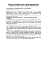
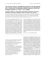
The AR gene is a protein-coding gene located at
Xq11.2-q12. It spans over 90 kb and codes for a protein
that functions as a steroid-hormone-act ivated transcrip-
tion factor (Figure 1). The androgen receptor (AR), like
other members of the nuclear receptor superfamily, has
three major functional domains. The AR is characterized
by a modular structure consisting of four functional
domains: an N-terminal domain (NTD), a DNA-binding
domain (DBD), a hinge region, and a ligand-binding
domain (LBD; in this case, the ligand being an andro-
gen) [3].
The complete form of androgen insensitivity syn-
drome (CAIS) is relat ively rare. In childhood, the most
common clinical presentation is the presence of bilateral
inguinal hernias. Individuals not diagnosed during child-
hood are detected after puberty because of primary
amenorrhea. Patients with the complete form of AIS
have female external genitalia, an absence or thinning of
pubic hair and the absence of a uterus [4].
The AR NTD is relativ ely long and displays the great-
est sequence variability among nuclear receptors. It is
very flexible and displays a high degree of intrinsic dis-
order. In addition, the NTD has a variable number of
homopolymeric repeats, the most important of which is
a polyglutamine repeat that ranges from eight to 31
repeat s in normal individuals, with an average length of
20 base pairs [5].
The DBD is centrally located in the AR and it is the
most conserved region within the nuclear receptor
family. This region has nine cysteine residues, eight of
which are involved in forming two zinc fingers, and a
C-terminal extension. The first zinc finger, most p roxi-
mal to the NTD, determines the specificity of DNA
recognition, whereas residues in the second zinc finger
are involved in AR dimerization. Two AR monomers in
a head-to-head conformation bind as a homodimer to
androgen response elements which are direct or indirect
repeats of the core consensus 5’-TGTTCT-3’ or more
complex response elements with diverse arrangements
of AREs. The C-terminal extension is important for the
three-dimensional structure of the DBD and it plays a
Figure 1 Cytogenetic location of the AR gene, Xq11.2-q12, and molecular location on the X chromosome, base pairs 66,680,598 to
66,860,843.
Melo et al. Journal of Medical Case Reports 2011, 5:446
/>Page 2 of 6
role in mediating the AR selectivity of DNA interacti on
[6].
The hinge region has long been considered to be a
flexible linker between the DBD and LBD in the AR.
More recently, however, this region was shown to be
involved in DNA binding as well as AR dimerization. It
was suggested that the hinge region also acts to attenu-
ate transcriptional activity of the AR gene [7].
Mutations in the AR gene cause X-linked androgen
insensitivity syndrome (AIS) characterized by androgen
unresponsiveness, which affects proper male sexual
development both at embry ogenesis and at puberty. As
a genetic disorder, AIS presents problems to affected
people and their families, and is a major medical chal-
lenge for health providers. This impaired response to
androgen re sults from an incapacity or reduced capacity
of the AR to transactivate androgen-responsiv e genes in
target cells, and leads to abnormal differentiation and
development of male internal and external genitalia,
leading to male pseudo-hermaphroditism [1].
Most mutations on the AR gene result in AIS (OMIN
#300068). Depending on the e xtent of the AR defect,
the phenotype can vary. Three forms are classified, com-
plete (CAIS), partial (PAIS), and mild (MAIS). All forms
of AIS are X-linked traits inherited as a recessive disor-
der. Despite a normal male karyotype (46, XY), indivi-
duals affected with CAIS (also known as testicular
feminization syndrome) have female externa l genitalia,
blind vaginas, an absent uterus and female adnexa,
female breast development, and abdominal or inguinal
testes. Partial androgen insensitivity results in hypo spa-
dias and micropenis with gynecomastia [8]. Patients
usually come to medical attention during the neonatal
period because of inguinal hernia and/or ambiguous
genitalia, or at puberty because of primary amenorrhea
associated with normal breast development and reduced
pubic h air. In contrast, P AIS is a heterogeneous condi-
tion and covers a wide spectrum of undervirilization
situations resulting in different degrees of ambiguous
external genitalia, ranging from an almost complete
form to phenotypic males with isolated azoospermia [9].
The aim of the investigation reported here was to pro-
vide a genetic diagnosis of a teenage girl with normal
male ka ryotype using fluorescence in si tu hybridization
(FISH) and PCR in order to determine the nature and
the extent of the mutation that affected the AR gene.
Case presentation
A 15-year-old Caucasian girl was referred t o a labora-
tory in Goiânia, Goiás, Brazil, for genetic testing due to
primary amenorrhea. Her medical history included
removal of an abdominal mass as a newborn. The tissue
removed was referred to as an umbilical hernia. Her G-
band karyotype revealed a diploid set of chromosomes,
including 22 pairs of homologous autosomes and one
pair of sex chromosomes, compatible with a 46, XY
male chromosome complement.
The geneticists at our laboratory concluded that the
mass withdrawal from the abdomen of our patient was,
in fact, testes, and that our patient had a condition
known as cryptorchidism; a reproductive change charac-
terized by a failure of the movement of one or both
testes from the abdominal cavity to the scrotum.
WeperformedPCRandFISHtoverifymutationsof
the ex ons 1, 4, 6, 7 and 8 of the AR gene a nd to detect
the AR gene, respectively. We prepared a culture of T
lympho cytes in RPMI 16 40 medium, supplemented with
20% fetal calf serum and 2% of phytohemagglutinin.
Metaphasic preparations were made by conventional
methodology. The slides for FISH were prepared with a
micropipette, with about 15 μL of the mate rial set. Only
slides of good quality (in terms of metaphase) were
selected by phase contrast microscope, and were sub-
jected to FISH using the LSI Androgen Receptor Spec-
trumOrange (Xq12) probe (Vysis, Abbott Park, IL,
USA).ForPCR,primerswereusedforexons1,4,6,7
and 8 of AR [10].
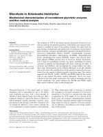
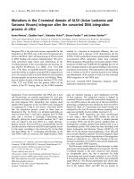
In si tu hybridization with the LSI AR probe indicated
the presence of the gene in all analyzed cells. However,
genomic DNA extracted from peripheral blood leuko-
cytes assessed by PCR revealed coding sequence
abnormalities for the AR gene, which lacked exons 1 to
7 indicating large de letion spanning the proximal region
of the gene. Figure 2 shows hybridization signals in both
interphase and metaphase nuclei.
Conclusions
The results reported in this paper underline the fact that
great care must be taken when selecting genetic testing
tools to be utilized to reach the proper diagnosis for
AIS. If a child reaches his or her teenage years undiag-
nosed due to clinical challenges presented by ambiguous
genitalia it further complicates matters, as there could
be several genetic events subjacent to that outcome, ran-
ging from total or partial deletion of the gene t o point
mutation, that efficiently silences the gene and therefore
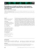
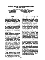
leads to AIS. Here, we report that FISH alone was not
able to pro perly diagnose our patient, despite the proxi-
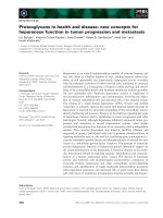
mal deletion within the AR observed on PCR (Figure 3).
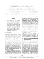
This r esult could be explained by the size of the probe
used (380 kb), which was bigger than the AR gene (90
kb), indicating that the deletion of some exons within
the gene was not large enough to prevent probe hybridi-
zation (Figure 4) [11]. Thus, in our patient’scase,the
PCR assay was able to confirm the diagnosis for our
patient with a history of abdominal mass removal as a
newborn who had a chromosomally normal male karyo-
type. However, due to complex chromosome aberrations
Melo et al. Journal of Medical Case Reports 2011, 5:446
/>Page 3 of 6
or other genomic mutations, other molecular tools to
detect and define DNA mutations, such as DNA
sequencing, may be required to properl y reach the con-
clusion of a diagnosis of AIS.
Sex determination and differentiation depend on a
cascade of events t hat begins with the establishment of
chromosomal sex at fertilization and ends with sexual
maturation at puberty subsequently leading to fertility.
Mutations affecting the AR gene may cause either com-
plete or partial AIS. The case of o ur patient reported
here is consistent with CAIS, misdiagnosed at birth, and
she was consequently raised socially and educationally
as a female. It is critical that health care providers
understand the importance of prope rly diagnosing a
newborn with ambiguous genitalia. Prompt evaluation of
both clinical and genetic findings is crucial to determine
proper gender assignment and the detection of life-
threatening conditions [12] . Furthermore, a child with a
pseudohermaphrodite phenotype should always undergo
adequate e ndocrine and genetic testing for a definitive
diagnosis before gender is a ssigned and surgical inter-
ventions are carried out. Inadequate investigation may
result in inappropriate gender assignment in infancy
with possible inferences on outcome [13].
A
B
Figure 2 Hybridization signals for the LSI androgen receptor probe, indicating the presence of the AR gene on the nuclei of a 15-
year-old patient affected with androgen insensitivity syndrome (AIS).
L
1
4
6
8
CN
L
1
7
4
6
7
8
CN
A
B
Figure 3 Images of polymerase chain reaction products on a 2% agarose gel. The first image (A) shows a gel from a normal patient with
the five exons analyzed. The second image (B) shows a gel from our patient, who had only exon 7. CN = negative control; L = ladder of 100
bp; 1 = exon 1 with a size of 528 bp; 4 = exon 4 with a size of 172 bp; 6 = exon 6 with a size of 378 bp; 7 = exon 7 with a size of 516 bp; 8 =
exon 8 with a size of 289 bp.
Melo et al. Journal of Medical Case Reports 2011, 5:446
/>Page 4 of 6
The presentation of a patient, and specifically a neo-
nate, with abnormal genital development represents a
difficult diagnostic and therapeutic challenge. Referral to
a center with experie nce in the diagnosis and manage-
ment of disorders of sexual development is advised,
where an emphasis should be placed on psychological
and genetic counseling [14,15].
Consent
Written informed consent was obtained from the
patient’s next-of-kin for publication o f this case report
and any accompanying images. A copy of the written
consent is available for review by the Editor-in-Chief of
this journal.
Author details
1
Pontifícia Universidade Católica de Goiás, Departamento de Biologia, Núcleo
de Pesquisas Replicon, Goiânia, Goiás, Brazil.
2
Pró-Reitoria de Pós-Graduação
e Pesquisa, Mestrado em Genética, Pontifícia Universidade Católica de Goiás,
Goiânia, Goiás, Brazil.
3
LaGene, Laboratório de Saúde Pública Dr Giovanni
Cysneiros (Lacen), Laboratório de Citogenética Humana e Genética
Molecular, Secretaria Estadual de Saúde de Goiás, Goiânia, Goiás, Brazil.
4
Departamento de Biologia Geral, Instituto de Ciências Biológicas,
Universidade Federal de Goiás.
Authors’ contributions
COAM was involved in collating the information, reviewing the literature,
and preparation of the manuscript. DMS was involved in collating
information regarding the case and getting informed consent from our
patient. ADC was involved in the review of literature and revising the
manuscript critically. All authors read and approved the final manus cript.
Competing interests
The authors declare that they have no competing interests.
Received: 23 March 2011 Accepted: 8 September 2011
Published: 8 September 2011
References
1. Melo KFS, Mendonça BB, Billerbeck AEC, Costa EMF, Latronico AC,
Arnhold IJP: Síndrome de Insensibilidade aos Andrógenos: Análise
Clínica, Hormonal e Molecular de 33 Casos. Arquivo Brasileiro de
Endocrinologia e Metabolismo 2005, 49:87-97.
2. Zouboulis CC, Chen WC, Thornton MJ, Qin K, Rosenfield R: Sexual
hormones in human skin. Horm Metab Res 2007, 39:85-95.
3. National Center for Biotechnology Information. [.
gov].
4. Grumbach MM, Hughes IA, Conte FA: Disorders of sex differentiation. In
Williams Textbook of Endocrinology. Edited by: Larsen PR, Kronenberg HM,
Melmed S, Polonsky KS. Philadelphia, PA: WB Saunders Company;
2002:842-1003.
5. Trapman J: Interaction of the Androgen Receptor Ligand Binding
Domain with the N-terminal Domain and with Coactivators. In Androgen
Action in Prostate Cancer. Edited by: Tindal D, Mohle. New York, USA:
Springer Science; 2009:375-384.
6. Verrijdt G, Tanner T, Moehren U, Callewaert L, Haelens A, Claessens F: The
androgen receptor DNA-binding domain determines androgen
selectivity of transcriptional response. Biochem Soc Trans 2006,
34:1089-1094.
7. Haelens A, Tanner T, Denayer S, Callewaert L, Claessens F: The hinge
region regulates DNA binding, nuclear translocation, and transactivation
of the androgen receptor. Cancer Res 2007, 67:4514-4523.
8. Kore S, Chippa S, Khot R, Sarodey G, Kelkar S, Puri K, Aagwane V: Testicular
feminization syndrome. Bombay Hosp J 2010, 52:126-128.
9. Gottlieb B, Rose L, Lenore KB, Mark AT: Molecular pathology of the
androgen receptor in male (in) fertility. Repr Biomed Online 2004, 10:42-48.
10. Rajender S, Selvi D, Sakhamuri M, Nalini JG, Baidyanath C, Lalji S,
Kumarasamy T: Male infertility: no evidence of involvement of androgen
receptor gene among indian men. J Androl 2006, 27:102-105.
11. Abbott Molecular Gateway. [ />oncology/fish/vysis-lsi-androgen-receptor-gene-xq12-spectrumorange-probe.
html].
12. Zdravković D, Milenković T, Sedlecki K, Guć-Sćekić M, Rajić V: Causes of
ambiguous external genitalia in neonates [in Serbian]. Banićević Srp Arh
Celok Lek 2001, 129:57-60.
13. Dati E, Baldinotti F, Conidi ME, Simi P, Baroncelli GI, Bertelloni S: A girl with
tomboy behavior: lesson from misdiagnosis in a baby with ambiguous
genitalia. Sexual
Dev 2009, 4:150-154.
14. Drop SL, Boehmer AL, Slijper FM, Nijman JM, Hazebroek FW, Niermeijer MF:
Differential diagnosis and treatment of girls with 46XY-karyotype and
androgen insensitivity syndrome [in Dutch]. Ned Tijdschr Geneeskd 2001,
145:665-669.
Figure 4 Map of probe and exons amplified by polymerase chain reaction showing the problem with fluorescence in situ
hybridization (FISH) and lack of exons 1 to 7 on the AR gene.
Melo et al. Journal of Medical Case Reports 2011, 5:446
/>Page 5 of 6
15. Koch CA: Androgen insensitivity syndrome.[scape.
com/article/924996-overview].
doi:10.1186/1752-1947-5-446
Cite this article as: Melo et al.: Challenges in clinical and laboratory
diagnosis of androgen insensitivity syndrome: a case report. Journal of
Medical Case Reports 2011 5:446.
Submit your next manuscript to BioMed Central
and take full advantage of:
• Convenient online submission
• Thorough peer review
• No space constraints or color figure charges
• Immediate publication on acceptance
• Inclusion in PubMed, CAS, Scopus and Google Scholar
• Research which is freely available for redistribution
Submit your manuscript at
www.biomedcentral.com/submit
Melo et al. Journal of Medical Case Reports 2011, 5:446
/>Page 6 of 6