báo cáo khoa học: "A challenging case of gastric outlet obstruction (Bouveret''''s syndrome): a case report" docx
Bạn đang xem bản rút gọn của tài liệu. Xem và tải ngay bản đầy đủ của tài liệu tại đây (1.08 MB, 4 trang )
JOURNAL OF MEDICAL
CASE REPORTS
A challenging case of gastric outlet obstruction
(Bouveret's syndrome): a case report
Gajendran et al.
Gajendran et al. Journal of Medical Case Reports 2011, 5:497
(4 October 2011)
CAS E REP O R T Open Access
A challenging case of gastric outlet obstruction
(Bouveret’s syndrome): a case report
Mahesh Gajendran
1*
, Thiruvengadam Muniraj
1
and Andres Gelrud
2
Abstract
Introduction: Bouveret’s syndrome is a clinically distinct form of gallstone ileus caused by the formation of a
fistula between the biliary tract and duodenum. This case reinforces the need for early recognition and treatment
of Bouveret’s syndrome, as it is associated with high morbidity and mortality rates.
Case presentation: An 82-year-old Caucasian woman presented with signs and symptoms of small bowel
obstruction. Her laboratory workup showed elevated alkaline phosphatase and amylase levels. Computed
tomography of her abdomen revealed pneumobilia, a choledochoduodenal fistula and a gallstone obstructing her
distal duodenum. The impacted gallstone could not be extracted endoscopically, so our patient underwent open
enterolithotomy successfully. However, the postoperative course was complicated by myocardial infarction,
respiratory failure and disseminated intravascular coagulation. She died 22 days after surgery, secondary to
cardiopulmonary arrest.
Conclusion: This case clearly highlights the considerable morbidity and mortality associated with Bouveret’s
syndrome.
Introduction
Bouveret’s syndrome is defined as a cholecystoduodenal
or choledochoduodenal fistula with the passage of a gall-
stone into the duodenum or pylorus leading to gastric
outlet obstruction [1]. There have been very few case
reports about this syndrome published in the last 100
years because of its rarity. It is a ssociated with high
morbidity and mortality rates. With the availability of
computed tomography (CT) scans, earlier diagnosis and
better management of these cases are possible. Our
patient had a typical presentation of the disease b ut
ended up having multiple postoperative complications.
Case presentation
An 82-year-old Caucasian woman, with a history of
hypertension, depression, hypothyroidism, dyslipidemia,
carotid endarterectomy and coronary artery disease, pre-
sented with a four-day history of nausea, bilious vomit-
ing and epigastric pain radiating to her left scapula. Her
home medications included sertraline, atenolol,
calci triol, levothyroxine, tolterodine, omeprazole, aspirin
and atorvastatin. She denied smoking, use of alcohol or
drug abuse. On examination, she appeared lethargi c but
not in acute distress. She was afebrile and had a blood
pressure of 157/64 mmHg. Examination of her abdomen
revealed abdominal distension, epigastric tenderness,
tympanic sounds on percussion and decreased bowel
sounds. Initial laboratory result s were as follows: hemo-
globin 11.4 g/dL, white blood cell count 6.2 × 10
9
/L,
platel ets 115 × 10
9
/L, creatinine 3.2 mg/dL (n ormal: < 1
mg/ dL), bloo d ure a nitr ogen 30 mg/dL, amylase 710 U/
L ( normal: 30 to 110 U/L), lipase 133 U/L (normal: 22
to 51 U/L), albumin 3.1 g/dL, serum alkaline phospha-
tase 146 U/L (normal: 35 to 100 U/L) with normal levels
of alanine transaminase (ALT), aspartate aminotransfe r-
ase (AST) and total bilirubin.
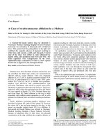
A CT scan of h er abdomen and pelvis with c ontrast
(Figure 1) showed pneumobilia with a choledochoduo-
denal fistula (common bile duct and second part of her
duodenum), significant wall thickening of the second
portion of her duodenum and a large 3.6 cm gallstone
obstructing her distal duodenum (Figures 2 and 3). Her
stomach and proxim al duodenum were dilated with
decompression of the distal small and large bowel loops.
* Correspondence:
1
Department of General Internal Medicine, University of Pittsburgh Medical
Center, 200 Lothrop Street, Suite 933W, Pittsburgh, PA 15213, USA
Full list of author information is available at the end of the article
Gajendran et al. Journal of Medical Case Reports 2011, 5:497
/>JOURNAL OF MEDICAL
CASE REPORTS
© 2011 Gajendran et al; licensee BioMed Central Ltd. This is an Open Access article dis tributed under the terms of the Creative
Commons Attribution License ( which permits unrestrict ed use, distribution, and
reproduction in any medium, provided the original work is properly cited.
These findings were consistent with gallstone ileus. In
addition there w as diffuse mesenteric stranding present
throughout her abdomen without bowel wall thickening.
An upper gastrointestinal (GI) endoscopy showed 1L of
bilious fluid in her stomach with an impacted gallstone
that could not be extracted with endoscopy (Figure 4) .
Our patient underwent an open jejunal enterolithotomy
for gallstone removal without cholecystectomy. Also, a
right hemicolectomy and ileotransverse colonic
anastomosis were performed because of an ischemic
ascending colon found intraoperatively. Pathology
results revealed a gallstone and colonic mucosal
ischemic changes. The postoperative course was compli-
cated by a non-ST elevation myocardial infarction, pul-
monary edema leading to respiratory failure requiring
mechanical ventilation and disseminated intravascular
coagulation m anifesting as hemoperitoneum. Over the
course of her hospital stay, her total bilirubin level
increased up to 35 mg/dL with the direct bilirubin level
being 19.8 mg/dL. Our patient had an international nor-
malized ratio of 2.6 on postoperativ e day 22. Her AST
and ALT levels were elevated at 203 U/L and 65 U/L,
respectively, but her alkaline phosphatase level was nor-
mal. An abdominal ultrasonogram did not show any
Figure 1 CT scan of the patient’ s abdomen showing
pneumobilia and a choledochoduodenal fistula.
Figure 2 CT scan of the patient’ s abdomen showing gastric
and duodenal dilatation (gallstone ileus).
Figure 4 Endoscopic view of the gallst one ob structing the
patient’s duodenum.
Figure 3 CT scan showing a gallstone that is completely
obstructing the patient’s duodenum.
Gajendran et al. Journal of Medical Case Reports 2011, 5:497
/>Page 2 of 3
biliary dilatation. Our patient died 22 days after surgery
secondary to cardiopulmonary arrest.
Discussion
Bouveret’s syndrome is a clinically distinct form of gall-
stone ileus (accounting for 1% to 3%), typically involving
the proximal small intestine, which was first described
by Leon Bouveret in 1896 [2]. It has a mortality rate of
4.5% to 25%. The major risk f actors for developing this
syndrome include age greater than 70 years, female gen-
der, gallstones larger than 2.5 cm and postsurgical
altered GI anatomy [3]. The presentation is similar to
that of small bowel obstruction (SBO). The first diag-
nostic test for suspected SBO would be an abdominal
radiograph; however, the classi c Rigler’s triad (pneumo-
bilia, SBO and gallstone) has been reported to be pre-
sent in only 30% to 35% of cases, since most of the
gallstones are radioluce nt. Contrast-enhanced CT eva-
luation of acute SBO offers prompt and rapid diagnosis
of gallstone ileus [4]. It has a high se nsitivity (93%), spe-
cificity (100%) and accuracy (99%) according to Yu et al.
[5]. The first line of treatment should be upper e ndo-
scopy with an attempt to retrieve the stone. Howe ver,
the success rate of this procedure has been only
approximately 30% to 40%. Other minimally invasive
techniques, such as laser lithotripsy and extracorporeal
shock w ave lithotripsy, are useful in high-risk patients
when it is prudent to avoid surgery. In most cases,
patients end up having su rgery, most commonly entero-
lithotomy with or without cholecystectomy and fistula
repair [6].
Conclusion
This case clearly illustrates the considerable morbidity
and mortality associated with Bouveret’s syndrome. Pre-
operatively, establishing the diagnosis is the challenge,
whereas postoperatively the management of complica-
tions can be even more challenging.
Consent
Written informed consent was obtained from the
patient’s daughter for publication of this case repo rt and
any accompanying images. A copy of the written con-
sent is available for review by the Editor-in-Ch ief of this
journal.
Abbreviations
ALT: alanine transaminase; AST: aspartate aminotransferase; CT: computed
tomography; GI: gastrointestinal; SBO: small bowel obstruction.
Author details
1
Department of General Internal Medicine, University of Pittsburgh Medical
Center, 200 Lothrop Street, Suite 933W, Pittsburgh, PA 15213, USA.
2
Department of Gastroenterology, Hepatology and Nutrition, University of
Pittsburgh Medical Center, 200 Lothrop Street M2, C Wing, Pittsburgh, PA
15213, USA.
Authors’ contributions
MG, TM, and AG analyzed and interpreted the patient data regarding our
patient’s presentation. MG was instrumental in obtaining informed consent
from our patient’s next of kin and also in the preparation of the manuscript.
All authors read and approved the final manuscript.
Competing interests
The authors report no financial relationships or conflicts of interest regarding
the content herein. All the radiologic and endoscopic images are original.
Received: 2 June 2011 Accepted: 4 October 2011
Published: 4 October 2011
References
1. Schweiger F, Shinder R: Duodenal obstruction by a gallstone (Bouveret’s
syndrome) managed by endoscopic stone extraction: a case report and
review. Can J Gastroenterol 1997, 11(6):493-496.
2. Cappell MS, Davis M: Characterization of Bouveret’s syndrome: a
comprehensive review of 128 cases. Am J Gastroenterol 2006,
101(9):2139-2146.
3. Koulaouzidis A, Moschos J: Bouveret’ s syndrome. Narrative review. Ann
Hepatol 2007, 6(2):89-91.
4. Lassandro F, Romano S, Ragozzino A, Rossi G, Valente T, Ferrara I,
Romano L, Grassi R: Role of helical CT in diagnosis of gallstone ileus and
related conditions. AJR Am J Roentgenol 2005, 185(5):1159-1165.
5. Yu CY, Lin CC, Shyu RY, Hsieh CB, Wu HS, Tyan YS, Hwang JI, Liou CH,
Chang WC, Chen CY: Value of CT in the diagnosis and management of
gallstone ileus. World J Gastroenterol 2005, 11(14):2142-2147.
6. Erlandson MD, Kim AW, Richter HMI, Myers JA: Roux-en-Y
duodenojejunostomy in the treatment of Bouveret syndrome. South Med
J 2009, 102(9):963-965.
doi:10.1186/1752-1947-5-497
Cite this article as: Gajendran et al.: A challenging case of gastric outlet
obstruction (Bouveret’s syndrome): a case report. Journal of Medical Case
Reports 2011 5:497.
Submit your next manuscript to BioMed Central
and take full advantage of:
• Convenient online submission
• Thorough peer review
• No space constraints or color figure charges
• Immediate publication on acceptance
• Inclusion in PubMed, CAS, Scopus and Google Scholar
• Research which is freely available for redistribution
Submit your manuscript at
www.biomedcentral.com/submit
Gajendran et al. Journal of Medical Case Reports 2011, 5:497
/>Page 3 of 3