Báo cáo y học: " Systemic Epstein-Barr-virus-positive T cell lymphoproliferative childhood disease in a 22-year-old Caucasian man: A case report and review of the literature" docx
Bạn đang xem bản rút gọn của tài liệu. Xem và tải ngay bản đầy đủ của tài liệu tại đây (2.94 MB, 5 trang )
CASE REP O R T Open Access
Systemic Epstein-Barr-virus-positive T cell
lymphoproliferative childhood disease in a
22-year-old Caucasian man: A case report and
review of the literature
Valentina Tabanelli
1
, Claudio Agostinelli
1
, Elena Sabattini
1
, Anna Gazzola
1
, Francesco Bacci
1
, Saveria Capria
2
,
Claudia Mannu
1
, Simona Righi
1
, Maria Teresa Sista
1
, Giovanna Meloni
2
, Stefano A Pileri
1
and Pier Paolo Piccaluga
1*
Abstract
Introduction: Systemic Epstein-Barr-virus-positive T cell lymphoproliferative disease of childhood is an extremely
rare disorder, characterized by clonal proliferation of Epstein-Barr-virus-infected T cells with an activated cytotoxic
phenotype. The disease is more frequent in Asia and South America, with only few cases reported in Western
countries. A prompt diagnosis, though often difficult, is a necessity due to the very aggressive clinical course of the
disease.
Case presentation: We report the clinicopathological features of fulminant T cell lymphoprolifer ative disease that
arose in the setting of acute primary Epstein-Barr virus infection. Our patient, a 23-year-old man, presented to our
facility with persisting fever, hepatosplenomegaly and severe pancytopenia. On bone marrow biopsy, an abundant
lymphoid infiltrate was observed. Immunophenotypic and molecular studies revealed that the atypical lymphoid
cells displayed a CD8
+
, Epstein-Barr-encoded-RNA-positive T cell phenotype with clonal rearrangement of the T cell
receptor genes, the final diagnosis being systemic Epstein-Barr-viru s-positive T cell lymphoproliferative disease. On
reviewing the literature we found only 14 similar cases, all presenting with very aggressive clinical courses and
requiring extensive phenotyping and molecular techniques for final diagnosis.
Conclusion: Though extremely rare, this disease can occur in Europe, and a comprehensive diagnostic approach is
thus recommended in all case of Epstein-Barr-viru s-positive lymphoproliferative disorders. Unfortunately, at present
no specific treatment is available; howev er, prompt administration of anti- Epstein-Barr virus treatment and rapid
attempts to control the hemophagocytic syndrome are indicated.
Introduction
Primary infection of Epstein-Barr virus (EBV) is com-
monly asymptomatic, but some children, adolescents and
young adults develop infec tious mononucleosis [1] (IM),
a benign f ebrile disease characterized by hepa tospleno-
megaly, lymphadenopathy, and increase of activated CD8
+
T lymphocytes in peripheral blood [1,2]. However,
exceptionally, younger patients can develop a very
aggressive form, referred to in the past as ‘fulminant
infectious mononucleosis’ or ‘fatal haemophagocytic syn-
drome’. The disorder is characterized by rapid deteriora-
tion in previously healthy c hildren, secondary to acute
primary EBV infection; this syndrome is accompanied by
high fever, skin rash, pulmonary infiltrate, jaundice, hepa-
tosplenomegaly, cytopenia, h aemophagocyti c syndrome,
and coagulopathy [3]. Unfortunately, patients commonly
die within a few weeks of diagnosis.
In addition, EBV is implicated in the pathogenesis o f
different types of lymphoproliferative diseases (LPD),
which are related to diverse immune alterations or
peculiar clinical backgrounds [4]. Typically, E BV-asso-
ciated lymphoproliferative disorders are derived from B
cells, such as Hodgkin disease and Burkitt lymphoma,
* Correspondence:
1
Department of Hematology and Oncological Sciences ‘L and A Seràgnoli’,
Hematopathology Section, S Orsola-Malpighi Hospital, University of Bologna,
Bologna, Italy
Full list of author information is available at the end of the article
Tabanelli et al. Journal of Medical Case Reports 2011, 5:218
/>JOURNAL OF MEDICAL
CASE REPORTS
© 2011 Tabanelli et al; licensee BioM ed Centra l Ltd. This is an Ope n Access article distributed under the terms of the Creative
Commons Attribution License ( y/2.0), which permits unrestricted use, distribution, and
reproduction in any medium, provided the original work is properly cited.
where memory B cells are the physiological reservoir of
latent EBV [1]. Nonetheless, rare EBV-driven T cell
tumors have been recognized.
In this regard, fulminant mononucleosis has recently been
demonstrated to be a monoclonal CD8
+
LPD, and is cur-
rently classified as systemic EBV+ T cell LPD of childhood
in the World Health Organization classification of tumors
of hematopoietic and lymphoid tissues [5]. This entity is a
rare clonal proliferation of EBV-infected T cells with an
activated cytotoxic phenotype [5]; the disease occurs with
increased frequency in immunocompetent children and
young adults, appears to be more common in Asians and
Native Americans, and is associated with r apid progression,
high morbidity and mortality. It can develop after primary
EBV infection or in association with chronic active EBV
infection (CAEBV). Despite the name, the disease occurs
not only in children but in adolescent and young adults as
well, the median age being around 20 years [5].
At morphology, neoplastic T cells are usually small and
lack significant cytological atypia [6]. However, cases
with pleomorphic medium- sized to large-sized lymphoid
cells, irregular nuclei and frequent mitoses have been
described. The most typical phenotype is CD2
+
,CD3
+
,
CD8
+
, CD56
-
, and TIA
+
[6-8]; conversely, cases arising in
the setting of severe CAEBV are CD4
+
. Neoplastic cells
have monoclonally rearranged T cell receptor (TCR)
genes, and consistent Epstein-Barr encoded RNA (EBER)
positivity at in situ hybridizat ion (ISH). Differential diag-
nosis mainly concerns reactive conditions as well as
aggressive natural killer (NK) cell leukemia.
Here, we report the clinicopathological features of ful-
minant T-LPD that arose in the setting of acute primary
EBV inf ection in our patient, characterized by a mono-
clonal proliferation of EBV-infected T cells.
Case presentation
A 23- year-old Caucasian man was hospita lized for per-
sisting fever resistant to conventional therapies. On phy-
sical examination, our patient presented with marked
hepatosplenomegaly and abnormal sounds at thoracic
auscultation. Laboratory findings consisted of severe
pancytopenia (hemoglobin 9.3 g/dL, platelets 93 × 10
9
cells/L, white blood cells 2.2 × 10
9
cells/L, neutrophils
410 × 10
9
cells/L, lymphocytes 1.570 × 10
9
cells/L),
increased LDH, signs of disseminated intra-vascular coa-
gulopathy (CID), and anti-EBV IgM positivity, while a
chest X-ray showed diffuse pulmonary infiltrates. No
prior immunological abnormalities were recorded.
For the suspicion of either massive bone marrow infil-
tration by leukemia/lymphoma or hemophagocytic syn-
drome a bone marrow biopsy was performed. Results
from the bi opsy showed the bone marrow was hypercel-
lular, with numerous atypical lymphoid cells and occa-
sional hemophagocytes (identified by positive staining
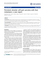
for CD68/PGM1) (Figure 1). Lymphocytes were more
often sm all and without significant atypia; a smaller per-
centage was represented by larger cells (Figures 1
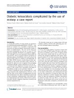
and 2). Immunohistochemistry (IHC) investigation
results show ed atypical l ymphocytes were CD79a
-
,
CD3
+
, CD2
+
, CD8
+
and TIA1
+
(Figure 2). ISH for EBER
demonstrated that the majority of lymphoid cells were
positive (Figure 2). Finally, polymerase chain reaction
(PCR) analysis revealed a monoclonal rearrangement of
the TCRg genes. IHC, ISH and molecular analyses were
carried out as previously described [9,10].
Based on the above findings, a final diagnosis of sys-
temic EBV+ T cell LPD of childhood was made. Our
patient was initially treated with two sequential doses of
VP16 with moderate improvement of his clinical and
laboratory data. In particular, the fever transiently
improved, hepatosplenomegaly was reduced, and coagu-
lation parameters were partially corrected; however,
severe pe ripheral blood cytopenia persisted. Soon after,
the patient developed a fever recrudescence in associa-
tion with pulmonary fungal infection.
Figure 1 Pathological findings on lymph node biopsy.Giemsa,
Ki67 and CD68 immunostains are shown. Arrows indicate atypical
cells (GM) as well as eritrophagocytic syndrome (CD68).
Tabanelli et al. Journal of Medical Case Reports 2011, 5:218
/>Page 2 of 5
A second bone marrow biopsy was performed, reveal-
ing (hypo)aplasia with a minimum percentage of CD79a
-
,CD3
+
,CD4
-
,CD8
+
,EBER
-
small lymphocytes and
absence of the previously observed CD8
+
large cells.
VP16 was then replaced with cyclosporine, obtaining a
white blood cell count increase and a further decline of
splenomegaly, but with no improvement in thrombocy-
topenia. A third bone marrow biopsy showed an
increased cellularity with reappearance of numerous
CD8
+
lymphocytes and evident hemophagocytosis.
Our patient then developed rectal hemorrhages only
treat able with surgery, which turned out to be sustained
by microvascular thrombosis on histological examina-
tion. Finally, after a short period of relative good health,
our patient had a rela pse of rectal bleeding and died
soon after with cerebral manifestations.
Discussion
Systemic EBV+ T cell LPD of childhood is a rare disor-
der characterized by an aggressive disease course and
dismal prognosis [5]. As death unfortunately often
occurs within a few weeks, and at present there is no
specific treatment, a prompt diagnosis is necessary. Our
case report highlights the fact that, though rare, such a
disease can occur also in Europe.
On reviewing the literature, we found only 14 cases
reported in Western countries [6,11-14], specifically
cases recorded in Europe and the USA (Table 1). Inter-
estingly, the majority of cases have been reported in
eastern Asia [5], specifically in Japan and Taiwan. The
geographical distri bution has been suggested to indicate
possible genetically determined defects in T cell
responses to EBV in certain populations.
Our review of Western cases showed that four of
those 14 patients developed a T cell LPD after CAEBV
infection [6,11], and 10 presented a fulminant EBV T
cell LPD following acute EBV infection. In the former
group, ethnic origin was specified only in one case
(white); in the latter g roup, one patient was of Cauca-
sian descent, four wer e Asian or Native American, while
in five cases the ethnic group was not specified. Mean
age at onset was 17 years and the male/fema le ratio was
2.3:1. Common symptoms were fever, hepatosplenome-
galy and hematophagocytic syndrome; the clinical
courses were fulminant in patients with T cell LPD after
acute IM. In particular, in the acute IM group three
cases had a CD8
+
phenotype, two a CD4
+
phenotype
and one showed double positivity for CD8 and CD4; in
one c ase phenotype was not interpretable and in three
casesitwasnotreported.TCRa presented with clonal
rearrangements in nine out of 10 patients and EBV gen-
ome was clonal in all but one case. In contrast, among
patients with T cell LPD after CAEBV infection two
were CD4
+
, one was CD45RO
+
and one presented with
an admixture of CD8
+
and CD4
+
lymphocytes; three
cases presented with monoclonal patterns with regard to
rearrangement of both TCRa genes and EBV genome.
In the remaining case, only the EBV positivity was
assessed.
In all described cases, an accurate diagnostic investiga-
tion including clinical, morphological, immunohisto-
chemical, and molecular analyses was necessary in order
to formulate a correct diagnosis. In particular, the differ-
ential diagnosis with aggressive NK cell leukemia was
based on surface sCD3 and CD8 positivity, CD56 nega-
tivity, and evidence of TCRa rearrangement in systemic
EBV
+
T cell LPDs, and also sCD3/CD8 negativity, CD56
positivity and germl ine TCRa patterns in aggressive NK
cell leukemia cases.
Conclusion
In conclusion, our case r eport underlines the impo r-
tance of a comprehensive diagnostic approach in t he
management of atypical EBV
+
LPDs. In fact, though, at
present, specific therapies are not available, the correct
Figure 2 Pathological findings on lymph node biopsy.CD3,
CD8, CD56, TIA1 immunostains and Epstein-Barr encoded RNA
(EBER) in situ hybridization are shown.
Tabanelli et al. Journal of Medical Case Reports 2011, 5:218
/>Page 3 of 5
description of rare disorders is essential for improving
current knowledge and possibly future therapeutic
approaches.
Consent
Written informed consent was obtained from the patient
for publication of this case report and any accompany-
ing images. A copy of the written consent is avail able
for review by the Editor-in-Chief of this journal.
Acknowledgements
This work was supported by Centro Interdipartimentale per la Ricerca sul
Cancro ‘G Prodi’, BolognAIL, AIRC (IG4987; IG1007; and 5xMille), RFO (to SAP
and PPP), Fondazione Cassa di Risparmio in Bologna, Fondazione della
Banca del Monte e Ravenna, Progetto Strategico di Ateneo 2006 (to SAP
and PPP).
Author details
1
Department of Hematology and Oncological Sciences ‘L and A Seràgnoli’,
Hematopathology Section, S Orsola-Malpighi Hospital, University of Bologna,
Bologna, Italy.
2
Hematology, Department of Cellular Biotechnologies and
Hematology, ‘Sapienza’ University, Rome, Italy.
Table 1 Cases of systemic Epstein-Barr virus positive (EBV
+
) T cell lymphoproliferative disease (LPD) of childhood
described in Western countries
Reference Age/
sex
Race Case description Time to
lymphoma
Histopathological
features
TCR
status
EBV
status
Jones et al.
[11]
two/M Unspecified Fever, generalized erythematous skin eruption,
hepatosplenomegaly, pancytopenia, hypoplastic
bone marrow, pulmonary infiltrates
six years Pulmonary large cell
lymphoma (phenotype:
CD4
+
, HLA-DR
+
)
TCR-b
rearranged
EBV
+
,
clonal
31/F Unspecified Fever, generalized lymphadenopathy,
hepatosplenomegaly, pancytopenia, diarrhea,
gastric pain
one year Lymphoblastic
lymphoma (phenotype:
CD4
+
, HLA-DR
+
)
TCR-bg
rearranged
EBV
+
,
clonal
55/M Unspecified Gluten enteropathy for 19 years; fever, persistent
diarrhea, nodular erythematous skin lesion
one year Peripheral T cell
lymphoma (phenotype:
UCHL1
+
)
- EBV
+
Gaillard et
al. [13]
seven/F Unspecified Infectious acute mononucleosis, persistent high-
grade fever, weight loss, adenopathy, necrotizing
skin lesions and VAHS
four
months
Fulminant EBV
+
T cell
LPD (phenotype: CD8
+
)
TCR-bg
rearranged
EBV
+
Craig et al.
[15]
20
months/
F
Unspecified Fever, generalized erythematous skin eruption,
hepatosplenomegaly
- T cell lymphoma NOS
(phenotype: not
interpretable)
TCR-b
rearranged
EBV
+
,
clonal
Quintanilla-
Martinez et
al.[6]
37/M White Fever, mental status of one week duration,
hepatosplenomegaly, pancytopenia, jaundice
- Fulminant EBV
+
T cell
LPD (phenotype: CD4
+
,
TIA1
+
)
TCR-g
rearranged
EBV
+
,
clonal
17/M Native
American
Symptoms of viral upper respiratory illness,
hepatosplenomegaly, pancytopenia, jaundice
- Fulminant EBV
+
T cell
LPD (phenotype: CD8
+
,
TIA1
+
)
TCR-g
rearranged
EBV
+
,
clonal
23/M Asian Fever, night sweats, weight loss,
hepatosplenomegaly, pancytopenia, jaundice,
generalized lymphadenopathy
- Fulminant EBV
+
T cell
LPD (phenotype: CD4
+
,
CD8
+
, TIA1
+
)
TCR-g
rearranged
EBV
+
,
clonal
22/F Native
American
Fever weight loss, hepatosplenomegaly, jaundice - Fulminant EBV
+
T cell
LPD (phenotype: CD4
+
,
TIA1
+
)
Polyclonal EBV
+
,
clonal
27
months/
M
Native
American
Fever, skin rash, hepatosplenomegaly,
pancytopenia
- Fulminant EBV
+
T cell
LPD (phenotype: CD8
+
,
TIA1
+
)
TCR-g
rearranged
EBV
+
,
clonal
15/F White IM at eight years old, followed by CAEBV. At 14
years old developed hepatosplenomegaly and
hemophagocytic syndrome.
- Fulminant EBV
+
T cell
LPD (phenotype: CD4
+
,
CD8
+
, TIA1
+
)
TCR-g
rearranged
EBV
+
,
clonal
Wick et al.
[14]
12/M Unspecified Hemophagocytic syndrome, FIM - Fulminant EBV
+
T cell
LPD (phenotype: not
reported)
TCR-bg
rearranged
EBV
+
,
clonal
three/F Unspecified Hemophagocytic syndrome, FIM - Fulminant EBV
+
T cell
LPD (phenotype: not
reported)
TCR-b
rearranged
EBV
+
,
clonal
nine/M Unspecified Hemophagocytic syndrome, FIM - Fulminant EBV
+
T cell
LPD (phenotype: not
reported)
TCR-b
rearranged
EBV
+
,
biclonal
CAEBV = chronic active EBV infevtion; FIM = fatal infectious mononucleosis; HLA = human leukocyte antigen; IM = infectious mononucleosis; NOS = not
otherwise specified; TCR = T cell receptor; VAHS = virus-associated hemophagocytic syndrome.
Tabanelli et al. Journal of Medical Case Reports 2011, 5:218
/>Page 4 of 5
Authors’ contributions
VT performed research, analyzed data and wrote the manuscript; CA
performed research and analyzed data; ES and FB analyzed data; SC and GM
were responsible for patient care and provided clinical information; AG, CM,
SR, and MTS analyzed data; SAP and PPP performed research, analyzed data
and wrote the manuscript. All authors read and approved the final
manuscript. VT and CA contributed equally to this work; SAP and PPP
contributed equally to this work.
Competing interests
The authors declare that the y have no competing interests.
Received: 18 August 2010 Accepted: 7 June 2011
Published: 7 June 2011
References
1. Straus SE, Cohen JI, Tosato G, Meier J: NIH conference. Epstein-Barr virus
infections: biology, pathogenesis, and management. Ann Intern Med
1993, 118:45-58.
2. Callan MF, Steven N, Krausa P, Wilson JD, Moss PA, Gillespie GM, Bell JI,
Rickinson AB, McMichael AJ: Large clonal expansions of CD8+ T cells in
acute infectious mononucleosis. Nat Med 1996, 2:906-911.
3. Ohshima K, Kimura H, Yoshino T, Kim CW, Ko YH, Lee SS, Peh SC, Chan JK:
Proposed categorization of pathological states of EBV-associated T/
natural killer-cell lymphoproliferative disorder (LPD) in children and
young adults: overlap with chronic active EBV infection and infantile
fulminant EBV T-LPD. Pathol Int 2008, 58:209-217.
4. Carbone A, Gloghini A, Dotti G: EBV-associated lymphoproliferative
disorders: classification and treatment. Oncologist 2008, 13:577-585.
5. Quintanilla-Martinez L, Kimura H, Jaffe E: EBV+ T-cell lymphoproliferative
disorders of childhood. In WHO Classification of Tumors of Hematopoietic
and Lymphoid Tissues 4 edition. Edited by: Swerdlow S, Campo E, Harris NL,
Jaffe ES, Pileri SA, Stein H, Thiele J, Vardiman J. Lyon: IARC; 2008:278-280.
6. Quintanilla-Martinez L, Kumar S, Fend F, Reyes E, Teruya-Feldstein J,
Kingma DW, Sorbara L, Raffeld M, Straus SE, Jaffe ES: Fulminant EBV(+) T-
cell lymphoproliferative disorder following acute/chronic EBV infection: a
distinct clinicopathologic syndrome. Blood 2000, 96:443-451.
7. Kasahara Y, Yachie A, Takei K, Kanegane C, Okada K, Ohta K, Seki H,
Igarashi N, Maruhashi K, Katayama K, Katoh E, Terao G, Sakiyama Y,
Koizumi S: Differential cellular targets of Epstein-Barr virus (EBV) infection
between acute EBV-associated hemophagocytic lymphohistiocytosis and
chronic active EBV infection. Blood 2001, 98:1882-1888.
8. Su IJ, Chen RL, Lin DT, Lin KS, Chen CC: Epstein-Barr virus (EBV) infects T
lymphocytes in childhood EBV-associated hemophagocytic syndrome in
Taiwan. Am J Pathol 1994, 144:1219-1225.
9. van Dongen JJ, Langerak AW, Brüggemann M, Evans PA, Hummel M,
Lavender FL, Delabesse E, Davi F, Schuuring E, García-Sanz R, van
Krieken JH, Droese J, González D, Bastard C, White HE, Spaargaren M,
González M, Parreira A, Smith JL, Morgan GJ, Kneba M, Macintyre EA:
Design and standardization of PCR primers and protocols for detection
of clonal immunoglobulin and T-cell receptor gene recombinations in
suspect lymphoproliferations: report of the BIOMED-2 Concerted Action
BMH4-CT98-3936. Leukemia 2003, 17:2257-2317.
10. Went P, Agostinelli C, Gallamini A, Piccaluga PP, Ascani S, Sabattini E,
Bacci F, Falini B, Motta T, Paulli M, Artusi T, Piccioli M, Zinzani PL, Pileri SA:
Marker expression in peripheral T-cell lymphoma: a proposed clinical-
pathologic prognostic score. J Clin Oncol 2006, 24:2472-2479.
11. Jones JF, Shurin S, Abramowsky C, Tubbs RR, Sciotto CG, Wahl R, Sands J,
Gottman D, Katz BZ, Sklar J: T-cell lymphomas containing Epstein-Barr
viral DNA in patients with chronic Epstein-Barr virus infections. New Engl
J Med 1988, 318:733-741.
12. Dolezal MV, Kamel OW, van de Rijn M, Cleary ML, Sibley RK, Warnke RA:
Virus-associated hemophagocytic syndrome characterized by clonal
Epstein-Barr virus genome. Am J Clin Pathol 1995, 103:189-194.
13. Gaillard F, Mechinaud-Lacroix F, Papin S, Moreau A, Mollat C, Fiche M,
Peltier S, De Faucal PJ, Rousselet MC, Praloran V, et al: Primary Epstein-Barr
virus infection with clonal T-cell lymphoproliferation. Am J Clin Pathol
1992, 98:324-333.
14. Wick MJ, Woronzoff-Dashkoff KP, McGlennen RC: The molecular
characterization of fatal infectious mononucleosis. Am J Clin Pathol 2002,
117:582-588.
15. Craig FE, Gulley ML, Banks PM: Posttransplantation lymphoproliferative
disorders. American journal of clinical pathology 1993, 99:265-276.
doi:10.1186/1752-1947-5-218
Cite this article as: Tabanelli et al.: Systemic Epstein-Barr-virus-positive T
cell lymphoproliferative childhood disease in a 22-year-old Caucasian
man: A case report and review of the literature. Journal of Medical Case
Reports 2011 5:218.
Submit your next manuscript to BioMed Central
and take full advantage of:
• Convenient online submission
• Thorough peer review
• No space constraints or color figure charges
• Immediate publication on acceptance
• Inclusion in PubMed, CAS, Scopus and Google Scholar
• Research which is freely available for redistribution
Submit your manuscript at
www.biomedcentral.com/submit
Tabanelli et al. Journal of Medical Case Reports 2011, 5:218
/>Page 5 of 5