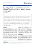
Báo cáo y học: "Opitz trigonocephaly syndrome presenting with sudden unexplained death in the operating room: a case report" docx
Bạn đang xem bản rút gọn của tài liệu. Xem và tải ngay bản đầy đủ của tài liệu tại đây (513.7 KB, 3 trang )
CAS E REP O R T Open Access
Opitz trigonocephaly syndrome presenting with
sudden unexplained death in the operating
room: a case report
Laura Travan
1*
, Vanna Pecile
2
, Mariacristina Fertz
1
, Antonella Fabretto
2
, Pierpaolo Brovedani
1
, Sergio Demarini
1
and
John M Opitz
3
Abstract
Introduction: Opitz trigonocephaly C syndrome (OTCS) is a rare malformation syndrome with the following
features: synostosis of metopic suture, craniofacial abnormalities, severe mental retardation and a multitude of
pathological findings affecting almost every organ system. OTCS is associated with a high mortality rate.
Case presentation: We describe the case of a Caucasian male baby who died at five months of age during
surgical correction of the craniofacial anomaly.
Conclusion: As previously reported, OTCS may have an increased mortality rate during craniofacial surgery. Careful
evaluation of surgery risk-be nefit ratio is warranted in such patients.
Introduction
Opitz trigonocephaly C sy ndr ome (OTCS) is a rare and
heterogeneous genetic disorder characterized by synos-
tosis of metopic suture, dysmorphic facial features, vari-
able mental retardation and other conge nital somatic
and cerebral anomalies. Morbidity and mortality are
very high. F ewer than 60 cases have been reported in
the literature, mostly as single case reports or small ser-
ies. We describe a white male baby who died at five
months of age during surgery performed to correct the
craniofacial anomaly.
Case Presentation
Our patient was a Caucasian baby, born to nonconsan-
guineous parents at 39 weeks of gestational age. This
was the first pregnancy of a 30-year-old mother with a
bicornuate uterus. Pregnancy was complicated by early
intrauterine growth retardation; antenatal ultrasound
assessment was otherwise reported as normal.
Labor and delivery were spontaneous. The Apgar
score was 9 and 10, respectively at one and five minutes.
Birth weight was 2470 g (< 3rd percentile, small for
gestational age), length was 46.7 cm (3
rd
to 10th percen-
tile), head circumference 33.1 cm (10th percentile).
At birth there was a marked trigonocephaly and other
dysmorphic craniofacial features: micro gnathia, upslant-
ing eyelids, hypotelorism, depressed nasal bridge, low set
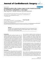
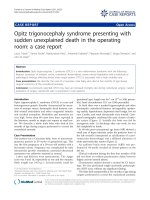
ears. Cardiac and renal ultrasounds were normal. Com-
puted tomography confirmed the early closure of meto-
picsuture(Figure1).Initiallythebabywasfedby
nasogastric tube. At discharge after one week, he was
fed completely by bottle.
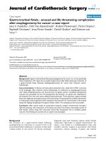
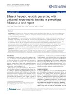
At 40 wks post-conceptional age brain MRI showed a
small area of hyper-intensity under the posterior horn of
the left ventricle (interpreted as calcification of a peri-
ventricular hemorrhage) and a diffused alteration of
white periventricular matter (Figure 2).
An auditory brain stem response (ABR) test per-
formed at 44 weeks revealed an absent pattern on the
left ear.
Clinical evaluation during the first four months of life
did not show an evident psychomotor delay; however
fidgety activity seemed absent.
Chromosome analysis showed a normal 46 XY karyo-
type. We also performed single-nucleotide polymorph-
ism (SNP) array without any significant finding.
The baby died unexpectedly at five months of age during
surgery performed to correct the craniofacial anomaly.
* Correspondence:
1
Neonatal Intensive Care Unit, Institute for Maternal and Child Health Burlo
Garofolo, Via dell’Istria 65/1, 34100, Trieste, Italy
Full list of author information is available at the end of the article
Travan et al. Journal of Medical Case Reports 2011, 5:222
/>JOURNAL OF MEDICAL
CASE REPORTS
© 2011 Travan et al; lic ense e BioMed Central Ltd. This is an Open Access article distributed under the te rms of the Creative Commons
Attribution License ( g/licenses/by/2.0), which permits unrestricted use, distribution, and reproduction in
any medium, provided the original work is properly cited.
Autopsy did not add anything to the clinical picture: speci-
fically, there were no additional anomalies except for a
double left renal artery. Some micro calcifications were
found around brain vessels.
Discussion
OTCS, first described in 1969 by Opitz [1] is character -
ized by trigonocephaly, m ental retardation, short neck,
typical facial appearance, joint and limb anomalies, up-
slanting palpebral fissures, epicantha l folds, a broad
depressed nasal bridge, small nose, abnormally low-set
ears, and central nervous system and visceral anomalies,
such as renal and heart anomalies.
OTCS is a heterogeneous genetic disorder which
occurs sporadically, although familial cases have also
been reported [2,3].
A very high mortality rate has been described: almost
50% o f patients with OTCS die within the first year of
life [3]. Some patients, ho wever, may have a good qual-
ity of life: Patient 2 of Lalatta [4] has normal IQ. She
underwent multiple c raniosynostectomies but she did
well at the University and was also able to play the
piano.
Our patient had many of the clinical and anatomic
findings typical of OTCS: the dysmorphic face, white
matter alteration, as described by Lalatta [4] and by
Azimi [5], cerebral hemorrhage [3] and hearing loss as
reportedbyNacarkucuketal.[6]andZampinoetal.
[7].
We did not find any genetic abnormality either in the
karyotype or in the region of CD96 gene, as r ecently
described by Kaname [8].
To the best o f our knowledge this is the second case
after patient 1 reported by Opitz [3] who died after sur-
gery for craniosyn ostosis repair. That patient, after the
skull reconstruction, developed hematuria, cardiac
arrhythmia and severe acidosis requiring cardiopulmon-
ary resuscitation. Twenty minutes later, he developed a
severe intra-vascular coagulation. After the autopsy,
experts in genetics, immunology and rheumatology con-
cluded that patient 1 of Optiz had a possible connective
tissue abnormal ity and increased vascular fragility that
started the catastrophic cascade that led to death.
Our patient died under the same circumstances as
patient 1 described by Optiz. Autopsy did not find vas-
cular malformation or connective tissue anomalies that
could have explained death during sur gery. However, as
in Opitz’ s patient 1 the cause of death was an unex-
pected massive bleeding.
Conclusion
OTCS is a complex and heterogeneous condition that is
still under-recognized and under-diagnosed. The fact
that two children died as a consequence of craniofaci al
Figure 1 Three-dimensional computerized tomography. See the fusion of metopic suture.
Travan et al. Journal of Medical Case Reports 2011, 5:222
/>Page 2 of 3
surgery may have clinical implications: diagnosing
OTCS in trigonocephalic patients before surgery, may
allow a better evaluation of risks and benefits of cranio-
synostosis repair.
Consent
Written informed consent was obtained from patient’ s
next-of-kin for publication of this case report and
accompanying images. A copy of the written consent is
available for review by the Editor-in-Chief of this
journal.
Author deta ils
1
Neonatal Intensive Care Unit, Institute for Maternal and Child Health Burlo
Garofolo, Via dell’Istria 65/1, 34100, Trieste, Italy.
2
Department of Genetics,
Institute for Maternal and Child Health Burlo Garofolo, Via dell’Istria 65/1,
34100, Trieste, Italy.
3
Departments of Pediatrics, Human Genetics, Obstetrics,
and Gynecology, University of Utah, Salt Lake City, UT, USA.
Authors’ contributions
LT made the patient diagnosis confirmed by JMO; they and SD were major
contributors in writing the manuscript. VP and FA performed and
interpreted the genetic analysis. MF and PB performed clinical evaluations.
All authors read and approved the final manuscript.
Competing interests
The authors declare that they have no competing interests.
Received: 17 November 2010 Accepted: 21 June 2011
Published: 21 June 2011
References
1. Opitz JM, Johnson RC, Mc Creadie SR, Smith DW: The C syndrome of
multiple congenital anomalies. In Birth Defects, Original Article Series.
Volume 2. Edited by: Bergsma D. New York: The National Foundation;
1969:161-166.
2. Antley RM, Hwang DS, Theopold W: Further delineation of the C
(trigonocephaly) syndrome. Am J Med Genet 1981, 9:147-163.
3. Opitz JM, Putnam AR, Comstock JM, Chin S, Byrne JL, Kennedy A, Frikke MJ,
Bernard C, Albrecht S, Der Kaloustian V, Szakacs JG: Mortality and
pathological findings in C (Opitz trigonocephaly) syndrome. Fetal Pediatr
Pathol 2006, 25:211-231.
4. Lalatta F, Clerici Bagozzi D, Salmoiraghi MG, Tagliabue P, Tischer C,
Zollino M, Di Rocco C, Neri G, Opitz JM: “C” trigonocephaly syndrome:
clinical variability and possibility of surgical treatment. Am J Med Genet
1990, 37:451-456.
5. Azimi C, Kennedy SJ, Chitayat D, Chakraborty P, Clarke JT, Forrest C,
Teebi AS: Clinical and genetic aspects of trigonocephaly: a study of 25
cases. Am J Med Genet A 2003, 117A:127-135.
6. Nacarküçük E, Okan M, Sarimehmet H, Ozer T: Opitz trigonocephaly C
syndrome associated with hearing loss. Pediatr Int 2003, 45:731-733.
7. Zampino G, Di Rocco C, Butera G: Opitz C trigonocephaly syndrome and
midline brain anomalies. Am J Med Genet 1997, 73:484-488.
8. Kaname T, Yanagi K, Chinen Y, Makita Y, Okamoto N, Maehara H, Owan I,
Kanaya F, Kubota Y, Oike Y, Yamamoto T, Kurosawa K, Fukushima Y,
Bohring A, Opitz JM, Yoshiura K, Niikawa N, Naritomi K: Mutations in CD96,
a Member of the Immunoglobulin Superfamily, Cause a Form of the C
(Opitz Trigonocephaly) Syndrome. Am J Med Genet A 2007, 81:835-841.
doi:10.1186/1752-1947-5-222
Cite this article as: Travan et al.: Opitz trigonocephaly syndrome
presenting with sudden unexplained death in the operating room: a
case report. Journal of Medical Case Reports 2011 5:222.
Submit your next manuscript to BioMed Central
and take full advantage of:
• Convenient online submission
• Thorough peer review
• No space constraints or color figure charges
• Immediate publication on acceptance
• Inclusion in PubMed, CAS, Scopus and Google Scholar
• Research which is freely available for redistribution
Submit your manuscript at
www.biomedcentral.com/submit
Figure 2 Cerebral MRI (whitened T2 sequences), showing
diffuse white matter periventricular hyperintensity
(hypointensity in T1 sequences).
Travan et al. Journal of Medical Case Reports 2011, 5:222
/>Page 3 of 3