Báo cáo y học: "Chylopericardium after cardiac surgery can be treated successfully by oral dietary manipulation: a case report" docx
Bạn đang xem bản rút gọn của tài liệu. Xem và tải ngay bản đầy đủ của tài liệu tại đây (209.36 KB, 3 trang )
BioMed Central
Page 1 of 3
(page number not for citation purposes)
Journal of Cardiothoracic Surgery
Open Access
Case report
Chylopericardium after cardiac surgery can be treated successfully
by oral dietary manipulation: a case report
Sing Yang Soon*, Sharath Hosmane and Paul Waterworth
Address: South Manchester University Hospital NHS Trust, Southmoor Road, Manchester, M23 9LT, UK
Email: Sing Yang Soon* - ; Sharath Hosmane - ;
Paul Waterworth -
* Corresponding author
Abstract
We report a case of chylopericardium after ascending aorta and aortic valve replacement, which
presented as late tamponade. We discuss the various treatment options in this rare condition
which can result in serious morbidity or death.
Introduction
Chylopericardium after intra-thoracic surgery is rare. Its
incidence is reported to be between 0.22% to 0.5% [1,2]
following paediatric cardiac surgery but is not quantified
following cardiac surgery in the adult population. A delay
in diagnosis can lead to serious consequences with tam-
ponade and death [3]. Chronic lymph leak can also lead
to immunosuppresion, hypoproteinemia and malnutri-
tion [3]. The majority of published literatures on this con-
dition after cardiac surgery are in children. There are few
reports of chylopericarium in adults following coronary
artery bypass surgery and valvular surgery [4-6], and these
advocate treatment with either total parenteral nutrition
or surgical intervention. We report on the first case of chy-
lopericardium after ascending aorta and aortic valve
replacement in an adult patient treated successfully by
oral dietary manipulation.
Case report
A 52 years old man who presented with an incidental
finding of an aortic regurgitant murmur underwent fur-
ther investigations which reveal a dilated ascending aorta
(5.1 cm at its widest point) and associated aortic regurgi-
tation. There was no other significant past medical his-
tory. He subsequently underwent aortic valve replacement
with a mechanical prosthesis and also ascending aorta
replacement with a PTFE interposition tube graft. The
thymic fat was divided in the midline. Cardiopulmonary
bypass was established with a single two-stage venous
cannula and aortic return was to left femoral artery. There
was no intra-operative complication and the patient made
an uneventful post-operative recovery. He was discharged
on the 8
th
post-operative day at which time he was well
and a chest x-ray did not show any signs of cardiomegaly.
The patient represented on the 12
th
post operative day
with increasing shortness of breath, accompanied by nau-
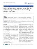
sea and vomiting. Chest x-ray showed gross cardiomegaly
(fig 1). Echocardiography demonstrated a 6.5 cm pericar-
dial effusion with diastolic right ventricular collapse. A
pericardial pigtail catheter was inserted for relief of tam-
ponade with drainage of 3.0 litres of milky white fluid.
Subsequent biochemical and microbiological analysis
confirmed sterile chyle.
Due to presence of the prosthetic aortic valve and Dacron
graft, our aim was to avoid total parenteral nutrition with
its attendant risk of prosthetic infection. Therefore, a deci-
sion was undertaken to treat the chylopericardium by a
trial of oral dietary manipulation with medium chain trig-
Published: 18 August 2009
Journal of Cardiothoracic Surgery 2009, 4:44 doi:10.1186/1749-8090-4-44
Received: 29 January 2009
Accepted: 18 August 2009
This article is available from: />© 2009 Soon et al; licensee BioMed Central Ltd.
This is an Open Access article distributed under the terms of the Creative Commons Attribution License ( />),
which permits unrestricted use, distribution, and reproduction in any medium, provided the original work is properly cited.
Journal of Cardiothoracic Surgery 2009, 4:44 />Page 2 of 3
(page number not for citation purposes)
lycerides/fat free diet. The second day after pericardiocen-
tesis was performed, the drainage was still substantial at
1.5 litres. However on the third day, the drainage tailed
dramatically to 150 ml. The patient was brought to theatre
for creation of a subxiphoid pericardial window with
insertion of 32F drain for more effective drainage. The
chyle leak continued to diminish in volume over the next
five days, without any drainage by day eight. However on
application of low pressure (10 cm of water) suction on
day nine, a small piece of debri was dislodged from the
drain and there was a sudden drainage of 450 ml of chyle.
Therefore thrice daily low pressure suction was instituted.
The patient spiked a temperature the following day to
39.5 degree Celsius. A full septic screen was performed
including blood cultures and the chyle was sent for micro-
biological analysis. Initially, the patient was commenced
on broad spectrum antibiotics. Subsequently, gram nega-
tive bacilli were found to be growing in both the blood
cultures and the chyle. Treatment with meropenem was
instituted. The patient responded to the antibiotics treat-
ment and became apyrexial after seven days.
By day 20 post readmission, the drainage had tailed off to
less than 20 ml per day. The patient was subsequently
commenced on a normal diet. The drain output was
observed closely for 5 days after reinstitution of normal
diet. There was no further chyle leak. An echocardiogram
confirmed no re-accumulation in the pericardial sac and
the drain was therefore removed and the patient dis-
charged. The white cell, lymphocyte and albumin count
remained within normal limits throughout the patient's
readmission even during the septic episode.
Discussion
Chylopericardium after cardiac surgery is rare and there-
fore a high index of suspicion is required for its diagnosis.
Its aetiology is usually due to disruptions of the tributaries
of the thoracic duct rather than to the main duct itself [2].
The thoracic duct originates as the cisterna chili adjacent
to the second lumbar vertebrae. It ascends anterior to the
vertebral bodies and enters the thorax through the aortic
hiatus. It is a predominantly right sided structure and
crosses over to the left at the level of the fourth and fifth
thoracic vertebrae. It empties the lymph that it transports
into the left jugulosubclavian venous junction. It has a
highly variable intra-thoracic course. There are also vari-
ous tributaries found in the pericardial reflections and
thymic tissues that confluences to the thoracic duct [1,7].
Therefore, one should ensure that division through the
thymic tissues and pericardium be conducted carefully to
prevent subsequent chyle leakage. Other causative factors
include caval obstruction, subclavian vein thrombosis,
congenital lymphangiectasia, filariosis and medistinal
tumors [8].
Chyle leak is suspected with the appearance of milky efflu-
ent in the chest drain. Confirmation comes with biochem-
ical analysis of the fluid that reveals presence of
chylomicrons, cholesterol, lactate dehydrogenase and
protein [8-10]. Cytology usually demonstrates a lym-
phocytic picture while microbiological culture is invaria-
bly sterile.
Upon diagnosis, there are various treatment options avail-
able. Although nutritional support with parenteral hyper-
alimentation has been advocated as the method of choice
[6,9,11], we advocate one of minimal intervention with a
dual strategy of decreasing lymph production and ensur-
ing adequate protein intake to counter any effects of the
potential hypoproteineamia. As first line management,
the patient should be commenced on a trial of enteral
nutrition with a fat free diet or a low-fat diet with medium
chain triglycerides, which are absorbed directly into the
portal system rather than through the lymphatics. This
would reduce the production of lymph and allow the
spontaneous closure of the fistula in the majority of cases.
This option is also more palatable for the patient and
avoids the potential complications of total parenteral
nutrition. It also has the added theoretical benefit of pro-
moting normal gut flora and preventing translocation of
pathogens in a patient that might be leukopenic. Care
must also be given to ensure that the patient has adequate
caloric and nitrogen intake in a highly catabolic state.
Pericardial decompression should be achieved with either
a pig-tail catheter inserted under echocardiography or the
insertion of a drain with the creation of a subxiphoid win-
dow
CXR showing patients enlarged mediastinal shadowing from chylopericardiumFigure 1
CXR showing patients enlarged mediastinal shadow-
ing from chylopericardium.
Journal of Cardiothoracic Surgery 2009, 4:44 />Page 3 of 3
(page number not for citation purposes)
The duration of treatment is variable, but typically lasts
for 7 to 21 days [1,7,12]. Regular monitoring of the albu-
min and leukocyte count should be carried out to assess
the nutritional and immunological status during the
length of enteral/parenteral treatment. Consistent fall of
both of these counts are relative indications for operative
intervention should the chyle drainage be small (less than
500 ml/day) yet persistent. Cessation of chyle drainage
usually indicates successful treatment. However, it is pru-
dent to request a repeat echocardiography to assess peri-
cardial effusion prior to drain removal in the event of
drain blockage with debri.
If the above measures do not result in the resolution of the
chyle leak, operative intervention needs to be considered.
There is no clear consensus about indications for surgery
but it has been recommended that if chyle drainage is
greater than 500 ml per day for 5 consecutive days or fail-
ure of conservative treatment after 14 days or if metabolic
complications developed [7,13,14].
The identification of the site of chyle leak can be problem-
atic. A lymphangiogram can be performed preoperatively
to give an indication of the area where the leakage is situ-
ated. Other measures to assist in the location of chyle leak
include asking the patient to consume methylene blue or
a high fat cream one hour prior to the surgery [9]. The area
involved would stain blue or exude thick milky fat at the
time of operation.
After localizing the culprit lesion, ligaclips or simple liga-
tures could be employed to deal with the problem. Prob-
lems arise when one fail to localize the site of drainage.
Mass ligature of the thymic tissues and diathermy of the
pericardial reflection should be carried out on a "best
guess" basis. Plication of all the tissues anterior to the ver-
tebral bodies from the level of the azygous vein to the
level of the proximal descending aorta has also been advo-
cated [8]. Other options of intervention include right
sided video assisted thoracoscopic ligation of the thoracic
duct [7] which has been reported to be without any rate of
recurrence at four years.
In conclusion, patients with chylopericardium after car-
diac surgery can potentially be treated effectively with oral
dietary manipulation with a medium chain triglyceride
diet and effective pericardial decompression. This
approach would reduce the complications associated with
total parenteral nutrition and the attendant morbidities of
surgical interventions. Monitoring of the rate of chyle
leakage will guide subsequent therapy.
Consent
Written informed consent was obtained from the patient
for publication of this case report and accompanying
images. A copy of the written consent is available for
review by the Editor-in-Chief of this journal.
Competing interests
The authors declare that they have no competing interests.
Authors' contributions
SYS – Manuscript writeup, SH – Carried out image scan-
ning, patient consent, manuscript upload and revision.
PW – Senior author, provided guidance and input on
manuscript writeup. All authors read and approved the
final manuscript.
References
1. Campbell RM, Benson LN, Williams WW, Adatia IA: Chylopericar-
dium after cardiac operations in children. Ann Thorac Surg
2001, 72:193-6.
2. Shanmugam G, Sundar P, Shukla V, Korula RJ: Chylopericardium
after atrial septal defect repair: an unusual entity. Int J Thorac
Cardiovasc Surg 2003, 19:124-125.
3. Patterson GA, Todd TRJ, Delarue NC, Ilves R, Pearson FG, Copper
JD: Supradiaphragmatic ligation of the thoracic duct in
intractable chylous fistula. Ann Thorac Surg 1981, 32:44-9.
4. Thomas CS Jr, McGoon : Isolated massive chylopericardium fol-
lowing cardiopulmonary bypass. J Thorac Cardiovasc Surg 1971,
61:945-7.
5. Pollard WM, Schuchmann GF, Bowen TE: Isolated chylopericar-
dium after cardiac operations. J thorac Cardiovasc Surg 1981,
81:943-6.
6. Sharpe DAC, Pullen MDM, McGoldrick JP: A minimally invasive
approach to Chylopericardium after coronary artery sur-
gery. Ann Thorac surg 1999, 68:1062-3.
7. Wurnig PN, Hollaus PH, Ohtsuka T, Flege JB, Wolf RK: Thoraco-
scopic direct clipping of the thoracic duct for chylopericar-
dium and chylothorax. Ann Thorac Surg 2000, 70:1662-5.
8. Sleilaty G, Rassi I, Alawi A: Primary isolated chronic chyloperi-
cardium. Int Cardiovasc Thorac Surg 2002, 1:86-7.
9. Furrer M, Hopf M, Ris HB: Isolated primary chylopericardium:
treatment by thoracoscopic thoracic duct ligation and peri-
cardial fenestration. J Thorac Cardiovasc Surg 1996, 112:1120-1.
10. Sakata S, Yoshida I, Otani Y, Ishikawa S, Morishita Y: Thoracoscopic
treatment of primary pericardium. Ann Thorac Surg 2000,
69:1581-2.
11. Hashim SA, Roholt HB, Babayan VK, Itallie TB: Treatment of chy-
luria and chylothorax with medium chain triglyceride.
N Engl
J Med 1964, 270:756-61.
12. Nruyen D, Shum-Tim D, Dobell AR, Tchervenkov CI: The manage-
ment of chylothorax/chylopericardium following pediatric
cardiac surgery: a 10 year experience. J Card Surg 1995,
10(4):302-8.
13. Selle JG, Snyder WH, Schreiber JT: Chylothorax: indications for
surgey. Ann Surg 1973, 177:245-9.
14. Crosthwaite GL, Joypaul BV, Cuschieri A: Thoracoscopic manage-
ment of thoracic duct injury. J R Coll Surg Edin 1995, 40:303-4.