Báo cáo y học: "Hemangioma related to Maffucci syndrome in a man: a case report" docx
Bạn đang xem bản rút gọn của tài liệu. Xem và tải ngay bản đầy đủ của tài liệu tại đây (682.75 KB, 2 trang )
CAS E REP O R T Open Access
Hemangioma related to Maffucci syndrome in a
man: a case report
Takeshi Kondo
Abstract
Introduction: Maffucci syndrome is a rare clinical entity (app roximately 200 cases have been reported in the
medical literature) with a combined occurrence of multiple enchondromas and vascular tumors.
Case presentation: The case of a 43-year-old Japanese man with multiple chondromas and hemangiomas
(Maffucci syndrome) is reported. One of the hemangiomas was removed and examined pathologically. The
morphological picture was an admixture of cavernous hemangioma and spindle cell hemangioma without
cytological atypia or mitosis. Sheets of vacuolated endothelial cells were also observed.
Conclusion: A rare case of hemangioma associated with Maffucci syndrome, focusing on the pathologic nature of
the submitted tissue, is reported.
Introduction
Maffucci syndrome is a rare clinical entity (approxi-
mately 200 cases have b een reported in the medical lit-
erature) [1]. It consists of combined occurrence of
multiple enchondromas and vascular tumors. This syn-
drome is not inherited and shows female predilection.
Case presentation
A 43-year-old Japanese man presented with multiple
chondromas and hemangiomas. His disease had been
diagnosed as Maffucci syndrome. His available clinical
information, however, was limited. One of the heman-
giomas was removed and examined pathologically.
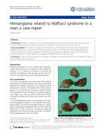
Macroscopically, the lesion showed a serpentin e appear-
ance (Figure 1A). The cut surface of the lesion showed
a blackish area filled with blood and a whitish area (Fig-
ure 1B).
The morphological picture was an admixture of caver-
nous hemangioma (Figure 2A) and spindle cell heman-
gioma (Figure 2B) without cytological atypia or mitosis.
Sheets of vacuolated endothelial cells were also observed
(Figure 2C). In the cavernous component, organized
thrombosis was observed (image not shown). No epithe-
lioid hemangiomatous area was found.
Correspondence:
Division of Legal Medicine, Department of Community Medicine and Social
Healthcare Science, Kobe University Graduate School of Medicine, 7-5-1
Kusunoki-cho, Chuo-ku, Kobe 650-0017, Japan
A
B
B
Figure 1 Macroscopic findings of the lesion. (A) The lesion had a
serpentine appearance. (B) The cut surface of the lesion. It had a
blackish area filled with blood and a whitish area.
Kondo Journal of Medical Case Reports 2011, 5:224
/>JOURNAL OF MEDICAL
CASE REPORTS
© 2011 Kondo; licensee BioMed Central Ltd. This is an Open Access article distri buted under the terms of the Creative Commons
Attribution License ( which permits unrestricted use, distribution, and reproductio n in
any medium, provided the original work is properl y cited.
Discussion
Most patients with Maffucci syndrome present at birth
or in early childhood with hemangiomas varying in siz e
from a few millimeters to several centimeters which are
typically located in t he dermis or subcutaneously on the
distal parts of the limbs. Hemangiomas, however, may
also be found in internal organs [2]. The most common
vascular lesions to occur in association with Maffucci
syndrome are spindle cell hemangiomas, although
occasional cases of lymphangiomas, arteriovenous mal-
formations, and angiosarcomas have also been reported
[3]. Thus, treatment for Maffucci syndrome should be
aimed at early detection of malignant transformation as
well as at symptom relief [4]. The problem could be
how to do the follow-up of multiple hemangiomas
located in the internal organs, how to analyze their his-
tology, and which lesions to biopsy. In this patient, a
histologically benign composite type hemangioma
(cavernous and spindle cell hemangioma) was found,
and no sarcomatous area was observed. Follow-up of
the patient has revealed no signs of malignant transfor-
mation for years. Careful follow-up is, however, needed.
Conclusion
In conclusion, a rare case of hemangioma associated
with Maffucci syndrome has been reported, with a focus
on the pathologic findings of the submitted tissue.
Consent
Written informed consent was obtained from the patient
for publication of this case report and any accompany-
ing images. A copy of the written consent is available
for review by the Editor-in-Chief of this journal.
Authors’ contributions
TK performed the histological examination, analyzed the case, and wrote the
manuscript.
Competing interests
The author declares that they have no competing interests.
Received: 29 July 2010 Accepted: 21 June 2011 Published: 21 June 2011
References
1. Amezyane T, Bassou D, Abouzahir A, Fatihi J, Akhaddar A, Mahassin F,
Ghafir D, Ohayon V: A young woman with Maffucci syndrome. Intern Med
2010, 49:85-86.
2. Mertens F, Unni K: Enchondromatosis: Ollier disease and Maffucci
syndrome. In Pathology and Genetics of Tumours of Soft Tissue and Bone.
Edited by: Fletcher CDM, Unni K, Mertens F. Lyon, France: IARC Press;
2002:356-357.
3. Fukunaga M, Suzuki K, Saegusa N, Folpe AL: Composite
hemangioendothelioma: report of 5 cases including one with associated
Maffucci syndrome. Am J Surg Pathol 2007, 31:1567-1572.
4. Lissa FC, Argente JS, Antunes GN, de Oliveira Basso F, Furtado J: Maffucci
syndrome and soft tissue sarcoma: a case report. Int Semin Surg Oncol
2009, 6:2.
doi:10.1186/1752-1947-5-224
Cite this article as: Kondo: Hemangioma related to Maffucci syndrome
in a man: a case report. Journal of Medical Case Reports 2011 5:224.
A
B
C
Figure 2 Microscopic image sho wing the lesion. (A) Cavernous
hemangiomatous component (hematoxylin and eosin stain; original
magnification, ×100). (B) Component of spindle cell hemangioma
(hematoxylin and eosin stain; original magnification, ×100). (C)
Sheets of vacuolated cells (hematoxylin and eosin stain; original
magnification, ×200).
Kondo Journal of Medical Case Reports 2011, 5:224
/>Page 2 of 2