Báo cáo y học: " A solitary primary subcutaneous hydatid cyst in the abdominal wall of a 70-year-old woman: a case report" pptx
Bạn đang xem bản rút gọn của tài liệu. Xem và tải ngay bản đầy đủ của tài liệu tại đây (1.33 MB, 3 trang )
CAS E REP O R T Open Access
A solitary primary subcutaneous hydatid cyst
in the abdominal wall of a 70-year-old woman:
a case report
Abdelmalek Ousadden
*
, Hicham Elbouhaddouti, Karim Hassani Ibnmajdoub, Khalid Mazaz and Khalid AitTaleb
Abstract
Introduction: A solitary primary hydatid cyst in the subcutaneous abdominal wall is an exceptional entity, even in
countries where the Echinococcus infestation is endemic.
Case presentation: We report a case of a 70-year-old Caucasian woman who presented to our hospital with a
subcutaneous mass in the para-umbilical area with a non-specific clinic al presentation. The diagnosis of
subcutaneous hydatid cyst was suspected on the basis of radiological findings. A complete surgical resection of
the mass was performed and the patient had an uneventful post-operative recovery. The histopathology confirmed
the suspected diagnosis.
Conclusion: Hydatid cyst should be considered in the differential diagnosis of every subcutaneous cystic mass,
especially in regions where the disease is endemic. The best treatment is the total excision of the cyst with an
intact wall.
Introduction
Hydatid disease is a parasitic infestation that is caused
by Echinococcus granulosis, the life cycle of which has
been well described [1]. Endemic areas are countries of
the t emperate zones, where the common intermediate
hosts, sheep, goats, and cattle, are raised, such as in
North Africa, the Middle East, Central Europe, Austra-
lia, and South America [1,2]. The liver is the most fre-
quently involved organ (75%), followed by the lung
(15%) [2,3]. The solitary primary subcutaneous localiza-
tion is extremely rare, and its incidence is unknown [2].
In our patient, the hydatid cyst was located in the abdo-
men anterior wall without any other involve ment, whic h
makes this an interesting case.
Case presentation
A 70-year-old Moroccan Caucasian woman presented to
our hospital with a subcutaneous cystic mass in the
right para-umbilical abdominal wall which had been
evolving for six months. Her physical examination
revealed an abdominal parietal mass 6 cm in diameter
that was palpated 5 cm to the rig ht of the umbilicu s. It
was cystic, fluctuant, mobile, and painless. The overlying
skin was normal. An abdominalultrasoundshoweda
rounded cystic mass that was limited within the right
para-umbilical abdominal wall and measured 60 mm.
No other abdominal cystic mass was found. The pre-
operative examinations (chest radiograph, complete
blood count, urine analysis, and blood biochemistry)
revealed no abnormalities. The hydatid serology was
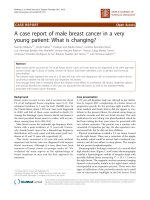
negative. Surgical exploration reveale d that the mass
was attached to the subcutaneous adipose tissue but was
not associated with any muscular or cutaneous structure
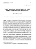
(Figure 1). The macroscopic appearance suggested a
hydatidcyst(Figure2).Perforationwasavoidedby
means of meticulous dissection. The histopathologic
examination of t he specimen revealed a hydatid cyst.
The patient has been followed for two years, and no
recurrence of hydatidosis has been detected.
Discussion
The mechanism of the primary subcutaneous localiza-
tion is unclear [2,4]. The ingested parasite’sovapene-
trate the intestinal wall, join the portal system, and
reach the liver, where most of them are caught in the
* Correspondence:
Service de Chirurgie Viscérale, Hôpital des Spécialités, CHU Hassan II, Route
de Sidi Harazem, Fès 30070, Morocco
Ousadden et al. Journal of Medical Case Reports 2011, 5:270
/>JOURNAL OF MEDICAL
CASE REPORTS
© 2011 Ousadden et al; licensee BioMed Central Ltd. This is an Open Access arti cle distributed under the terms of the Creative
Commons Attribution Lice nse ( which permits unrestricted use, distribution, and
reproduction in any medium, provided the original work is properly cited.
hepatic sinusoids [2]. A few ova may pass through the
liver (first filter) and reach the lung (second filter) and
the systemic circulation, causing hydatid disease in
other organs [1,2]. A possible dissemination through
lymphatic channels has also been reported. This
accounts for cases with solitary cysts in uncommon sites
[3-5]. The direct spread from adjacent sites may be
another mechanism of infection [6].
In our case, the hydatid cyst was located subcuta-
neously. The patient had not undergone previou s sur-
gery for any hydatid cysts, which were never found in
other organs. Therefore, our patient was diagnosed as
having a primary subcutaneous hydatid cyst.
In a large series of patients from Greece, the fre-
quency of extra-hepatic and extra-pulmonary hydatido-
sis was 9% [5]. However, in different series, the
frequency of subcutaneous tissue involvement, which is
usually associ ated with involvement of other solid
organs, has been re ported to be approximately 2%
[1,7,8]. Primary isolated hydatid cysts located in the
abdo minal wall rem ain extremely rare, however, even in
geographic areas in which echinococcal infestation is
frequent [3,4].
The clinical course is non-specific and depends on the
site of involvement, the size of the cyst, and the pressure
caused b y the enlarged cyst [1]. Usually, it presents as
an inert, painless, non-inflammatory mass without any
deterioration of the patient’ s general condition [4,9].
However, if super-infected or cracked, the cyst can
simulate an abscess or a cancer [8,9].
Radiological imaging (ultrasonography, computed
tomography, and MRI) is useful in rendering the diag-
nosis, showing the size, localization, relationship to adja-
cent organs, and type of the cyst. It can also be used to
search for another hydatid location [1,4] . The radiolog i-
cal findin gs of a thick cyst wall, calcif ications, daughter
cysts, and a germinative membrane separated from the
cyst wall are all specific to hydatid cysts [1-4]. Enhance-
ment of the peri-cystic soft tissues can be considered an
MRI feature suggestive of soft- tissue hydatid disease [9].
Serology is a useful tool that confirms the diagnosis,
although it is rarely positive for cysts in extra-hepatic
and extra-pulmonary locations (25%) [1,4,8]. It is
furthermore associated with fa lse-negative and false-
positive results [4].
The best treatment option is complete surgical excision
of the intact cyst, which avoids leakage of cyst content
that can cause anaphylaxis and local recurrence [1,2,8]. If
the ideal surgery is impossible, the cyst content (fluid,
membrane, and daughter cysts) has to be removed intra-
operatively and the cyst pouch has to be irrigated with
scolicidal solutions [1,2]. Other options include percuta-
neous treatment under ultrasound guidance with needle
aspiration irrigation of scolicidal solutions, as well as
medical treatment with the use of albendazole [2,8].
Conclusion
Hydatid cyst should be considered in the differential
diagnosis o f every subcutaneous c ystic mass, especially
in regions where the disease is endemic. The best treat-
ment is the total excision of the cyst with an intact wall.
Consent
Written informed consent was obtained from the patient
for publication of this case report and any accompany-
ing images. A copy o f the written consent is available
for review by the Editor-in-Chief of this journal.
Acknowledgements
The authors thank the patient for providing her written consent for the
publication of this case report. We also thank IbnMajdoub Hassani Soukaina
Figure 1 Peri-operative view of the subcutaneous hydatid cyst.
Figure 2 Image of the totally excised hydatid cyst.
Ousadden et al. Journal of Medical Case Reports 2011, 5:270
/>Page 2 of 3
(Faculté des lettre Saiss/Université Sidi Mohamed Ben Abdellah) for her help
in correcting this manuscript.
Authors’ contributions
AO, KA, and HE operated on the patient. KHI took the photos. KM
participated in follow-up. All authors participated in writing the case report
and revising the draft. All authors read and approved the final manuscript.
Competing interests
The authors declare that they have no competing interests.
Received: 23 February 2010 Accepted: 2 July 2011
Published: 2 July 2011
References
1. Orhan Z, Kara H, Tuzuner T, Sencan I, Alper M: Primary subcutaneous cyst
hydatic disease in proximal thigh: an unusual localisation: a case report.
BMC Musculoskelet Disord 2003, 7:25.
2. Dirican A, Unal B, Kayaalp C, Kirimlioglu V: Subcutaneous hydatid cysts
occurring in the palm and the thigh: two case reports. J Med Case
Reports 2008, 13:273.
3. Ok E, Sözüer EM: Solitary subcutaneous hydatid cyst: a case report. Am J
Trop Med Hyg 2000, 62:583-584.
4. Bedioui H, Makni A, Nouira K, Mekni A, Daghfous A, Ayadi S, Rebai W,
Ksantini R, Chebbi E, Fteriche F, Ammous A, Jouini M, Kacem M, Ben
Safta Z: [Subcutaneous hydatid cyst: case report of an exceptional
location] [in French]. Med Trop (Mars) 2007, 67:181-182.
5. Prousalidis J, Tzardioglou K, Sgouradis L, Katsohis C, Aletras H: Uncommon
sites of hydatid disease. World J Surg 1998, 22:17-22.
6. Safioleas M, Nikiteas N, Stamatakos M, Safioleas C, Manti CH, Revenas C,
Safioleas P: Echinococcal cyst of the subcutaneous tissue: a rare case
report. Parasitol Int 2008, 57:236-238.
7. Chevalier X, Rhamouni A, Bretagne S, Martigny J, Larget-Piet B: Hydatid
cyst of the subcutaneous tissue without other involvement: MR imaging
features. AJR Am J Roentgenol 1994, 163:645-646.
8. Rezakhaniha B, Sirosbakht S: Coincidental hydatid cyst of skin and kidney:
a very rare case report. Iran J Pathol 2010, 5:47-50.
9. Alouini Mekki R, Mhiri Souei M, Allani M, Bahri M, Arifa N, Jemni Gharbi H,
Kochtali H, Tlili Graiess K: [Hydatid cyst of soft tissues: MR imaging
findings. Report of three cases] [in French]. J Radiol 2005, 86:421-425.
doi:10.1186/1752-1947-5-270
Cite this article as: Ousadden et al.: A solitary primary subcutaneous
hydatid cyst in the abdominal wall of a 70-year-old woman: a case
report. Journal of Medical Case Reports 2011 5:270.
Submit your next manuscript to BioMed Central
and take full advantage of:
• Convenient online submission
• Thorough peer review
• No space constraints or color figure charges
• Immediate publication on acceptance
• Inclusion in PubMed, CAS, Scopus and Google Scholar
• Research which is freely available for redistribution
Submit your manuscript at
www.biomedcentral.com/submit
Ousadden et al. Journal of Medical Case Reports 2011, 5:270
/>Page 3 of 3