Báo cáo y học: " Collet-Sicard syndrome as an initial presentation of prostate cancer: a case report" doc
Bạn đang xem bản rút gọn của tài liệu. Xem và tải ngay bản đầy đủ của tài liệu tại đây (1.25 MB, 3 trang )
CAS E REP O R T Open Access
Collet-Sicard syndrome as an initial presentation
of prostate cancer: a case report
Rosa Villatoro
1*
, Carlos Romero
2
and Antonio Rueda
1
Abstract
Background: Collet-Sicard syndrome is caused by lesions at the base of the skull affecting the lower cranial nerves.
It is associated with various etiologies of tumoral and other origin. Although this syndrome has been reported
previously in the literature, most cases are diagnosed as part of primary disease follow-up. This case is unusual
because of the diagnosis of bone metastasis secondary to prostate cancer.
Case presentation: We present the case of a 70-year-old Caucasian man with a three-week history of headache
and maxillary pain on the right side together with paresis of the low cranial nerves. This study was carried out with
a computed tomography (CT) scan of the larynx and neck and MRI, which revealed a bone lesion at the base of
the skull affecting the right occipital condyle and part of the right side of the basilar bone. On the basis of
differential diagnosis, a fibrous dysplasia, Paget’s disease or metastasis was considered. Finally, and after other
studies were performed, a diagnosis of bone metastasis secondary to prostate cancer was established.
Conclusions: We think that this case is curious because it involved an initial presentation of metastatic prostate
cancer. It is important this should be considered in the differential diagnosis when a patient with unusual clinical
findings is first seen in view of the fact that first-line hormonal treatment may control the disease for months or
years.
Background
Collet-Sicard syndrome is caused by lesions at the base of
the skull affecting the lower cranial nerves, which pro-
duces dysphonia, displacement of the palate, and atony of
the trapezius muscle and sternocleidomastoid, as well as
anesthesia of the larynx, pharynx and soft palate. It is asso-
ciated with various etiologies of tumoral and other origins.
The differential diagnosis is important. Among the non-
tumoral factors causing Collet-Sicard syndrome, the most
common are traumatic events (fractures at the base of the
skull, aneurisms, and so on), inflammatory processes
(osteomyelitis, Paget’s disease, and so on) or other altera-
tions such as diabetes mellitus or porphyrias [1]. However,
considering a potential tumor cause in the differential
diagnosis is important.
Collet-Sicard syndrome may be diagnosed based on
clinical history, a physical examination or imaging studies
such as computed tomography (CT) and MRI scans [1].
The site mos t frequently affected is the petrous apex,
although the external auditory canal, t he middle ear and
the mastoid apophysis can also be involved [2]. The
symptoms vary depending on the location of occurrence,
producing effects ranging from loss of hearing to tinnit us
or disord ers of cranial nerve VIII, the jugular foramen or
the anterior condylar canal. The latter is the site
described in our patient’s case [3].
Case presentation
We report the case of a 70-year-old Caucasian man, with
no significa nt clinical back ground, who presented to our
casualty department with a three-week history of head-
ache and maxillary pain on the right side, together with
the recent appearance of dysphonia and dysphagia for
solids. No urinary disorder was reported. A neurological
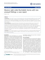
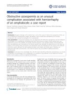
examination revealed a paresis of cranial nerves IX and
X, manifested by the displacement of the soft palate to
the right and difficulty swallowing. Paresis of cranial
nerves XI and XII was also observed, indicated by the
lowe ring of the right shoulder and hypotonia of the right
trapezius muscle, and was accompanied by displacement
* Correspondence:
1
Unidad Oncologia Médica, Autovia A-7, km 187, Hospital Costa del Sol,
Marbella, 29603, Spain
Full list of author information is available at the end of the article
Villatoro et al. Journal of Medical Case Reports 2011, 5:315
/>JOURNAL OF MEDICAL
CASE REPORTS
© 2011 Villatoro et a l; licensee BioMed Central Ltd. This is an Open Access article distributed under the terms of the Creative Commons
Attribution License ( which permits unrestricted use, dis tribution, and reproduction in
any medium, provided the original work is properly cited.
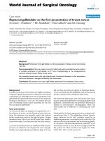
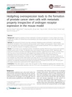
of the tongue toward the right, right-side hypotonia and
muscle twitching (Figures 1 and 2). Results of the rest of
the physical examination were normal.
Blood analysis results revealed an alkaline phosphatase
level of 350 UI/L (normal range 44 to 147 UI/L) but no
other significant alterations. In view of the paresis of the
four lower cranial nerves, a CT scan of the lary nx and
neck was performed; the CT scan revealed an asymmetri-
cal union between the clivus and the right occipital con-
dyle, adjacent to the jugula r foramen, with increa sed
ground-glass bone density. There was no visible lesion to
the bone cortex or soft tissues. This study was comple-
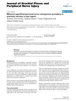
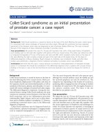
mented with an MRI scan, which revealed a bone lesion
producing a hypointense signal at sequences T1 and T2.
Administration of a gadolinium contrast agent produced
a moderate d egree of enhan cement at the base of the
skull, affecting the right occipital condyle and part of the
right side of the basilar bone (Figure 3). The image corre-
sponded to a moderately space-occupying blastic lesion,
visible in the CT scan, which slightly decreased the cali-
ber of the jugular foramen and the condylar canal. There-
fore, fibrous dysplasia, Paget ’s disease and metastasis
were considered in the differential diagnosis.
Subsequently, an additional radiographic examination of
the lumbar column and pelvis was carried out; this exami-
nation did not reveal any lesions suggestive of Paget’s dis-
ease. A bone gammagraphy was then requested, and
images showed multiple pathological foci of tracer uptake
in the right maxilla, the rib cage, right scapula, spine and
pelvis. These foci were compatible with disseminated bone
metastases. The blood analysis was repeated, and tumor
markers were studied. The initial prostate-specific antigen
(PSA) value was 21.30 ng/mL.
A physical examination revealed an enlarged prostate
with a hard consistency, d estructured in the left lobe.
Because there was a strong suspicion of prostate
neoplasm, a biopsy was performed. The anatomic
pathology findings were bilateral common adenocarci-
noma, with a Gleason grade of 8 (4+4), affecting 60% of
the tissue. There was no presence in the periprostatic
adipose tissue and no perineural infiltration.
Following the diagnosis of stage IV prostate adenocar-
cinoma by metastatic bone dissemination with Collet-
Sicard or jugular foramen syndrome, hormone treatment
was begun with an antiandrogen. Then, 15 days later, a
luteinizing hormone-releasing hormone (LHRH) analog
Figure 1 Paresis of cranial nerve XI.
Figure 2 Paresis of cranial nerve XII.
Figure 3 MRI scan with moderate degree of enhancement at
the base of the skull, affecting the right occipital condyle and
part of the right side of the basilar bone.
Villatoro et al. Journal of Medical Case Reports 2011, 5:315
/>Page 2 of 3
was added and a monthly dose of zoledronic acid was
subsequently included. The PSA value during the diag-
nostic process, prior to the start of antiandrogen ther-
apy, was 71.9 ng/mL.
After three months of treatment, our patient was able
to swallow normally, but the dysphonia remained. The
rightward displace ment of the palate, the lowering of
the right shoulder and the atony of the right side of the
tongue (paresis of cranial nerves XI and XII) remained
unaltered. The latest PSA value was 0.11 ng/mL.
Discussion
The clinical presentation of metastasis to the temporal
bone is uncommon, and few cases have been reported.
Nevertheless, its incidence is probably greater than com-
monly estimated because of the number of cases that
remain undiagnosed. The multi-symptom nature of
metastatic bone disease tends to produce more incapaci-
tating symptoms than those associated with diseases of
the temporal bone.
Various retrospective series of patients presenting
with this syndrome have been reported in the literature.
Vázquez et al. described 21 cases, of which 71% were
secondary to neoplasia (57% from paraganglioma and
14% by the direct exten sion of carcinoma of the cavum)
[1]. Imamura et al. reviewed the potential mechanisms
responsible for metastatic dissemination to the temporal
bone. Of the six patients studied, three cases presented
hem atogenous dissemination (hepatocellular car cinoma,
non-microcytic lung cancer and adenocarcinoma of
unknown origin), two cases were the consequence of
direct invasion by carcinoma of the head and neck, and
one case was caused by leptomeningeal carcinomatosis
(carcinoma of transitional cell carcinoma of the renal
pelvis) [4]. Gloria-Cruz et al. sele cted 212 corpses of
patients with non-disseminated neoplasias for an autop-
sical study of the temporal bone. These authors identi-
fied 47 patients with metastasis in the temporal bone,
and the involvement was bilateral in 62% of these cases.
The most frequently occurring site was the petrous
apex, and the hematogenous pathway was the normal
route of dissemination [5,6].
The management of Collet-Sicard s yndrome consists
of treating the cause that originates. In this case, therapy
over primary tumor, followed by other measures such as
the use of steroids or radiotherapy to help reduce
edema and, thus, alleviate symptoms that can be limiting
for the patient [7].
Conclusions
The medical literature contains various descriptions of
patients with disseminated prostate cancer who pr e-
sented with Collet-S icard syn drome; however, in almost
every case, this diagnosis was already known when
neurological symptoms began [7-12]. Apart from our
patient, only one other case has been reporte d where
metastasis to the temporal bone was the first recognized
symptom of the disease [12]. It is important to consider
the possibility of the existence of prostate cancer when
a patient with an unusual clinical presentation is first
seen, in view of the fact that first-line hormonal treat-
ment may control the disease for months or years.
Consent
Written informed consent was obtained from the patient
for publication of this case report and any accompany-
ing images. A copy of the written consent is available
for review by the Editor-in-Chief of this journal.
Author details
1
Unidad Oncologia Médica, Autovia A-7, km 187, Hospital Costa del Sol,
Marbella, 29603, Spain.
2
Servicio Medicina Interna, Autovia A-7, Km 187,
Hospital Costa del Sol, Marbella, 29603, Spain.
Authors’ contributions
CR made substantial contributions to the design, and the acquisition and
interpretation of data. AR revised the manuscript critically for important
intellectual content. RV was a major contributor in writing the manuscript.
All authors read and approved the final manuscript.
Competing interests
The authors declare that they have no competing interests.
Received: 7 October 2010 Accepted: 14 July 2011
Published: 14 July 2011
References
1. Vázquez BV, Saynes MFJ, Hernández VG: Sindrome de Agujero Rasgado
posterior. Casuística y manejo. An Orl Mex 2002, 47:4-8.
2. Belal A Jr: Metastatic tumours of the temporal bone. A histopathological
report. J Laryngol Otol 1985, 99:839-846.
3. Syms MJ, Singson MT, Burgess LP: Evaluation of lower cranial nerve
deficits. Otolaryngol Clin North Am 1997, 30:489-463.
4. Imamura S, Murakami Y: Secondary malignant tumor of the temporal
bone. A histopathologic study and review of the world literature. Nippon
Jibiinkoka Gakkai Kaiho 1991, 94:924-937.
5. Gloria-Cruz TI, Schachern PA, Paparella MM, Adams GL, Fulton SE:
Metastases to temporal bones from primary nonsystemic malignant
neoplasms. Arch Otolaryngol Head Neck Surg 2000, 126:209-214.
6. Nelson EG, Hinojosa R: Histopathology of metastatic temporal bone
tumors. Arch Otolaryngol Head Neck Surg 1991, 117:189-193.
7. Chacon G, Alexandraki I, Palacio C: Collet-Sicard syndrome: an uncommon
manifestation of metastatic prostate cancer. South Med J 2006, 99:898-899.
8. Shine NP, O’Sullivan P: Collet-Sicard syndrome: a rare presentation of
metastatic prostate adenocarcinoma. Auris Naus Larynx 2005, 32:315-318.
9. Prashant R, Franks A: Collet-Sicard syndrome–a report and review. Lancet
Oncol 2003, 4:376-377.
10. Satoh H, Nishiyama T, Horiguchi A, Nakashima J, Saito S, Murai M: A case of
Collet-Sicard syndrome caused by skull base metastasis of prostate
carcinoma. Nippon Hinyokika Gakkai Zasshi 2000, 91:562-564.
11. Wilson H, Johnson DH: Jugular foramen syndrome as a complication of
metastatic cancer of the prostate. South Med J 1984, 77:92-93.
12. Onishi A, Shida K: Case of Collet-Sicard syndrome due to metastasis of
prostatic cancer. Naika 1970, 26:755-757.
doi:10.1186/1752-1947-5-315
Cite this article as: Villatoro et al.: Collet-Sicard syndrome as an initial
presentation of prostate cancer: a case report. Journal of Medical Case
Reports 2011 5:315.
Villatoro et al. Journal of Medical Case Reports 2011, 5:315
/>Page 3 of 3