Báo cáo y học: " Infective endocarditis with Lactococcus garvieae in Japan: a case report" doc
Bạn đang xem bản rút gọn của tài liệu. Xem và tải ngay bản đầy đủ của tài liệu tại đây (1.08 MB, 4 trang )
CAS E REP O R T Open Access
Infective endocarditis with Lactococcus garvieae
in Japan: a case report
Yukiko Watanabe
1,2
, Toshio Naito
1,2*
, Ken Kikuchi
1,2
, Yu Amari
1
, Yuki Uehara
1,2
, Hiroshi Isonuma
1
, Teruhiko Hisaoka
1
,
Terutoyo Yoshida
4
, Kenji Yaginuma
3
, Norihide Takaya
3
, Hiroyuki Daida
3
and Keiichi Hiramatsu
2
Abstract
Introduction: Lactococcus garvieae is a well-recognized fish pathogen, and it is considered a rare pathogen with
low virulence in human infection. We describe the 11th case of L. garvieae infective endocarditis reported in the
literature, and the first reported case in Japan.
Case presentation: We report a case of a 55-year-old Japanese woman who had native valve endocarditis with L.
garvieae. The case was complicated by renal infarction, cerebral infarction, and mycot ic aneurysms. After anti-
microbial treatment, she was discharged from the hospital and is now well while being monitored in the out-
patient clinic.
Conclusion: We encountered a case of L. garvieae endocarditis that occurred in a native valve of a healthy
woman. The 16S ribosomal RNA gene sequencing was useful for the identification of this pathogen. Although
infective endocarditis with L. garvieae is uncommon, it is possible to treat high virulence clinically.
Introduction
Lactococcus garvieae was first isolated from cases of
bovine mastitis [1], and it is rarely reported in human
infection. The first hum an infection was documented as
infective endocarditis (IE) in 1991 [2]. Since then, it has
been identified as a pathogen i n IE [3-9], liver abscess
[10], and osteomyelitis [4]. We describe the 11th case of
L. garvieae IE reported in the literature, and the first
reported case in Japan.
Case presentation
A 55-year-old healthy Japanese woman presented to our
hospita l with a two -month history of malaise a nd myal-
gia, and she had had a fever for one month. The patient
had no significant medical history and no relevant his-
tory regarding pets, travel, dental treatment, or drug
abuse. Her occupational history was that she worked at
an import grocery store. During her physical examina-
tion, we noted her body temperature of 37.6°C, a grade
2 systolic murmur at the apex, and a painful 1 mm
black induration on her right forefinger. Trans-thoracic
echocardiography showed a 1 cm mobile mass on the
mitral valve with mo derate mitral r egurgitation. Her
laboratory tests showed a white blood cell count of
8200/μL, a C-reactive protein level of 6.6 mg/dL, and a
hemoglobin level of 9.9 g/dL. Gram-positive cocci form-
ing pairs or short chains were shown by Gram staining
of three sets of blo od cultur es drawn at the time of
admission.
IE was diagnosed on the basis of the Duke criteria for
the diagnosis of infective endocarditis [11] (t wo major
and two minor criteria) and antibiotic treatment with
benzylpenicillin 3 million units every four hours and
gentamicin 60 mg every eight hours was started, as
Streptococcus sp. w as suspected as the causative patho-
gen. Subsequently, the rapid ID32 Strep (bioMérieux,
Marcy-l’ Etoile, France) profile (code 34323501010)
revealed L. garvieae. We also performed molecular spe-
cies identification by 16S ribosomal RNA gene (16S
rRNA) sequenci ng, and the almost full- length 16S
rDNA matched L. garvieae (99.9%). The results of an
anti-microbial susceptibility test using Etest strips (AB
Biodisk, Dalvagen, Solna, Sweden) showed the following
values: erythromycin 0.25 mg/ L, clindamycin ≥ 256 mg/
L, vancomycin 0.38 mg/L, linezolid 2.0 mg/L, penicillin
0.5 mg/L, ceftriaxone 0.38 mg/L, gentamicin 1.5 mg/L,
* Correspondence:
1
Department of General Medicine, Juntendo University School of Medicine,
2-1-1 Bunkyo-ku, Hongo, Tokyo 113-8421, Japan
Full list of author information is available at the end of the article
Watanabe et al. Journal of Medical Case Reports 2011, 5:356
/>JOURNAL OF MEDICAL
CASE REPORTS
© 2011 Watanabe et al; licensee Bi oMed Central Ltd. This is an Open Acce ss article distributed under the terms of the Creative
Commons Attribution License (http://creat ivecommons.org/licenses/by/2.0), which permits unrestricted use, distribution, and
reproduction in any medium, provided the original work is properly cit ed.
and streptomycin 64 mg/L. Consider ing these results,
we changed th e anti-microbial treatment to intravenous
ceftriaxone 2 g every 12 hours and gentamicin 80 mg
every eight hours intraveneously.
Meanwhile, her body temperature rose to 39°C with
left lower back pain on her sixth day in the hospital.
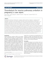
Left-sided renal infarction was diagnosed on the basis of
contrast-enhanced computed tomography (CT) (Figure
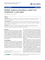
1). On the 12th hospital day, cerebral infarction
occurred, followed by a sudden onset of right-sided
hemiparesis and slurred speech (National Institutes of
Health Stroke Scale (NIHSS) score 12). Diffusion-
weighted MRI showed a cerebral infarction of t he mid-
dle cerebral artery area (Figure 2). Furthermore, cerebral
mycotic aneurysms were detected by three-dimensional
CT angiography on the 24th hospital day (Figure 3).
The patient also developed aspiration pneumonia after
the cerebral infarction complication.
Anti-microbial treatment was complet ed in n ine
weeks, including the treatment for pneumonia. Several
repeat blood cultures thereafter were all negative. The
vegetation on the mitral valve was no longer detected by
trans-esophageal echocardiography performed just
before she was disch arged from the hospital. We fo und
by CT that her left-sided renal infarction had improved,
and the consequences of her cerebral infarction had also
improved with only right-sided muscular weakness
(NIHSS score 2 ). She was dis charged from the hospital,
and she is now well while being monitored in the out-
patient clinic.
Discussion
Genus Lactococcus was separated from genus Streptococ-
cus in 1985 based on DNA-DNA relatedness and 16S
rRNA sequencing data published by Ludwig et al. [12].
The genus Lactococcus was classified into eight species
and sub-sp ecies by Facklam and Elliott in 1995 [13] and
Pu et al. in 2002 [14]: L. lactis ssp. lactis,L.lactisssp.
cremoris,L.lactisssp. hordniae, L. garvieae, L. piscium,
L. plantarum, L. raffinolactis,andL. xyloses.Cases
involving the L. garvieae pathogen in human infection
are rarely reported [2-10]. The major clinical presenta-
tion is IE, which has been reported in 10 cases i n the
Figure 1 Contrast-enhanced abdominal computed tomography.
Yellow arrow indicates left renal infarction.
Figure 2 Diffusion-weighted MRI scan. Yellow arrow indicates the
middle cerebral artery areas of high signal intensity.
Figure 3 Three-dimensional computed tomography
angiography. The two yellow circles indicate cerebral mycotic
aneurysms.
Watanabe et al. Journal of Medical Case Reports 2011, 5:356
/>Page 2 of 4
literature [2-9]. Five of ten ca ses involv ed native valv es
[3,6-9], and four cases required surgery for valve repla-
cement [3,6,8,9]. In addition, two cases were compli-
cated by cerebral inf arction or hemorrhage [3,9]. L.
garvieae IE is uncommon; however, the virulence of L.
garvieae might be underestimated. The treatment for L.
garvieae IE is not standardized, bec ause th e exact criter-
ion of the susceptibility test has not been established. In
the published cases, penicillin combined with gentami-
cin, amoxicillin combined with netilmicin, ampicillin,
ceftriaxone, and vancomycin were used as anti-microbial
treatment. In our case, the results of the anti-microbial
susceptibility test of the isolate using the Etest method
was susceptibility to ceftriaxone (0.38 mg/L), erythromy-
cin (0.25 mg/L), vancomycin (0.38 mg/L), and linezolid
(2 mg/L), less susceptibility to penicillin (0.5 mg/L), and
resistance to clindamycin (> 256 mg/L).
Elliott and Facklam [15] reported that clindamycin
resistance is a feature of L. garvieae, and this feature can
be us ed to dis tinguish it from other la ctococci. To con-
firm the precise identification, various molecular techni-
ques are available: whole-cell protein analysis, 16S rRNA
gene sequencing, and sodA
int
(an internal fragment of
the sodA gene encoding the manganese-dependent
superoxide dismutase) gene sequencing.
L. garvieae has pathogenicity for several fish species,
ruminants, and humans. Therefore, it could be a poten-
tially zoonotic infection. Investigators in one recent
report hypothesized that eating infected raw fis h carrying
L. garvieae caused its infection [7]. To investigate the
relationship between our isolate and fish isolates, the
immunological property w as examined by using several
anti-sera against L. garvieae that have previously been
produced by fish in Japan. The results showed that our
isolate did not have a capsule detected by any anti-cap-
sule antibody for fish isolates. Pulsed-field gel electro-
phoresis also showed that the pat tern of our isolate was
quite different from that of fish-derived strains (data not
shown). Vela et al. [16] found great phenotypic heteroge-
neity and genetic diversity among the L. garvieae isolates
from fish, cows, water buffalo, and humans, with a gener-
ally good correlation between the phenotypic and genetic
properties of L. garvieae. As the origin of the L. garvieae
strain in our patient could not be clarified by the investi-
gations used to detect J apanese fish p athogens, other
causes of her infection, such as cow’s milk or cheeses, are
possible. To confirm the etiology of the transmission of
L. garvieae, larger molecular epidemiological studies
including investigating causes from the environment, ani-
mals, and humans must be carried out.
Conclusion
Herein we report the first case of L. garvieae IE in
Japan. Molecular biological investigation techniques
such as 16S ribosomal RNA gene sequencing are useful
for the identification of L. garvieae. Although L. gar-
vieae is considered a rare pathogen with low virulence
in human infection, it could cause native valve endo-
carditis in healthy people. It might be necessary
to review the precise estimation of the pathogenicity of
L. garvieae.
Consent
Written informed consent was obtained from the patient
for publication of this case report and any accompany-
ing images. A co py of the written consent is available
for review by the Editor-in-Chief of this journal.
Acknowledgements
We thank Dr Shigeki Misawa, Clinical Laboratory, Juntendo University
Hospital, Tokyo, Japan, for phenotypic characterization of the bacterial
isolate.
Author details
1
Department of General Medicine, Juntendo University School of Medicine,
2-1-1 Bunkyo-ku, Hongo, Tokyo 113-8421, Japan.
2
Department of Infection
Control Science, Juntendo University School of Medicine, 2-1-1 Bunkyo-ku,
Hongo, Tokyo 113-8421, Japan.
3
Department of Cardiology, Juntendo
University School of Medicine, 2-1-1 Bunkyo-ku, Hongo, Tokyo 113-8421,
Japan.
4
Department of Fisheries, Faculty of Agriculture, Miyazaki University,
Miyazaki City, Miyazaki 889-2192, Japan.
Authors’ contributions
YW drafted and edited the manuscript. TN, KK, YA, YU, HI, TH, and KH
helped draft the manuscript. KY, NT, and HD helped in the patient’s
treatment after the diagnosis was made. TY helped in the investigation of L.
garvieae as a fish pathogen. All authors read and approved the final
manuscript.
Competing interests
The authors declare that they have no competing interests.
Received: 13 December 2010 Accepted: 9 August 2011
Published: 9 August 2011
References
1. Collins MD, Farrow JAE, Phillips BA, Kandler O: Streptococcus garvieae sp.
nov. and Streptococcus plantarum sp. nov. J Gen Microbiol 1983,
129:3427-3431.
2. Furutan NP, Breiman RF, Fischer MA, Facklam RR: Lactococcus garvieae
infections in humans: a cause of prosthetic valve endocarditis [abstract
C297]. Proceedings of the 91st General Meeting of the American Society for
Microbiology Dallas: American Society of Microbiology; 1991, 109.
3. Fefer JJ, Ratzan KR, Sharp SE, Saiz E: Lactococcus garvieae endocarditis:
report of a case and review of the literature. Diagn Microbiol Infect Dis
1998, 32:127-130.
4. James PR, Hardman SM, Patterson DL: Osteomyelitis and possible
endocarditis secondary to Lactococcus garvieae: a first case report.
Postgrad Med J 2000, 76:301-303.
5. Fihman V, Raskine L, Barrou Z, Kiffel C, Riahi J, Berçot B, Sanson-Le Pors MJ:
Lactococcus garvieae endocarditis: identification by 16S rRNA and sodA
sequence analysis. J Infect 2006, 52:e3-e6.
6. Vinh DC, Nichol KA, Rand F, Embil JM: Native-valve bacterial endocarditis
caused by Lactococcus garvieae. Diagn Microbiol Infect Dis 2006, 56:91-94.
7. Wang CY, Shie HS, Chen SC, Huang JP, Hsieh IC, Wen MS, Lin FC, Wu D:
Lactococcus garvieae infections in humans: possible association with
aquaculture outbreaks. Int J Clin Pract 2007, 61:68-73.
8. Yiu KH, Siu CW, To KK, Jim MH, Lee KL, Lau CP, Tse HF: A rare cause of
infective endocarditis: Lactococcus garvieae. Int J Cardiol 2007,
114:286-287.
Watanabe et al. Journal of Medical Case Reports 2011, 5:356
/>Page 3 of 4
9. Li WK, Chen YS, Wann SR, Liu YC, Tsai HC: Lactococcus garvieae
endocarditis with initial presentation of acute cerebral infarction in a
healthy immunocompetent man. Intern Med 2008, 47:1143-1146.
10. Mofredj A, Baraka D, Kloeti G, Dumont JL: Lactococcus garvieae septicemia
with liver abscess in an immunosuppressed patient. Am J Med 2000,
109:513-514.
11. Durack D, Lukes A, Bright D, Duke Endocarditis Service: New criteria for
diagnosis of infective endocarditis: utilization of specific
echocardiographic findings. Am J Med 1994, 96:200-209.
12. Ludwig W, Seewaldt E, Kilpper-Bälz R, Schleifer KH, Magrum L, Woese CR,
Fox GE, Stackebrandt E: The phylogenetic position of Streptococcus and
Enterococcus. J Gen Microbiol 1985, 131:543-551.
13. Facklam R, Elliott JA: Identification, classification, and clinical relevance of
catalase-negative, gram-positive cocci, excluding the streptococci and
enterococci. Clin Microbiol Rev 1995, 8:479-495.
14. Pu ZY, Dobos M, Limsowtin GK, Powell IB: Integrated polymerase chain
reaction-based procedures for the detection and identification of
species and subspecies of the Gram-positive bacterial genus
Lactococcus. J Appl Microbiol 2002, 93:353-361.
15. Elliott JA, Facklam RR: Antimicrobial susceptibilities of Lactococcus lactis
and Lactococcus garvieae and a proposed method to discriminate
between them. J Clin Microbiol 1996, 34:1296-1298.
16. Vela AI, Vázquez J, Gibello A, Blanco MM, Moreno MA, Liébana P,
Albendea C, Alcalá B, Mendez A, Domínguez L, Fernández-Garayzábal JF:
Phenotypic and genetic characterization of Lactococcus garvieae isolated
in Spain from lactococcosis outbreaks and comparison with isolates of
other countries and sources. J Clin Microbiol 2000, 38:3791-3795.
doi:10.1186/1752-1947-5-356
Cite this article as: Watanabe et al.: Infective endocarditis with
Lactococcus garvieae in Japan: a case report. Journal of Medical Case
Reports 2011 5:356.
Submit your next manuscript to BioMed Central
and take full advantage of:
• Convenient online submission
• Thorough peer review
• No space constraints or color figure charges
• Immediate publication on acceptance
• Inclusion in PubMed, CAS, Scopus and Google Scholar
• Research which is freely available for redistribution
Submit your manuscript at
www.biomedcentral.com/submit
Watanabe et al. Journal of Medical Case Reports 2011, 5:356
/>Page 4 of 4