Báo cáo y học: "A 64-year old man presenting with carotid artery occlusion and corticobasal syndrome: a case report" pps
Bạn đang xem bản rút gọn của tài liệu. Xem và tải ngay bản đầy đủ của tài liệu tại đây (347.73 KB, 3 trang )
CAS E REP O R T Open Access
A 64-year old man presenting with carotid artery
occlusion and corticobasal syndrome: a case
report
Marc Engelen, Dunja Westhoff, Jan de Gans and Paul J Nederkoorn
*
Abstract
Introduction: Magnetic resonance imaging of the brain in patients with corticobasal degeneration typically shows
focal or asymmetric atrophy, usually maximal in the frontoparietal cortex. Many patients who are diagnosed with
corticobasal degeneration using current diagnostic criteria do not have classical corticobasal degeneration
pathology. Our case is remarkable for the fact that the symptoms and the characteristic magnetic resonance
imaging appearance were typical for corticobasal degeneration. However, we were quite convinced that the
clinical picture had a vascular etiology. Only a few cases have been reporte d where the presumed cause for the
corticobasal syndrome was multiple brain infarctions bilaterally.
Case presentation: A 64-year-old Caucasian man visited a neurologist because of profound asymmetric sensory
and motor disturbances. A magnetic resonance imaging scan of his brain revealed occlusion of his internal carotid
artery on the left side with multiple vascular lesions in his left hemisphere and notable atrophy of mainly the left
parietal and frontal cortex.
Conclusion: We describe a patient with corticobasal syndrome caused by multiple infarctions, probably caused by
emboli of the carotid stenosis. This patient illustrates the fact that the word ‘syndrome’ should be preferred above
‘degeneration’ in the name of this disease.
Introduction
Corticobasal degeneration (CBD) was formerly consid-
ere d to be a well-defined clinicopathological entity. The
classic description of CBD includes clumsiness and loss
of function of one hand due to a combination of fronto-
parietal and basal ganglia sensorimotor dysfunction [1].
However, many patients who are diagnosed using cur-
rent diagnostic criteria do not have classical corticobasal
degeneration pathology [2]. Therefore it is now custom-
ary to diagnose corticobasal syndrome (CBS) during life,
and refer to the classical pathology as CBD. CBS can be
caused by classical CBD pathology, but also by the
pathology of progressive supranuclear palsy, frontotem-
poral lobe degeneration or even Alzheimer’s [3]. A few
cases have been reported where the presumed cause of
CBS was multiple brain infarctions bilaterally [4]. Mag-
netic resonance imaging (MRI) of the brain in patients
with CBS typica lly shows f ocal or asymmetric atrophy,
usually maximal in the frontoparietal cortex.
Case presentation
A 64-year-old Caucasian man experienced sudden
cramping of the toes of his right foot, and si multa neous
weakness and numbness of his right leg. This lasted for
approximately 20 minutes, after which he completely
recovered. These incidents recurred, increasing in fre-
quency for several weeks. At first, he fully recovered
after each episode. Some w eeks later, he noticed a per-
sisting numbness of both his right leg and hand. Walk-
ing became more difficult because of roaming and
clumsiness of his right leg. About 10 months later, he
visited a general practitioner and was referred to a neu-
rologist.Atthattimeheexperienced gait difficulty and
numbness of his right arm and leg. His previous medical
history was remarkable for hypertension and he is a
cigarette smoker. He uses metoprolol but no other
* Correspondence:
Department of Neurology, H2.216, Academic Medical Center, University of
Amsterdam, PO Box 22660, 1100 DD Amsterdam, The Netherlands
Engelen et al. Journal of Medical Case Reports 2011, 5:357
/>JOURNAL OF MEDICAL
CASE REPORTS
© 2011 Engelen et al; licensee BioMed Central Ltd. This is an Open Access article distributed under the terms of the Creati ve Commons
Attribution License ( which permits unrestr icted use, distribution, and reproduction in
any medium, provided the original work is properly cited.
medication. He had no significant family history of neu-
rological disease.
On examination t here was flattening of the nasolabial
fold on the right side of his face. The fine motor skills of
his right arm were impaired. There was clearly impaired
two-point discrimination on both his right arm and leg,
while position-, movement-, and vibration-sense were
intact. There was hyperpathia of his right leg a nd arm.
Ataxia of his right leg was noted, not improving with
visual correction. Deep tendon reflexes were higher in his
right leg, wit h a Babinski sign. There was a hen’sgaiton
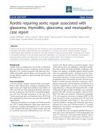
his right side. An MRI scan of his brain performed in the
referring hospital revealed occlusion of his internal caro-
tid artery on the left side with multiple vascular lesions in
the left hemisphere and notable atrophy of mainly the left
parietal and frontal cortex (Figure 1). We started prophy-
lactic treatment with aspirin, dipyridamole and simvas ta-
tin. We urged our patient to stop smoking.
Discussion
Our patient fulfilled the criteria for CBS with a pro-
found asymmetric presentation, with dystonia of the
right foot, cortical sensory disturbance (with profoundly
impaired two-point discrimination) and pyramidal tract
syndrome. MRI of the brain showed left frontoparietal
atrophy with multiple subcortical hyperintensities. Revi-
sion of the MRI scan by our neuroradiologist revealed
occlusion of the left internal carotid artery.
Usually, the onset of CBS is insidious. This patient
described an acute onset of symptoms, suggesting a vas-
cular origin. After that there were a few instances of
fluctuating deficits, but eventually there was residual
impairment as described. There does not appear to have
been any further progression over the last few mont hs.
Theclinicalpictureandtheevolutionofsymptoms
seem compatible with a presumed vascular cause in this
patient.
A few cases of a presumed vascular origin of CBS
have been reported [4,5] (Table 1), however our case is
remarkable for the fact that it is associated with
marked frontoparietal atrophy on brain imaging, there-
fore also mimicking the characteristic MRI appearance.
It is likely that multiple strokes resulted in atrophy
and gliosis.
Figure 1 Atrophy of t he left frontoparietal lobe, with extensive gliosis (A, B, C; T2-weighted MRI). The left internal carotid artery is
occluded, since there is no flow void (D; T1-weighted MRI).
Engelen et al. Journal of Medical Case Reports 2011, 5:357
/>Page 2 of 3
Conclusion
We describe a patient with CBS cause d by multiple
infarctions, probably caused by emboli of his carotid ste-
nosis. This patient illustrates the fact that the word ‘syn-
drome’ should be preferred above ‘degeneration’ in the
name of this disease.
Consent
Written informed consent was obtained from the patient
for publication of this case report and any accompany-
ing images. A copy of the written consent is avail able
for review by the Editor-in-Chief of this journal.
Authors’ contributions
DW and ME wrote this case report. PJN and JdG revised it critically. All
authors read and approved the final manuscript.
Competing interests
The authors declare that they have no competing interests.
Received: 27 January 2011 Accepted: 9 August 2011
Published: 9 August 2011
References
1. Ling H, O’Sullivan SS, Holton JL, Revesz T, Massey LA, Williams DR,
Paviour DC, Lees AJ: Does corticobasal degeneration exist? A
clinicopathological re-evaluation. Brain 2010, 133(Pt 7):2045-2057.
2. Litvan I, Agid Y, Goetz C, Jankovic J, Wenning GK, Brandel JP, Verny M, Ray-
Chaudhuri K, Pearce RK, Bartko JJ, Agid Y: Accuracy of the clinical
diagnosis of corticobasal degeneration: a clinicopathologic study.
Neurology 1997, 48(1):119-125.
3. Wadia PM, Lang AE: The many faces of corticobasal degeneration.
Parkinsonism Relat Disord 2007, 13(Suppl 3):S336-S340.
4. Kim YD, Kim JS, Lee ES, Yang DW, Lee KS, Kim YI: Progressive “vascular”
corticobasal syndrome due to bilateral ischemic hemispheric lesions.
Intern Med 2009, 48(18):1699-1702.
5. Kreisler A, Mastain B, Tison F, Fenelon G, Destee A: [Multi-infarct disorder
presenting as corticobasal degeneration (DCB): vascular pseudo-
corticobasal degeneration?]. Rev Neurol (Paris) 2007, 163(12):1191-1199.
doi:10.1186/1752-1947-5-357
Cite this article as: Engelen et al.: A 64-year old man presenting with
carotid artery occlusion and corticobasal syndrome: a case report.
Journal of Medical Case Reports 2011 5:357.
Submit your next manuscript to BioMed Central
and take full advantage of:
• Convenient online submission
• Thorough peer review
• No space constraints or color figure charges
• Immediate publication on acceptance
• Inclusion in PubMed, CAS, Scopus and Google Scholar
• Research which is freely available for redistribution
Submit your manuscript at
www.biomedcentral.com/submit
Table 1 Previously published cases
Article Patient Diagnosis Symptoms
Kim et al. [4] 75-year-old woman extensive cortical vascular-ischemic lesions Progressive symptoms of:
- dementia
- asymmetric parkinsonism
- apraxia
- action myoclonus
- focal hand dystonia
Kreisler et al. [5] 5 women, aged between 64 and 77 years extensive vascular lesions Progressive symptoms of:
- asymmetric parkinsonism
- apraxia
- focal action myoclonus
- focal dystonia
- cortical sensory loss
- alien limb phenomenon
Engelen et al. Journal of Medical Case Reports 2011, 5:357
/>Page 3 of 3