Pathology and Laboratory Medicine - part 4 docx
Bạn đang xem bản rút gọn của tài liệu. Xem và tải ngay bản đầy đủ của tài liệu tại đây (744.75 KB, 49 trang )
Disease-Induced Protein Modifications 129
prior to release into the blood. These forms of cardiac troponins (intact as well as mod-
ified) display a characteristic “rising and falling” pattern after the onset of symptoms,
producing a continuum of changing troponin profiles. The ability to distinguish between
different forms of circulating troponins would offer more precise information about
severity of damage, time of onset, or even type of disease, assisting in the triage of indi-
vidual patients. This is not possible with the currently available diagnostic assays.
In addition to the advantage of providing information about certain modification
states of the analyte, WB-DSA has a high analytical sensitivity. In fact, it enabled us to
detect serum cTnI in patients undergoing CABG surgery at levels below the LLD of a
routinely used commercial assay (0.1 ng/mL) (42). This enhanced sensitivity most likely
is due to the denaturing conditions used in WB-DSA, which would result in the com-
plete exposure of linear epitopes, thereby increasing the probability of detection by
various antibodies. cTnI in serum may be “hidden” owing to its ternary structure and/or
the formation of three-dimensional complexes with other proteins. To test this method
for its clinical application, serum from patients presenting at the emergency depart-
ment early after onset of symptoms of ACS (£4 h) was analyzed by WB-DSA and the
results compared to routine clinical testing (66). A subset of the patients enrolled in
this study with nondiagnostic electrocardiogram for ACS and nonsignificantly elevated
routine biochemical markers (cTnI, CK, and CK-MB) showed detectable amounts of
cTnI when retrospectively analyzed by WB-DSA (n = 6/10). These patients were diag-
nosed for unstable angina (n = 3), second-degree heart block (n = 1), or discharged from
the ED as “chest pain not yet diagnosed” (n = 2). Three of the six patients revisited the
ED within 3 mo complaining about chest pain.
ONE ANALYTE = ONE DISEASE = ONE ASSAY?
The exact release pattern of the various forms of cTnI (whether free or complexed
with other proteins) after an ischemic insult and the correlation between the severity of
the insult and the released forms in humans remains unknown, although it has been shown
in Langendorff perfused rat hearts that cTnI undergoes selective and progressive modi-
fication with increased severity of ischemia/reperfusion injury (38,40). Complex for-
mation between cTnI and other troponin subunits influences its three-dimensional
structure, having various consequences for the susceptibility of cTnI to enzymatic and
chemical modification (61). Certainly the same will apply to cTnI bound to any serum
protein. cTnI is highly charged and insoluble in its free form at neutral pH. It has been
proposed that only a small amount of free cTnI is detectable in blood of MI patients
(61), although the proportions of free cTnI varied among the patients analyzed and the
severity of MI.
By comparing data from the three studies undertaken in our laboratory applying
WB-DSA to identify serum cTnI (42,59,66), the differences in the modification states
of the analyte become apparent (Fig. 1). This is by no means surprising, as three very
different cohorts of patients were enrolled in these studies, representing different stages
of myocardial disease. While degradation seems preferably to occur in cases of MI,
patients presenting with unstable angina show only intact cTnI. Patients undergoing
thrombolytic therapy showed only intact cTnI before, but a variety of degradation prod-
ucts shortly after thrombolysis, again indicating that cTnI modifications occur prior to
release from the myocardium (unpublished data). Extensive proteolytic degradation of
130 Labugger et al.
cTnI and prolonged detectability in blood indicate longer ischemic periods and there-
fore cellular necrosis with disintegration of the troponin complex. On the other hand,
intact cTnI as the only detectable form in angina patients, with faster clearance from
the blood, may represent unassembled cytosolic troponin and resemble the first phase
of a biphasic release as shown for cTnT on revascularization after MI (67).
In their consensus document ESC and ACC suggest the use of a cutoff concentration
for cardiac troponins at the 99th percentile (CV £ 10%) for the diagnosis of MI (8,9).
An increase in sensitivity of troponin assays can principally be appreciated because ele-
vated troponins are associated with a risk of adverse clinical events (13–17), even though
the majority of manufacturers cannot yet meet these recommendations (68). Automati-
cally labeling an increase of cardiac troponin above this level as MI, in cases in which
other causes of cardiac damage cannot be found, could be misleading. For example, a
possible non-necrotic release (or a potential release with apoptosis) of troponins from
the myocardium does not meet the criteria for MI. This leads to an important question.
Is a single diagnostic assay adequate to diagnose all forms of cardiac disease that involve
the release of troponins, or is it necessary to design specific assays for different patient
cohorts to ensure precise diagnosis? The three studies mentioned above (42,59,66)
must be confirmed for larger cohorts and other forms of cardiac disease. Regardless of
the small groups of patients enrolled in these studies, our findings indicate significant
variability of cTnI in patients with cardiac diseases. Different diseases or disease states
may lead to different troponin modifications, creating disease-specific “troponin finger-
prints” for MI, unstable angina, heart failure, and so on, as well as for damage caused
by interventional procedures or cardiac surgery. Consequently, the use of antibodies
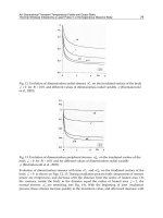
Fig. 1. Different cTnI modifications found at different stages of ischemic heart disease.
WB-DSA using mAb 8I-7 (Spectral Diagnostics) on serum from four patients with the corre-
sponding values for total CK (Beckman CX7), CK-MB, and cTnI (both Bayer Immuno1) indi-
cated underneath and the relative molecular mass to the left. ND, Not determined. Lane 1: Patient
with history of CHF and symptoms of chest pain, sample drawn 2 h after admission, discharged
from the ED with “chest pain not yet diagnosed”. Lane 2: Patient with history of myocardial
infarction (6 mo prior) and symptoms of chest pain, sample drawn 2 h after admission, dis-
charged from the hospital with unstable angina. Lanes 3 and 4: Patient undergoing CABG sur-
gery with samples drawn 30 min and 24 h after removal of cross-clap respectively. Lane 5: Patient
with AMI, sample drawn at admission.
Disease-Induced Protein Modifications 131
raised against specific disease-induced troponin modification products would provide
greater qualitative information than the simple determination of elevated troponin levels,
and would in turn lead to the development of disease-specific diagnostic assays.
Although WB-DSA, for the first time, allows us to visualize modifications to serum
troponins without any concentration or extraction step prior to analysis, it is still limited
in scope. It can separate the analyte only by relative molecular mass, and the number of
identified modification products depends on the antibodies used for western blotting
(59). This method is therefore inherently biased in the same manner as other immuno-
assays, and thus care in the selection of antibodies is paramount. To think that avoiding
antibodies binding to epitopes within the very N- as well as C-terminus of cTnI, re-
gions known to be proteolytically cleaved after MI (57–61,65), would be enough to
guarantee the detection of all cTnI modification products is far too simplistic. Katrukha
et al. (61) very thoroughly listed possible modifications to cTnI that can influence immu-
nogenicity and as a consequence detectability by a diagnostic assay. Within the region of
cTnI that is supposed to be relatively resistant to proteolysis (between amino acids 30
and 110) there are at least two protein kinase C phosphorylation sites (69) and two Cys
residues (61) that may be oxidized. Western blot analysis on native as well as on dephos-
phorylated human cardiac tissue (42) and serum from MI patients (59) showed that the
affinity of some antibodies to cTnI is indeed altered by phosphorylation. The same may
hold true for other posttranslational modifications (such as Cys oxidation) and might
explain the differing performance of various commercial diagnostic cTnI assays. The
impact of disease-induced posttranslational modifications to cTnT (59) on the two exist-
ing cTnT assays is more difficult to assess. There is no reason to believe that antibody
binding to cTnT and, consequently, assay performance would be unaffected by such
modifications. Standardization issues surrounding cTnI assays are not applicable to the
currently marketed cTnT assays, as both use the same pair of antibodies (70). Of course,
similar issues undoubtedly will arise when additional assays using different antibodies
are introduced in the future.
The identification and characterization of all myocardial-derived disease-induced
forms of the analyte, be it cTnI, cTnT, or any other protein, must be the first step in the
development of diagnostic assays capable of specifically detecting one or more of these
modification products present in a particular patient’s blood. To achieve this, we are well
advised to endorse the tools of proteomics to investigate disease-induced protein modifi-
cations, elucidating the potential of these modifications to serve as diagnostic markers.
APPLICATION OF PROTEOMICS
TO DIAGNOSTIC MARKER DEVELOPMENT
In one way or another, as the cause or a symptom, proteins are involved in virtually
every disease, with cardiac diseases being no exception. It is therefore inevitable that a
disease or disease state will manifest itself as a change to the proteome. Whether these
changes are restricted to posttranslational modifications or also involve genetic alter-
ations in protein levels depends on the particular disease state. For example, during
ACS one will primarily observe the first group of modifications, whereas a subsequent
development of CHF will also likely be accompanied by the latter. Therefore, various
stages in the progression of a disease will be expected to reflect unique protein profiles.
Not every protein change will automatically, but very well may, result in an altered
132 Labugger et al.
physiological function. Understanding the functional consequences, if any, of a certain
modification increases the value of an assay specific for this very modification. Thus,
when a particular protein modification can be correlated with a certain disease or disease
state and this modification is detectable, it serves as an invaluable diagnostic marker. It
is in this venue that proteomics has the potential to revolutionize the development and
application of future diagnostics.
The power of proteomics to identify disease-induced protein change is widely recog-
nized, with an ever burgeoning number of publications dealing with this approach as a
means to unravel the importance of protein modifications in the development, progres-
sion, treatment, and diagnosis of disease. Thus far, the majority of this work has focused
on neurological and infectious diseases, cancer, and, to a lesser degree, cardiovascular
diseases (for reviews see 6,71–76). Collections of detailed proteomic techniques have
extensively been described elsewhere (77–79), and we focus instead on the application
of these techniques to the development of diagnostics.
A systematic use of the full armamentarium of proteomics will facilitate a much more
detailed description of pathological processes in terms of protein modifications. The
rapid development of proteomics, in both methods of protein separation and identifica-
tion, now has the potential to guide the development of diagnostic assays as well as
therapeutics in a number of ways. First, it may be used to identify and characterize spe-
cific disease-induced protein modifications associated with current biomarkers. Second,
it may facilitate the identification of useful new biomarkers, which may stand alone for
diagnostic purposes, or perhaps be used in combination with additional proteins to pro-
vide greater confidence in diagnosis or risk stratification. Finally, there is the possibility
of the direct incorporation of proteomic techniques into diagnostic assays themselves.
The current tools of proteomics already allow us to improve the design of diagnostics
through the identification and characterization of specific protein modifications. Unlike
in cancer diagnosis, where biopsies and smears from patients are taken on a regular
basis (and can be used for basic research purposes), proteomic analysis of human cardiac
material at stages of onset or disease development is more difficult because of limited
access. This is often possible only with samples from cadaveric donors or explanted
hearts from end-stage heart failure patients, but such hearts will show overlapping dis-
ease- or drug-induced and postmortem changes to the proteome. Biopsies taken during
heart surgery do provide a viable source of human myocardial material for determina-
tion of specific cardiac disease-induced protein modifications, but only recent techno-
logical advances in analytical proteomics are capable of the reproducible analysis of
such minuscule samples.
Unfortunately, the dearth of available tissue samples, as well as a lack of immortal-
ized cell lines (as are available for cancer research), has led to the widespread use of ani-
mal models to study cardiac/cardiovascular diseases. The majority of broad screening
studies for proteome changes associated with these animal models employed the tradi-
tional proteomic separation method, two-dimensional gel electrophoresis (2-DE). While
a number of proteins showed disease-induced changes (mainly in expression levels;
for reviews see 71,76), few have the potential to serve as specific biomarkers due to
their ubiquitous distribution in the body.
One recent development in the field of proteomics is the concept of simplifying the
task of identifying protein modifications through a subproteomic approach, whereby only
Disease-Induced Protein Modifications 133
a fraction of the proteome is studied at any one time. Unlike the broad-based screening
method mentioned above, partitioning the proteome into manageable portions facilitates
identification of modifications to many proteins that would not otherwise be identified,
by increasing the ability to detect both lower abundance proteins and protein modifica-
tions that are very subtle in nature, such as a shift in the extent of a protein posttransla-
tional modification.
Using a subproteomic approach, we recently identified two ventricular myosin light
chain 1 (vMLC1) posttranslational modifications. An analysis of isolated rabbit ven-
tricular myocytes revealed that vMLC1 was phosphorylated at one serine and one threo-
nine residue, which was quite remarkable considering that vMLC1 has also been called
the unphosphorylatable light chain. Furthermore, we found that the extent of vMLC1
phosphorylation increased significantly following treatment with adenosine (7), at levels
previously demonstrated to be sufficient to precondition the cells, thereby protecting the
heart from further ischemic injury (80). Mass spectrometry was used not only to iden-
tify the presence of phosphorylated vMLC1 peptides, but also to map the actual modified
amino acids. This is the type of information that is vital for the design of antibodies
capable of binding and specifically detecting a modified protein.
While vMLC1 posttranslational modification is associated with early ischemic dam-
age, vMLC1 was also found to be specifically degraded at its N-terminus in a rat model
of extreme ischemia (39). Therefore, differentiating between intact vMLC1 and its
phosphorylated and its degraded forms may yield insights into the duration of an ische-
mic insult. To detect a specific modification and observe its change over time, rather
than simply to look at the presence or absence of a protein, also offers the possibility of
using marker proteins as surrogates for the progression of a disease or the success of a
therapy. Species differences in protein sequences and, consequently, the possibility of
different changes due to disease, make it difficult to draw direct conclusions from ani-
mal models for the identification of biomarkers in humans. These animal studies do,
however, narrow down candidate proteins for a targeted approach using human tissue
specimens. In parallel, the search for such proteins could be extended to blood and urine,
sample sources more suitable for routine clinical testing. Ideally, the disease-induced
modification will result in a change to a certain characteristic of the protein, that enables
the modified and native forms to be easily distinguished by means such as specific anti-
bodies or chromatographic and electrophoretic separation techniques.
In some instances it might be necessary to use more than one biomarker for a defini-
tive diagnosis of a disease, or to distinguish between different diseases of the same
organ or organ system. This is already routinely practiced when elevated CK or CK-MB
levels are confirmed by an elevated troponin level to diagnose MI, or in the case of non-
elevated troponin to rule out MI. A traditional proteomic approach may allow the iden-
tification of multiple proteins that show a specific pattern that can be correlated with a
certain disease or disease state, while only a single one of these proteins might be non-
indicative for disease. This is reiterating the concept of a “protein fingerprint” for a
certain disease (as proposed for cardiac troponins earlier in this text), and the possibility
that a specific protein can be used in the diagnosis of different diseases when combined
with other proteins. An example of such an approach is mentioned below.
Protein separation by 2-DE followed by mass spectrometric identification and char-
acterization of proteins and their disease-induced modifications are the main tools in
134 Labugger et al.
identifying these “protein fingerprints.” Despite this, the application of 2-DE to rou-
tine diagnosis is limited, for it is both labor intensive and time consuming. Ideally, it is
desirable to incorporate information obtained from proteomic analysis on a platform
suitable for routine diagnosis. This can be done by raising antibodies against specific
disease-induced modifications, that can then be used on immunoassay platforms. This
concept can even be extended to the design of antibody arrays for high-throughput
screening of multiple proteins at the same time, as demonstrated by de Wildt et al. (81).
Taken one step further, proteomic techniques may directly be used as future diag-
nostic tools. The development of methods such as laser capture microdissection (LCM),
ProteinChip technology, and surface-enhanced laser desorption/ionization (SELDI)
mass spectrometry may allow high-throughput analysis of very small samples to com-
pare the entire protein profile between control and patient samples. Wright et al. applied
ProteinChip proteomics to search for prostate cancer biomarkers (82). The authors ana-
lyzed tissue and body fluid, and found expression level changes to a number of pro-
teins. Interestingly, a combination of several proteins, and no single protein alone, was
required to distinguish between cancer and non-cancer patient groups. Once again, the
identification of one specific marker protein may well be insufficient, and instead the
“protein fingerprint” concept may be required for accurate diagnoses. Furthermore, in
combination, ProteinChip and SELDI technologies may also allow the development of
quantitative immunoassays (83). To our knowledge, however, LCM, ProteinChip, and
SELDI have not yet been used to investigate cardiovascular diseases. As for any immuno-
assay, care has to be taken in antibody selection to guarantee that all forms of the ana-
lyte can be captured. This closes the loop to the initial determination of disease-induced
protein modification as the key step in the improvement of diagnostic assays. How
applicable these methods will be to the everyday routine in a clinical laboratory has to
be evaluated, but their usefulness in the search for potential markers is unparalleled.
Because cardiac troponins are essential components of the contractile apparatus—
the force generating part of the cardiomyocytes—it is no surprise that they outperform
other biomarkers in sensitivity and specificity for the diagnosis of ACS, but we have not
yet taken full advantage of their multiple forms that can exist in a patient. The moment
we know the exact nature of the analyte we are really looking for, sensitivity and speci-
ficity of diagnostic assays will no doubt increase and differentiate potential diagnoses.
On the other hand, like their predecessors, cTnI and cTnT may eventually be replaced
by a better biomarker for certain cardiac conditions, or be used in combination with
other proteins to provide superior diagnostic capability.
ACKNOWLEDGMENT
This work was supported by funding from the Canadian Institutes of Heatlh Research
(grant-in-aid 49843) and the Ontario Heart and Stroke Foundation (grant-in-aid T-3759).
ABBREVIATIONS
ACC, American College of Cardiology; ACS, acute coronary syndromes; AHA,
American Heart Association; AST, aspartate aminotransferase; CABG, cardiopulmo-
nary bypass graft; CHF, congestive heart failure; CK, creatine kinase; CK-MB, MB iso-
enzyme of CK; cTnT and cTnI, cardiac troponin T and I; CV, coefficient of variance;
Disease-Induced Protein Modifications 135
2-DE; two dimensional gel electrophoresis; ESC, European Society of Cardiology;
LCM, laser capture microdissection; LDH, lactate dehydrogenase; LLD, lower limit of
detection; MI, myocardial infarction; ROC, receiver operating characteristic; SELDI,
surface-enhanced laser desoprtion/ionization; vMLC1, ventricular myosin light chain
1; WB-DSA, Western Blot–Direct Serum Analysis.
REFERENCES
1. International Human Genome Sequencing Consortium. Initial sequencing and analysis of
the human genome. Nature 2001;409:860–921.
2. Venter JC, Adams MD, Myers EW. The sequence of the human genome. Science 2001;291:
1304–1351.
3. Lenfant C. Cardiovascular research: a look into tomorrow. Circ Res 2001;88:253–255.
4. Pleissner KP, Soding P, Sander S, et al. Dilated cardiomyopathy-associated proteins and
their presentation in a WWW-accessible two-dimensional gel protein database. Electro-
phoresis 1997;18:802–808.
5. Weekes J, Wheeler CH, Yan JX, et al. Bovine dilated cardiomyopathy: proteomic analysis
of an animal model of human dilated cardiomyopathy. Electrophoresis 1999;20:898–906.
6. Dunn MJ. Studying heart disease using the proteomic approach. Drug Discov Today 2000;
5:76–84.
7. Arrell DK, Neverova I, Fraser H, et al. Proteomic analysis of pharmacologically precondi-
tioned cardiomyocytes reveals novel phosphorylation of myosin light chain 1. Circ Res 2001;
89:480–487.
8. Alpert JS, Thygesen K, Antman E, et al. Myocardial infarction redefined—a consensus doc-
ument of the Joint European Society of Cardiology/American College of Cardiology Com-
mittee for the Redefinition of Myocardial Infarction. J Am Coll Cardiol 2000;36:959–969.
9. Joint European Society of Cardiology/American College of Cardiology Committee. Myo-
cardial infarction redefined—a consensus document of the Joint European Society of Car-
diology/American College of Cardiology Committee for the Redefinition of Myocardial
Infarction. Eur Heart J 2000;21:1502–1513.
10. Braunwald E, Antman EM, Beasley JW, et al. ACC/AHA guidelines for the management
of patients with unstable angina and non-ST segment elevation myocardial infarction: exe-
cutive summary and recommendations. A report of the American College of Cardiology/
American Heart Association Task Force on Practice Guidelines (Committee on the Manage-
ment of Patients with Unstable Angina). Circulation 2000;102:1193–1209.
11. Storrow AB, Gibler WB. The role of cardiac markers in the emergency department. Clin
Chim Acta 1999;284:187–196.
12. Hudson MP, Cristenson RH, Newby LK, et al. Cardiac markers: point of care testing. Clin
Chim Acta 1999;284:223–237.
13. Stubbs P, Collinson P, Moseley D, et al. Prognostic significance of admission troponin T
concentrations in patients with myocardial infarction. Circulation 1996;94:1291–1297.
14. Hamm CW, Goldmann BU, Heeschen C, et al. Emergency room triage of patients with
acute chest pain by means of rapid testing for cardiac troponin T or troponin I. N Engl J
Med 1997;337:1648–1653.
15. Ottani F, Galvani M, Ferrini D, et al. Direct comparison of early elevations of cardiac tro-
ponin T and I in patients with clinical unstable angina. Am Heart J 1999;137:284–291.
16. Hamm CW, Heeschen C, Goldmann B, et al. Benefit of abciximab in patients with refrac-
tory unstable angina in relation to serum troponin T levels: c7E3 Fab Antiplatelet Therapy
in Unstable Refractory Angina (CAPTURE) study investigators. N Engl J Med 1999;340:
1623–1629.
136 Labugger et al.
17. Lindahl B, Diderhohn E, Lagerqvist B, et al. Troponin T 0.1 µg/L is an inappropriate cut-
off value for risk stratification in unstable coronary artery disease using the new third gen-
eration troponin T assay (abstract). Circulation 2000;102(Suppl II):S22.
18. Apple FS, Murakami M, Panteghini M, et al. International survey on the use of cardiac
markers. Clin Chem 2001;47:587–588.
19. Voss EM, Sharkey SW, Gernert AE, et al. Human and canine cardiac troponin T and creatine
kinase-MB distribution in normal and diseased myocardium: infarct sizing using serum pro-
files. Arch Pathol Lab Med 1995;119:799–806.
20. Ricchiuti V, Sharkey SW, Murakami MM, et al. Cardiac troponin I and T alterations in dog
hearts with myocardial infarction: correlation with infarct size. Am J Clin Pathol 1998;
110:241–247.
21. Remppis A, Ehlermann P, Giannitsis E, et al. Cardiac troponin T levels at 96 hours reflect
myocardial infarct size: a pathoanatomical study. Cardiology 2000;93:249–253.
22. Solaro RJ, Rarick HM. Troponin and tropomyosin: proteins that switch on and tune in the
activity of cardiac myofilaments. Circ Res 1998;83:471–480.
23. Filatov VL, Katrukha AG, Bulargina TV, et al. Troponin: structure, properties, and mecha-
nism of functioning. Biochemistry (Moscow) 1999;64:969–985.
24. Perry SV. Troponin I: inhibitor or facilitator. Mol Cell Biochem 1999;190:9–32.
25. Chalovich JM. Actin mediated regulation of muscle contraction. Pharmacol Ther 1992;55:
95–148.
26. Van Eyk JE, Hodges RS. The use of synthetic peptides to unravel the mechanism of muscle
regulation. Methods: A Companion to Methods in Enzymology 1993;5:264–280.
27. Lehrer SS. The regulatory switch of the muscle thin filament: Ca
2+
or myosin heads? J Mus-
cle Res Cell Motility 1994;15:232–236.
28. Tobacman LS. Thin filament-mediated regulation of cardiac contraction. Annu Rev Physiol
1996;58:447–481.
29. Solaro RJ, Van Eyk JE. Altered interactions among thin filament proteins modulate car-
diac function. J Mol Cell Cardiol 1996;28:217–230.
30. Lehrer SS, Geeves MA. The muscle thin filament as a classical cooperative/allosteric regu-
latory system. J Mol Biol 1998;277:1081–1089.
31. Murphy AM, Kogler H, Georgakopolous D, et al. Transgenic mouse model of stunned myo-
cardium. Science 2000;287:488–491.
32. Tardiff JC, Factor SM, Tompkins BD, et al. A truncated cardiac troponin T molecule in
transgenic mice suggests multiple cellular mechanisms for familial hypertrophic cardiomy-
opathy. J Clin Invest 1998;101:2800–2811.
33. Tardiff JC, Hewett TE, Palmer BM, et al. Cardiac troponin T mutations result in allele-spe-
cific phenotypes in a mouse model for hypertrophic cardiomyopathy. J Clin Invest 1999;
104:469–481.
34. Bolli R, Marban E. Molecular and cellular mechanisms of myocardial stunning. Physiol Rev
1999;79:609–634.
35. Lamers JM. Preconditioning and limitation of stunning: one step closer to the protected
protein(s)? Cardiovasc Res 1999;42:571–575.
36. Solaro RJ. Troponin I, stunning, hypertrophy, and failure of the heart. Circ Res 1999;84:
122–124.
37. Foster DB, Van Eyk JE. In search of the proteins that cause myocardial stunning. Circ Res
1999;85:470–472.
38. Gao WD, Atar D, Liu Y, et al. Role of troponin I proteolysis in the pathogenesis of stunned
myocardium. Circ Res 1997;80:393–399.
39. Van Eyk JE, Powers F, Law W, et al. Breakdown and release of myofilament proteins dur-
ing ischemia and ischemia/reperfusion in rat hearts: identification of degradation products
and effects on the pCa-force relation. Circ Res 1998;82:261–271.
Disease-Induced Protein Modifications 137
40. McDonough JL, Arrell DK, Van Eyk JE. Troponin I degradation and covalent complex
formation accompanies myocardial ischemia/reperfusion injury. Circ Res 1999;84:9–20.
41. Van Eyk JE, Organ LR, Buscemi N, et al. Cardiac disease-induced post-translational modi-
fications of troponin I: differential proteolysis, phosphorylation and covalent complex for-
mation. Biophys J 2000;78:107A.
42. McDonough JL, Labugger R, Pickett W, et al. Cardiac troponin I is modified in the myo-
cardium of bypass patients. Circulation 2001;103:58–64.
43. Missov ED, Calzolari C, Pau B. Circulating cardiac troponin I in severe congestive heart
failure. Circulation 1997;96:2953–2958.
44. Missov ED, Mair J. A novel biochemical approach to congestive heart failure: cardiac
troponin T. Am Heart J 1999;138:95–99.
45. Giannitsis E, Muller-Bardorff M, Kurowski V, et al. Independent prognostic value of cardiac
troponin T in patients with confirmed pulmonary embolism. Circulation 2000;102:211–217.
46. Lauer B, Niederau C, Kuhl U, et al. Cardiac troponin T in patients with clinically suspected
myocarditis. J Am Coll Cardiol 1997;30:1354–1359.
47. ver Elst KM, Spapen HD, Nam Nguyen D, et al. Cardiac troponins I and T are biological
markers of left ventricular dysfunction in septic shock. Clin Chem 2000;46:650–657.
48. Ammann P, Fehr T, Minder EI, et al. Elevation of troponin I in sepsis and septic shock.
Intensive Care Med 2001;27:965–969.
49. Tardiff BE, Califf RM, Tcheng JE, et al. for the IMPACT-II Investigators. Clinical out-
comes after detection of elevated enzymes in patients undergoing percutaneous interven-
tion: IMPACT-II trial (Integrilin [eptifibatide] to Minimize Platelet Aggregation and Coro-
nary Thrombosis-II). J Am Coll Cardiol 1999;33:88–96.
50. Schluter T, Baum H, Plewan A, et al. Effects of implantable cardioverter defibrillator implan-
tation and shock application on biochemical markers of myocardial damage. Clin Chem
2001;47:459–463.
51. Wu AHB, Ford L. Release of cardiac troponin in acute coronary syndromes: ischemia or
necrosis? Clin Chim Acta 1999;284:161–171.
52. Wu AHB. Increased troponin in patients with sepsis and septic shock: myocardial necrosis
or reversible myocardial depression? Inten Care Med 2001;27:959–961.
53. Adams JE. Clinical application of markers of cardiac injury: basic concepts and new con-
siderations. Clin Chim Acta 1999;284:127–134.
54. Mair J. Tissue release of cardiac markers: from physiology to clinical application. Clin
Chem Lab Med 1999;37:1077–1084.
55. Apple FS, Adams JE, Wu AHB, et al. Report on a survey of analytical and clinical charac-
teristics of commercial cardiac troponin assays. In: Markers in Cardiology: Current and
Future Clinical Applications. Adams JE III, Apple FS, Jaffe AS, Wu AHB, eds. Armonk,
NY: Futura, 2001, pp. 31–34.
56. Tate JR, Heathcote D, Rayfield J, et al. The lack of standardization of cardiac troponin I
assay systems. Clin Chim Acta 1999;284:141–149.
57. Katrukha AG, Bereznikova AV, Filatov VL, et al. Degradation of cardiac troponin I: impli-
cation for reliable immunodetection. Clin Chem 1998;44:2433–2440.
58. Shi Q, Ling M, Zhang X, et al. Degradation of cardiac troponin I in serum complicates
comparisons of cardiac troponin I assays. Clin Chem 1999;45:1018–1025.
59. Labugger R, Organ L, Collier C, et al. Extensive troponin I and T modification detected in
serum from patients with acute myocardial infarction. Circulation 2000;102:1221–1226.
60. Katus HA, Remppis A, Neumann FJ, et al. Diagnostic efficiency of troponin T measure-
ments in acute myocardial infarction. Circulation 1991;83:902–912.
61. Katrukha AG, Bereznikova AV, Esakova TV, et al. Troponin I is released in blood stream
of patients with acute myocardial infarction not in free form but as a complex. Clin Chem
1997;43:1379–1385.
138 Labugger et al.
62. Wu AHB, Feng YJ, Moore R, et al. Characterization of cardiac troponin subunit release
into serum after acute myocardial infarction and comparison of assays for troponin T and
I. American Association for Clinical Chemistry Subcommittee on cTnI Standardization.
Clin Chem 1998;44:1198–1208.
63. Wu AHB, Feng YJ. Biochemical differences between cTnT and cTnI and its significance
for the diagnosis of acute coronary syndromes. Eur Heart J 1998;19(Suppl N):25–29.
64. Giuliani I, Bertinchant JP, Granier C, et al. Determination of cardiac troponin I forms in
the blood of patients with acute myocardial infarction and patients receiving crystalloid or
cold blood cardioplegia. Clin Chem 1999;45:213–222.
65. Morjana NA. Degradation of human cardiac troponin I after myocardial infarction. Bio-
technol Appl Biochem 1998;8:105–111.
66. Colantonio DA, Pickett W, Brison RJ, et al. Detection of cardiac troponin I early after onset
of symptoms in patients with acute coronary syndromes. Clin Chem 2002;48:668–671.
67. Katus HA, Remppis A, Scheffold T, et al. Intracellular compartmentation of cardiac tropo-
nin T and its release kinetics in patients with reperfused and nonreperfused myocardial
infarction. Am J Cardiol 1991;67:1360–1367.
68. Apple FS, Wu AHB. Myocardial infarction redefined: role of cardiac troponin testing. Clin
Chem 2001;47:377–379.
69. Noland TS Jr, Gao X, Raynor AL, et al. Cardiac troponin I mutants. Phosphorylation by pro-
tein kinases C and A and regulation of Ca
2+
-stimulated MgATPase of reconstituted acto-
myosin S-1. J Biol Chem 1995;43:25445–25454.
70. Giannitsis E, Weidtmann B, Muller-Bardorff M, et al. Cardiac troponin T in coronary artery dis-
ease: where do we stand? In: Markers in Cardiology: Current and Future Clinical Applications.
Adams JE III, Apple FS, Jaffe AS, Wu AHB, eds. Armonk, NY: Futura, 2001, pp. 117–130.
71. Jungblut PR, Zimny-Arndt U, Zeindl-Eberhart E, et al. Proteomics in human disease: can-
cer, heart and infectious diseases. Electrophoresis 1999;20:2100–2110.
72. Fung ET, Wright GL, Dalmasso EA. Proteomic strategies for biomarker identification:
progress and challenges. Curr Opin Mol Ther 2000;2:643–650.
73. Rohlff C. Proteomics in molecular medicine: applications in central nervous systems dis-
orders. Electrophoresis 2000;21:1227–1234.
74. Jain KK. Applications of proteomics in oncology. Pharmacogen 2000;1:385–393.
75. Banks RE, Dunn MJ, Hochstrasser DF, et al. Proteomics: new perspectives, new biomedi-
cal opportunities. Lancet 2000;356:1749–1756.
76. Arrell DK, Neverova I, Van Eyk JE. Cardiovascular proteomics: evolution and potential.
Circ Res 2001;88:763–773.
77. Wilkins MR, Williams KL, Appel RD, Hochstrasser DF, eds. Proteome Research: New
Frontiers in Functional Genomics. New York: Springer, 1998.
78. Link AJ, ed. Methods in Molecular Biology: 2-D Proteome Analysis Protocols. Totowa, NJ:
Humana Press, 1999.
79. Rabilloud T, ed. Proteome Research: Two-Dimensional Gel Electrophoresis and Identifi-
cation Methods. New York: Springer, 2000.
80. Sato T, Sasaki N, O’Rourke B, et al. Adenosine primes the opening of mitochondrial ATP-
sensitive potassium channels: a key step in ischemic preconditioning? Circulation 2000;
102:800–805.
81. de Wildt RM, Mundy CR, Gorick BD, Tomlinson IM. Antibody arrays for high-through-
put screening of antibody–antigen interactions. Nat Biotechnol 2000;18:989–994.
82. Wright GL, Cazares LH, Leung S-M, et al. ProteinChip surface enhanced laser desorption/
ionization (SELDI) mass spectrometry: a novel protein biochip technology for detection
of prostate cancer biomarkers in complex protein mixtures. Prostate Cancer Prostate Dis
2000;2:264–276.
83. Xiao Z, Jiang X, Beckett ML, et al. Generation of a baculovirus recombinant prostate-
specific membrane antigen and its use in the development of a novel protein biochip quan-
titative immunoassay. Protein Exp Purif 2000;19:12–21.
cTn in Renal and Muscle Disease 139
139
From: Cardiac Markers, Second Edition
Edited by: Alan H. B. Wu @ Humana Press Inc., Totowa, NJ
8
Cardiac Troponin Testing in Renal Failure
and Skeletal Muscle Disease Patients
Fred S. Apple
INTRODUCTION
Cardiac disease is a major cause of death in patients with end-stage renal disease
(ESRD), responsible for up to 45% of overall mortality (1,2). Approximately 25% of
deaths from cardiac causes are due to acute myocardial infarction (AMI). The overall
mortality after AMI among 34,000 patients on long-term hemodialysis, identified form
the US Renal Data System database, was 59% at 1 yr, 73% at 2 yr, and 89% at 5 yr (1).
Furthermore, the mortality rate after AMI was substantially greater for patients on long-
term dialysis than for renal transplant recipients. Thus, sudden death and cardiac death
are common occurrences in chronic hemodialysis patients. Based on data for approx
325,000 deaths from 1977 through 1997 recorded by the US Renal Data System data-
base, Mondays and Tuesdays were the most common days for sudden and cardiac death
for hemodialysis patients (3). Approximately 20% of deaths occurred on Mondays and
Tuesdays, compared to 14% on Wednesdays through Saturdays. The intermittent nature
of hemodialysis, accompanied by large weight gains, increased potassium concentra-
tions, and post-dialysis hypotension on Mondays and Tuesdays likely contributed to the
differences in cardiac death rates. These data support the hypothesis that more aggres-
sive strategies may be beneficial for the prevention of AMI in patients on dialysis.
Cardiac symptoms are seldom the presenting complaint in patients with muscular skel-
etal diseases, such as dermatomyositis (DM), polymyositis (PM), and muscular dystro-
phy (MD). However, 30–50% of DM/PM patients have cardiac manifestations coincident
with the degenerative and inflammatory changes present in skeletal muscle in postmor-
tem examination (4). The diagnosis of Duchenne’s muscular dystrophy is usually uncom-
plicated; the affected patient is usually a boy with proximal muscle weakness and has
serum creatine kinase (CK) levels >20-fold the upper reference limit, and a muscle
biopsy finding typical of the disease. The clinical criteria for the diagnosis of the less
severe form of Duchenne’s dystrophy, Becker’s dystrophy, tend to vary. Furthermore,
there are cases of intermediate severity that are difficult to assign to either category.
Cardiac involvement occurs in the large majority of all dystrophy patients. Electrocar-
diogram (ECG) abnormalities may occur, tachycardia is common, and sudden death
from myocardial failure occurs. However, distinguishing patients with severe serum CK
and the MB isoenzyme of CK (CK-MB) increases between heart and skeletal muscle
140 Apple
etiology early in the course of the disease without clinical features of myocardial involve-
ment can be challenging.
The purpose of this chapter is to review (1) cardiac troponin T (cTnT) and I (cTnI)
expression in diseased myocardial and skeletal muscle, (2) the role of cardiac troponin
testing for detecting myocardial injury in ESRD and diseases of skeletal muscle, and (3)
the role of cardiac troponin testing in the assessment of long-term mortality in ESRD.
cTNT AND cTNI EXPRESSION
IN MYOCARDIAL AND SKELETAL MUSCLE
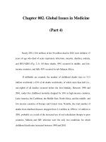
Three to four isoforms of cTnT, as shown in Fig. 1, have been shown to be expressed
in developing cardiac muscle as well as in human fetal skeletal muscle and diseased
human skeletal muscle (5). A developmental down-regulation of cTnT and up-regula-
tion of skeletal isoforms of TnT occurs in normal developing skeletal muscle, which
leads to the absence of cTnT in nondiseased adult skeletal muscle. For cTnI, however,
human cardiac muscle contains a single cTnI, and healthy and diseased human fetal and
adult skeletal muscle have never been shown to express cTnI (6,7).
One proposed explanation for the increase of cTnT in the blood of patients with ESRD
(and patients with diseases of skeletal muscle) was the possibility of extracardiac expres-
sion of cTnT observed in diseased skeletal muscle. Several studies have now addressed
cTnT expression in noncardiac tissues, specifically skeletal muscle tissues obtained
from patients with varied underlying pathologies. Utilizing the cTnT-specific antibodies
(M7, M11.7) used in the Roche cTnT immunoassay, no studies to date have demon-
strated a cTnT isoform that would cause a false-positive cTnT circulating in serum,
plasma, or whole blood. Skeletal muscle specimens obtained from patients with ESRD,
Duchenne muscular dystrophy, polymyositis, and dermatomyositis as well as in kid-
ney specimens from patients with varied renal diseases showed no expression of the
cTnT isoform found in human hearts (8–14). In a preliminary report, cTnT expression
by Western blot analysis in skeletal muscles from ESRD patients was postulated (15).
However, the antibodies utilized in this study were not as cardiac specific as those found
in the second- or third-generation cTnT immunoassay kit marketed by Roche. This
initial report contrasted with a second report that showed no evidence of the expression
of either cTnT mRNA or protein in truncal skeletal muscle from five ESRD patients
(16). Again, the antibodies used in the immunoblot experiments were not the same as
those found in the cTnT Roche assay kit. In a more definite report, the expression of
Fig. 1. Western blot of (1) failing human heart and (2) fetal human heart probed with mono-
clonal anti-cTnT antibody detecting 3 cTnT isoforms. (Adapted from ref. 5.)
cTn in Renal and Muscle Disease 141
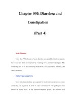
cTnT isoforms in ESRD skeletal muscles using both the capture antibody (M11.7) and
detection antibody (M7) from the Roche cTnT assay kit was addressed (8,9). As shown
in Fig. 2, the M7 antibody detected a 39-kDa cTnT isoform similar to that expressed in
human heart tissue, in 2 of 45 skeletal muscle biopsies. In contrast, the M11.7 antibody
detected two or three cTnT isoforms at 34–36 kDa, and no cTnT at 39 kDa in 20 of 45
skeletal muscle biopsies. Given the differences in epitopes recognized by the M7 and
M11.7 antibodies, it was concluded that the cTnT isoforms expressed in ESRD muscle
would not be detected by the Roche cTnT assay if released into the circulation and were
not the heart isoform of cTnT. Similar findings have been demonstrated in Western blots
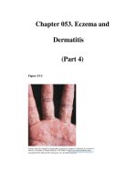
of skeletal muscle tissues obtained from DM, PM, and MD patients as shown in Fig. 3
(9–14). These data support the claim that the tissue source of circulating cTnT in these
patients is from the heart and are indicative of myocardial damage. Furthermore, West-
ern blot analysis of skeletal muscle has never demonstrated expression of cTnI (7,8).
Fig. 2. Western blots of normal human heart and normal and ESRD skeletal muscle detect-
ing cTnT and non-cTnT proteins using the cardiac-specific M11.7 and M7 antibodies. (Adapted
from ref. 9.)
Fig. 3. Western blot of (1) normal human heart and (2) skeletal muscle from dermatomyosi-
tis patient using the Roche anti-cTnT monoclonal M7 antibody demonstrating lack of expres-
sion of cardiac specific troponin T in diseased skeletal muscle.
142 Apple
ROLE OF CARDIAC TROPONIN TESTING
FOR DETECTING MYOCARDIAL INJURY IN ESRD
Numerous studies have shown that both cTnI and cTnT are increased in serum and
plasma in ESRD patients without clinical evidence of myocardial damage. From 1993
through 1998 a review of studies that measured cTnI involving more than 350 ESRD
patients and studies which measured cTnT involving more than 500 ESRD patients
showed 2–10% and 10–30% of patients had increased cTnI and cTnT values, respec-
tively (17–34). Explanations for these substantial differences are not clear at present.
Both cTnI and cTnT assay manufacturers show data in their package inserts regarding
the low percentage of ESRD patients who were found to have increased cardiac tro-
ponins. It should be noted that the studies reviewed in this chapter for cTnT involved
only the second- or third-generation cTnT assays, which have eliminated any interfer-
ence by skeletal muscle troponins. Explanations put forth for the cause of these increased
troponin concentrations include expression of cTnT in skeletal muscle (without evi-
dence), as well as detection of subclinical myocardial damage. The cause of the differ-
ences in positive rates between cTnT and cTnI also is not known. Possible mechanisms
have been proposed for differences between cTnI and cTnT increases. First, circulating
cTnT may reflect left ventricular hypertrophy with a different release pattern vs cTnI
(35). Second, a longer circulating half-life for cTnT may occur, possibly due to advanced
glycosylated end products of cTnT known to accumulate in diabetics with ESRD (36).
Third, cTnT may be more sensitive due to accumulation in the circulation from lack of
removal during the dialysis process compared to cTnI. Studies have emphasized the
problems of using CK-MB as a marker for myocardial damage in ESRD. Falsely increased
concentrations of CK-MB in up to 75% of ESRD patients have been demonstrated (17,
18), likely due to the reexpression of CK-MB in myopathic skeletal muscles (Fig. 4) (37)
in ESRD patients (38). In one study addressing the role of biomarkers for ruling out
AMI, serum cTnI monitoring was performed in 84 patients with renal insufficiency hos-
pitalized to rule out AMI (39). Clinical parameters (echocardiography, ECG) were used
to diagnose AMI. The clinical sensitivity of cTnI was 77%, which was significantly better
than CK-MB at 68%. Both markers showed >90% specificities. However, both sensitiv-
ity and specificity were lower in the renal diseased patients than observed in nonrenal
disease ACS patients presenting to rule in/rule out AMI. No mechanisms are provided
Fig. 4. Western blot of CK-MB standard, normal (N) human heart, normal human skeletal
muscle, and nine skeletal muscle samples from ESRD patients probed with monoclonal anti-
CK-B monoclonal antibody detecting substantial CK-MB expression in diseased skeletal mus-
cles. (Adapted from ref. 37.)
cTn in Renal and Muscle Disease 143
to explain this variance. Thus, the evidence supports cTnI and cTnT as markers with high
specificity for cardiac damage, and can be used to distinguish whether increases in CK-MB
are due to myocardial or skeletal muscle injury.
ROLE OF CARDIAC TROPONIN TESTING
IN THE ASSESSMENT OF MORTALITY IN ESRD
The presence of increased cTnI and cTnT concentrations identify acute coronary syn-
drome patients at significantly higher risk of developing cardiac events, such as car-
diac death and nonfatal AMI, both during hospitalization and at long-term follow-up
(40,41). Several studies have now also demonstrated that ESRD patients with increases
in cTnI and cTnT concentrations tend to have a poor prognostic outcome. First, in a pre-
liminary 1 yr follow-up study of 16 randomly selected ESRD patients, the cardiac event
rate (n = 4 fatal AMIs) was correlated to patients who displayed the higher increases of
serum cTnT and cTnI (23). Second, serum cTnT concentrations measured in 49 ESRD
patients who presented with no complaints of chest pain and in 83 renal insufficiency
patients (serum creatinine >2 mg/dL and not on dialysis) were clinically followed for
6 mo after entry into the study (42). Of the 25 ESRD patients with increased cTnT con-
centrations at entry, six had cardiac events. Thus, cTnT demonstrated 100% sensitivity
and 56% specificity. In comparison, all three patients with an increased cTnI had car-
diac events, demonstrating a 50% sensitivity and a 100% specificity for cTnI. Patients
with diabetes were more likely to have increased cardiac troponin concentrations. In con-
trast, only three patients in the entire renal insufficiency group had an increased cTnI or
cTnT. In the 6-mo follow-up, two patients suffered an AMI, but neither of these patients
had increased troponins. These data suggest that cardiac troponin testing may be effec-
tive in elucidating cardiac risks of patients undergoing chronic dialysis.
Third, measurement of cTnT in the blood of 97 ESRD patients showed that cTnT was
detectable in 29% of patients (16). The prevalence of increased cTnT concentrations
correlated with cardiac risk. Fifty percent (11 of 22) of known coronary artery disease
(CAD) patients had an increased cTnT concentration (median 1.6 ng/mL), compared to
31% (15 of 33) of patients with ³2 risk factors (median 0.1 ng/mL) and 11% (3 of 28) of
patients with 0 or 1 risk factors; p < 0.05 vs known CAD patients. Thus, a positive rela-
tionship existed between increased risk of CAD and increases in cTnT concentrations.
A fourth study investigated the use of monitoring cTnT and cTnI concentrations for
predicting cardiac outcomes by 6 mo in patients presenting with suspected acute coro-
nary syndromes (ACS) and renal insufficiency (creatinine >2.0 mg/dL) (n = 51) rela-
tive to ACS patients without renal disease (n = 102) (43). Thirty-five percent of patients
in the renal group and 45% in the nonrenal group experienced an adverse outcome dur-
ing initial hospitalization. However, at 6 mos, both groups had experienced >50% adverse
outcomes. The areas under the receiver operating characteristic (ROC) curves for both
cTnT and cTnI, used as predictors of initial and long-term outcomes, were significantly
lower in the renal group than the nonrenal group (0.56, 0.75, respectively). No mecha-
nisms were given to explain these findings.
In addition to these studies, four recent studies now clearly demonstrate the prognos-
tic power of cardiac troponin for predicting mortality in patients with ESRD. First, in
30 hemodialysis patients followed over 2 yr, cTnI (Dade Stratus II and Abbott AxSYM)
144 Apple
and cTnT (second- and third-generation Roche assay) concentrations were measured
at baseline (44). At 2 yr, an increased cTnT demonstrated 90% mortality vs 11% in
patients with a normal cTnT. In comparison, both increased and normal cTnI patients
had 40% mortality. Second (Study A, Table 1), in 102 ESRD patients with a baseline
cTnT (Elecsys) measured 24-mo outcomes were assessed (45). cTnT was a strong pre-
dictor of mortality, with 7-fold greater risk of death at cTnT >0.1 ng/mL. An increased
cTnT resulted in a 3.6-fold greater hazard ratio for overall mortality. Even at the low
cutoff concentration of 0.01 ng/mL, 22% of patients died at 24 mo. Third (Study B,
Table 1), 244 ESRD patients with baseline cTnT (Elecsys) values were followed for 34
mo (46). A significant correlation between increasing cTnT, all causes of death, and
cardiac death was found. In addition, increasing cTnT over a 6-mo period showed an
increasing death rate; risk ratio = 2.0. Fourth (Study C, Table 1), a preliminary study
from the author’s institution examined 18-mo mortality in 441 ESRD based on baseline
cTnT (Elecsys) and cTnI (Dade Dimension RxL) values (47). Both cardiac troponins
demonstrated significant increases in mortality in the troponin-positive vs negative
groups: cTnT 26% vs 13%; cTnI 41% vs 19%, respectively (p < 0.01). However, there
were a substantially greater number of patients with increased cTnT (n = 238) com-
pared to cTnI (n = 24).
CLINICAL IMPLICATIONS
The clinical chore for predicting the role of cardiac troponin testing in the ESRD
population will be (1) to distinguish the mechanistic type of increase an individual patient
has and (2) to determine whether it is best to (i) detect a larger number of patients with
Table 1
Risk Associated with Cardiac Troponin in ESRD Patients
Study A (ref. 45, n = 102)
cTnT (ng/mL) n Deaths %
<0.01 17 0 0
0.01–0.04 45 10 22
>0.04–0.1 28 8 28
>0.1 12 10 83
Study B (ref. 46, n = 244)
cTnT (ng/mL) Deaths (%) Cardiac deaths (%)
<0.01 6 0
0.01–0.09 43 14
³0.1 59 24
Study C (ref. 47, n = 441)
cTnT (ng/mL) cTnI,ng/mL
<0.04 ³0.04 <0.1 ³0.1
n 193 248 417 24
(%) (44) (56) (94.5) (5.5)
% Mortality 13 26 19 41
cTn in Renal and Muscle Disease 145
increased troponin T with the possibility of confounding increases with fewer outcomes,
(ii) detect fewer increased troponins I with a possible higher predictive mortality rate
(assay dependent), with the higher probability of missing patients who go on to have a
poor outcome. As is generally true for all diagnostic tests, the price of higher sensitivity
will usually be lower specificity.
The rationale for cardiac troponin testing in the management of ACS has been clearly
established. The ultimate role of cardiac troponin testing for risk stratification in the out-
patient dialysis unit is speculative, but attractive. The initiation of dialysis is temporally
associated with the occurrence of AMI, as about half of myocardial infarcts occurring
in these ESRD patients are clustered within the first 2 yr after dialysis initiation. There
are a host of conceivable strategies for the identification of the highest cardiac risk dialy-
sis patients after initiation of renal replacement therapy, but one plausible, potentially
cost-effective scenario is the developing role of outpatient cardiac troponin testing.
In conclusion, the use of cardiac tissue specific cTnT and cTnI testing in blood to
assist in ruling in and ruling out AMI in patients with renal disease and skeletal muscle
pathologies, and as a tool for cardiac risk assessment in ESRD patients now appears to
be evidence based. However, larger trial studies would be useful to increase the evidence
base to determine the overall incidences and differences between cTnT and cTnI moni-
toring in ESRD patients for cardiac risk assessment. Incorporation of cardiac troponin
testing in ESRD patients may assist in initiating more aggressive treatment of coronary
artery disease, detection of subclinical myocardial injury, and correlate increases in car-
diac troponin testing with increased cardiac risk. Further, monitoring blood cardiac tro-
ponin concentrations should assist in the detection and treatment of CAD before renal
transplantation, potentially reducing the risk of adverse cardiac events. However, each
cardiac troponin assay will need to be validated independently, as not all cardiac tropo-
nin assays are equivalent (48).
ABBREVIATIONS
ACS, acute coronary syndrome(s); AMI, acute myocardial infarction; CAD, coronary
artery disease; CK, creatine kinase; CK-MB, MB isoenzyme of CK; cTnT, cTnI, cardiac
troponins T and I; DM, dermatomyositis; ECG, electrocardiogram; ERSD, end-stage
renal disease; MD, muscular dystrophy; PM, polymyositis; ROC, receiver operating
characteristic.
REFERENCES
1. Herzog CA, Ma JZ, Collins AJ. Poor long-term survival after acute myocardial infraction
among patients on long-term dialysis. N Engl J Med 1998;339:799–805.
2. Herzog CA. Diagnosis and treatment of ischemic heart disease in dialysis patients. Curr
Opin Neph Hyper 1997;6:558–565.
3. Bleyer AJ, Russell GB, Satko SG. Sudden and cardiac death rates in hemodialysis patients.
Kid Int 1999;55:1553–1559.
4. Hoffman EP, Fishbeck KH, Brown RH, et al. Characterization of dystrophy in muscle biopsy
specimens from patient’s with Duchenne’s or Becker’s muscular dystrophy. N Engl J Med
1988;318:1363–1368.
5. Anderson PAW, Malouf NN, Oakley AE, Pagani ED, Allen PD. Troponin T isoform expres-
sion in humans: a comparison among normal and failing adult heart, fetal heart, and adult
and fetal skeletal muscle. Circ Res 1991;69:1226–1233.
146 Apple
6. Wilkinson JM, Grand JA. Comparison of amino acid sequence of troponin I from different
striated muscles. Nature 1978;271:31–35.
7. Bodor GS, Porterfield D, Voss EM, Smith S, Apple FS. Cardiac troponin I is not expressed
in fetal and healthy or diseased adult human skeletal tissue. Clin Chem 1995;41:1710–1715.
8. Ricchiuti V, Voss EM, Ney A, Odland M, Anderson PAW, Apple FS. Cardiac troponin T
isoforms expressed in renal diseased skeletal muscle will not cause false-positive results by
the second generation cardiac troponin T assay by Boehringer Mannheim. Clin Chem 1998;
44:1919–1924.
9. Ricchiuti V, Apple FS. RNA expression of cardiac troponin T isoforms in diseased human
skeletal muscle. Clin Chem 1999;45:2129–2135.
10. Hammer-Lercher A, Erlacher P, Bittner R, et al. Clinical and experimental results on car-
diac troponin expression in Duchenne muscular dystrophy. Clin Chem 2001;47:451–458.
11. Erlacher P, Lercher A, Falkensammer J, et al. Cardiac troponin and beta-type myosin heavy
chain concentrations in patients with polymyositis or dermatomyositis. Clin Chim Acta
2001;306:27–33.
12. Fredricks S, Murray JF, Bewick M, et al. Cardiac troponin T and creatine kinase MB are
not increased in exterior oblique muscle of patients with renal failure. Clin Chem 2001;47:
1023–1030.
13. Davis GK, Labugger R, Van Eyk JE, Apple FS. Cardiac troponin T is not detected in
Western blots of diseased renal disease. Clin Chem 2001;47:782–784.
14. Davis GK, Labugger R, Van Eyk JE, Apple FS. Cardiac troponin T is not detected in Western
blot of diseased renal tissue. Clin Chem 2001;47:782–784.
15. Bodor GS, Survant L, Voss EM, Smith S, Porterfield D, Apple FS. Cardiac troponin T
composition in normal and regenerating skeletal muscle. Clin Chem 1997;43:476–484.
16. Haller C, Zehelein J, Remppis A, Muller-Bardorff M, Katus HA. Cardiac troponin T in
patients with end-stage renal disease: absence of expression in truncal skeletal muscle. Clin
Chem 1998;44:930–938.
17. McLaurin MD, Apple FS, Voss EM, Herzog CA, Sharkey SW. Cardiac troponin I, cardiac
troponin T, and creatine kinase MB in dialysis patients without ischemic heart disease: evi-
dence of cardiac troponin T expression in skeletal muscle. Clin Chem 1997;43:976–982.
18. Adams J, Bodor G, Davila-Roman V, et al. Cardiac troponin I: a marker with high specific-
ity for cardiac injury. Circulation 1993;88:101–106.
19. Trinquier S, Flecheux O, Bullenger M, Castex F. Highly specific immunoassay for cardiac
troponin I assessed in noninfarct patients with chronic renal failure or severe polytrauma.
Clin Chem 1995:41:1675–1676.
20. Li D, Jialal I, Keffer J. Greater frequency of increased cardiac troponin T than increased
cardiac troponin I in patients with chronic renal failure. Clin Chem 1996;42:114–115.
21. Porter GA, Norton T, Bennett WB. Troponin T, a predictor of death in chronic haemodi-
alysis patients. Eur Heart J 1998;19(Suppl):N34–37.
22. Bhayana V, Gougoulias T, Cohee S. Discordance between results for serum troponin T and
I in renal disease. Clin Chem 1995;41:312–317.
23. Apple FS, Sharkey SW, Hoeft P, et al. Prognostic value of serum cardiac troponin I and T in
chronic dialysis patients: a one year outcomes analysis. Am J Kidney Dis 1997;29:399–403.
24. Hafner G, Thome-Kromer B, Schaube J, Kupferwasser I, Ehrenthal W, Cummins P. Car-
diac troponins in serum in chronic renal failure. Clin Chem 1994;40:1790–1791.
25. Ishii J, Ishikawa T, Yukitake J. Clinical specificity of a second generation cardiac troponin
T assay in patients with chronic renal failure. Clin Chim Acta 1998;270:183–188.
26. Akagi M, Nagake Y, Makino H, Ota Z. A comparative study of myocardial troponin T
levels in patients undergoing hemodialysis. Jpn J Nephrol 1995;37:639–643.
27. Frankel WL, Herold DA, Zregler TW, Fitzgerald RL. Cardiac troponin T is elevated in
asymptomatic patients with chronic renal failure. Am J Clin Pathol 1996;106:118–123.
cTn in Renal and Muscle Disease 147
28. Ooi DS, House AA. Cardiac troponin T in hemodialyzed patients. Clin Chem 1998;44:
1410–1416.
29. Bhayana V, Gougoulias T, Cohoe S, Henderson AR. Discordance between results for
serum troponin T and troponin I in renal disease. Clin Chem 1995;41:312–317.
30. Haller C, Stevanovich A, Katus HA. Are cardiac troponins reliable serodiagnostic markers
of cardiac ischemia in end-stage renal disease? Nephrol Dial Transplant 1996;11:941–944.
31. Katus HA, Haller C, Müller-Bardorff M, Scheffold T, Remppis A. Cardiac troponin T in
end-stage renal disease patients undergoing chronic maintenance hemodialysis. Clin Chem
1995;41:1201–1202.
32. Haller C, Katus HA. Cardiac troponin T in dialysis patients. Clin Chem 1998;44:358.
33. McNeil AR, Marshall M, Ellis CJ, Hawkins RC. Why is troponin T increased in the serum
of patients with end-stage renal disease? Clin Chem 1998;44:2377–2378.
34. Collinson PO, Stubbs PJ, Rosalki SB. Cardiac troponin T in renal disease. Clin Chem 1995;
41:1671–1673.
35. Lowbeer C, Deeberger AO, Gustafsson SA, Norrman R, Hulting J, Gutierrez A. Increased
cardiac T and endothelin-1 concentrations in dialysis patients may indicate heart disease.
Nephrol Dial Transplant 1999;14:1948–1955.
36. Kinchi K, Nejima J, Takano T, Ohta M, Hashimoto H. Increased serum concentrations of
advanced glycation end products: a marker of coronary artery disease activity in type 2 dia-
betic patients. Heart 2001;85:87–91.
37. Ricchiuti V, Apple FS. Regulation of muscle gene and protein expression of creatine kinase
B in chronic renal disease (abstract). Clin Chem 2000;46:A90.
38. Diesel W, Emms M, Knight B, Noakes TD, Swanepoel CR, Smit R. Morphology features
on the myopathy associated with chronic renal failure. Am J Kidney Dis 1993;22:677–684.
39. McLaurin MD, Apple FS, Falahati A, Murakami M, Miller EA, Sharkey SW. Cardiac tro-
ponin I and creatine kinase MB mass to rule out myocardial injury in hospitalized patients
with renal insufficiency. Am J Cardiol 1998;82:973–975.
40. Ottani F, Galvani M, Nicolini FA, Ferrini D, Pozzati A, DiPasquale G, Jaffe AS. Elevated
cardiac troponin levels predict the risk of adverse outcome in patients with acute coronary
syndromes. Am Heart J 2000;40:917–927.
41. Heidenreich PA, Alloggiamento T, Melsop K, McDonald KM, Go AS, Hlatky MA. The
prognostic value of troponin in patients with non-ST-elevation acute coronary syndromes:
a meta-analysis. J Am Coll Cardiol 2001;38:478–488.
42. Roppolo LP, Fitzgerald R, Dillow J, Ziegler T, Rice M, Maisel A. A comparison of tropo-
nin T and troponin I as predictors of cardiac events in patients undergoing chronic dialysis
at a Veteran’s hospital: a pilot study. J Am Coll Cardiol 1999;34:448–454.
43. VanLente F, McErlean ES, DeLuca SA, Rao JS, Nissen SE. Ability of troponins to predict
adverse outcomes in patients with renal insufficiency and suspected acute coronary syn-
dromes: a case matched study. J Am Coll Cardiol 1999;33:471–478.
44. Porter GA, Norton T, Bennett WM. Long term follow-up of the utility of troponin T to assess
cardiac risk in stable chronic hemodialysis patients. Clin Lab 2000;46:469–476.
45. Dierkes J, Domrose U, Westphal S, et al. Cardiac troponin T predicts mortality in patients
with end stage renal disease. Circulation 2000;102:1964–1969.
46. Ooi DS, Zimmerman D, Graham J, Wells GA. Cardiac troponin T predicts long-term out-
comes in hemodialysis patients. Clin Chem 2001;47:412–417.
47. Apple FS, Murakami MM, Pearce LA, Herzog CA. Predictive value of cardiac troponin I
and T for subsequent death in end stage renal disease. Circulation 2002;106:2941–2945.
48. Apple FS, Wu AHB. Myocardial infarction redefined: role of cardiac troponin testing. Clin
Chem 2001;47:377–379.
148 Apple
Beyond Ischemic Heart Disease 149
149
From: Cardiac Markers, Second Edition
Edited by: Alan H. B. Wu @ Humana Press Inc., Totowa, NJ
9
Cardiac-Specific Troponins
Beyond Ischemic Heart Disease
David Morrow
INTRODUCTION
Cardiac biomarkers play a pivotal role in the clinical evaluation of patients present-
ing with chest discomfort. Owing to their tissue specificity and high clinical sensitivity
for detecting myocyte injury, the cardiac troponins have become the preferred biomarker
for the diagnosis of myocardial infarction (MI) (1) and risk assessment of patients with
suspected acute coronary syndromes (ACS) (2,3). Nevertheless, clinicians often encoun-
ter increased concentrations of cardiac troponin in patients without overt coronary artery
disease or low clinical probability of myocardial ischemia, leading some to concerns over
biologic false-positive troponin results (4). A critical distinction must be made, however,
between the high specificity of cardiac troponin for myocardial injury and the lack of
specificity for coronary atherothrombosis and ischemia as the mechanism of injury. As
troponin assays with increasing analytic sensitivity have been developed, our ability to
detect minor degrees of myocardial injury in a variety of conditions has expanded, and
led to a growing list of clinical settings in which increased concentrations of cardiac tro-
ponin has been detected without evidence of overt ischemic heart disease (Table 1).
While the presence of myocyte damage and the mechanism of injury are well defined
(e.g., myocarditis) in several of these conditions, the availability of assays for cardiac
troponin has revealed unexpectedly prevalent myocardial injury in others, and led to
important new directions for research, as well as to possibilities for novel clinical applica-
tions in nonischemic cardiac disease. This chapter describes several of these clinical set-
tings in detail, starting with those in which some degree of myocardial damage is expected
and moving toward those in the etiology of myocardial injury less well explained. Other
important etiologies of troponin release are discussed individually in separate chapters
(chemotherapeutic agents, cardiac surgery, angioplasty, renal failure, skeletal muscle dis-
ease, and congestive heart failure).
MYOCARDITIS
Myocarditis connotes inflammatory involvement of the myocardium that may occur
in a wide range of infectious and noninfectious conditions (5). Pathologic evidence from
routine autopsies suggests that unrecognized myocardial involvement occurs more fre-
quently than expected in the setting of systemic infection (6). Myocarditis is an established
150 Morrow
cause of release of the MB isoenzyme of creatine kinase (CK-MB) in the absence of
coronary artery obstruction and myocardial ischemia (5). Given the difficulty in defini-
tively establishing the presence of myocarditis in the appropriate clinical setting, sen-
sitive and specific indicators of myocardial injury, such as the cardiac troponins, have
been viewed as a potential valuable aid to diagnosis (5). Viewed from an alternative per-
spective, abnormal concentrations of troponins resulting from unsuspected myocarditis
may also be an important source of diagnostic confusion in patients with undifferentiated
chest pain.
Clinical Presentation and Pathophysiology
Myocarditis is characterized by a leukocytic infiltrate and necrosis or degeneration
of cardiac myocytes that may be focal or diffuse and is generally randomly distributed
in the heart (7,8). It is common for endomyocardial biopsies to sample relatively normal
areas of myocardium when neighboring areas have significant inflammatory infiltrates
(9,10). Furthermore, the diverse etiologies of myocarditis may contribute to varied sen-
sitivity of histologic findings (11). In some series, histologic confirmation is made in only
10% of biopsies from patients with a clinically suspected myocarditis (12). This leaves
substantial room for improvement on current diagnostic techniques through the explor-
ation of alternative markers of cellular involvement (13).
Although most cases of myocarditis are viral in origin, numerous other microbiologic
agents, noninfectious immunologic conditions, hypersensitivity reactions, and physi-
cal agents (e.g., radiation) may all cause clinically important myocardial inflamma-
tion. The acute phase of myocarditis may be completely asymptomatic or may result in
fulminant congestive heart failure (8). Long-term outcomes are similarly variable, rang-
Table 1
Nonischemic
a
Conditions with Elevation of Troponin
Myocarditis
Direct cardiac trauma (cardiac surgery, ablation, endomyocardial biopsy, internal defibrillator
discharge, external cardioversion)
Blunt chest trauma
Chemotherapeutic agents (anthracyclines)
Infiltrative diseases with cardiac involvement (e.g., amyloidosis) (116,117)
Pericarditis (118)
Rhabdomyolysis with cardiac involvement (119)
Uncomplicated percutaneous coronary intervention
Pulmonary embolism
Hypertensive emergency (e.g., preeclampsia) (120)
Congestive heart failure
Uncomplicated noncardiac surgery
Acute neurologic disease, including subarachnoid hemorrhage (121,122)
Sepsis and related syndromes
Renal failure
Alcoholic cirrhosis (123)
Vital exhaustion (124,125)
a
Primary etiology is not due to coronary atherothrombosis.
Beyond Ischemic Heart Disease 151
ing from no recognizable impact, to increased risk of lethal arrhythmias, and possibly to
the development of postviral dilated cardiomyopathy (14,15). The symptoms are typi-
cally nonspecific, including fatigue, dyspnea, palpitations, and chest discomfort (8), and
may be difficult to discriminate from those related to acute myocardial ischemia (16).
Role of Cardiac Troponins in the Evaluation of Myocarditis
CK activities are increased in some but not all cases of myocarditis (5,17), and thus
the cardiac troponins have been investigated as a potential aid to the diagnosis of myo-
carditis (13,18–20). Studies of animal models of myocarditis have shown a time-depen-
dent rise in cardiac troponins after the onset of autoimmune myocarditis documented
by histopathologic examination (13,19). Human studies have extended these findings to
the clinical setting and supported the use of troponins to confirm the presence of injury
of cardiac myocytes and improve on the sensitivity of endomyocardial biopsy for estab-
lishing the diagnosis of myocarditis (13,20).
In a population of 80 patients with clinically suspected myocarditis, increased concen-
trations of cardiac troponin T (cTnT) were a strong predictor of histologic or immuno-
histologic evidence of myocarditis on endomyocardial biopsy (Fig. 1) (20). However,
44% of patients without increased concentrations of cTnT also had pathologic evidence
of myocarditis, indicating that measurement of cTnT alone is not sufficient to exclude a
Fig. 1. Predictive performance of cTnT at a threshold of 0.1 ng/mL for histologic evidence
of myocarditis. Serum levels of cTnT in 80 patients with clinically suspected myocarditis. Solid
squares are for patients with histologic or immunohistologic criteria for myocarditis. Open
squares indicate those with no evidence of myocarditis on biopsy. (Adapted from Lauer B,
et al. J Am Coll Cardiol 1997;30:1354, with permission.)
152 Morrow
diagnosis of myocarditis. CK-MB was increased in only one patient. Similar data are avail-
able for cardiac troponin I (cTnI) (13). Among 53 patients with biopsy-proven myocar-
ditis, 34% had increased concentrations of cTnI (³3.1 ng/mL, Dade Stratus) compared
with only 6% who had increased concentrations of CK-MB. Although it is a plausible
hypothesis that patients with detectable concentrations of cardiac troponins had more
active inflammation in the myocardium, no correlation between troponin elevation and
the histologic severity of myocarditis was observed (13,20).
The reasons for the absence of detectable troponin elevation in a large proportion of
patients with histologic evidence of myocyte necrosis are likely multifactorial (13). In
several studies, the timing of sample acquisition relative to symptom onset was seen to
be important. Specifically, patients with samples drawn earlier after the clinical onset
of the syndrome were substantially more likely to have increased concentrations of cTnI,
consistent with the theory that the majority of inflammatory injury occurs early in the course
of myocarditis (13). Depending on the specific etiology of myocardial inflammation, the
timing and duration of active myocyte injury and thus increases in troponin may vary.
As such, serial sampling over various stages of the illness may prove to improve sensitivity
of detection of active myocarditis. It is also possible that in some cases the degree of myo-
cardial injury was not sufficient to result in detection with the assays tested in these studies.
The availability of more sensitive current generation assays for cTnI and cTnT may there-
fore increase the proportion of patients with suspected myocarditis who have detectable
myocyte damage.
Current Use and Future Directions
Cardiac troponins frequently provide evidence of ongoing myocyte damage in patients
with suspected myocarditis when CK-MB concentrations are within the normal range.
However, cardiac troponins are not increased in all cases, and cannot be used to reliably
exclude the disease. Nevertheless, owing to the similarly poor sensitivity of endomyo-
cardial biopsy, some experts have recommended measurement of cardiac troponins and
correlation with the results of histologic assessment in all patients with suspected myo-
carditis (5). When increased concentrations of troponins are detected in the absence of
histologic and/or immunohistologic evidence of myocarditis, sampling error is a strong
possibility; however, other nonischemic and ischemic causes of myocyte necrosis should
be considered. Conversely, myocarditis should be among the diagnoses considered for
patients presenting with chest symptoms and increased troponins who subsequently
are shown to be free of significant epicardial coronary disease (21). Additional research
will likely clarify the impact of timing of sampling and the cause of myocarditis as deter-
minants of troponin elevation during the clinical course of the disease, and it is hoped
will guide strategies for testing that will assist in the challenge of discerning an inflam-
matory vs ischemic cause of myocardial injury in patient with chest pain.
BLUNT CARDIAC TRAUMA
Blunt cardiac trauma is a syndrome that includes a broad spectrum of nonpenetrating
traumatic cardiac injuries, ranging from mild pericardial inflammation to rupture of the
cardiac wall (22). Although the most severe cases generally demand immediate thoracot-
omy for diagnosis and treatment, cardiac biomarkers have traditionally been used to facil-
Beyond Ischemic Heart Disease 153
itate the detection of myocardial injury or “cardiac contusion” among patients who are
hemodynamically stable. However, measurement of CK and CK-MB for this purpose has
significant limitations due to the extensive skeletal muscle injury that usually occurs in
association with this syndrome. Because of superior tissue specificity, the cardiac tro-
ponins have offered potential to distinguish cardiac from skeletal muscle damage in the
setting of blunt chest trauma (23,24).
Clinical Presentation and Pathophysiology
Blunt cardiac injury typically results from direct compression of the heart or deceler-
ating forces delivered to the chest (25). Cardiac injury may occur even after relatively
low-energy chest trauma without other obvious injuries (26,27). The pathologic corre-
lates of such injury vary considerably and range from small areas of subepicardial or sub-
endocardial hemorrhage to full thickness myocardial necrosis with or without cardiac
rupture (28). The relationships between the less severe pathologic findings and the risk
of significant clinical manifestations of blunt cardiac trauma (e.g., severe arrhythmias)
are not well characterized. Rarely, epicardial coronary thrombosis may be induced by
chest trauma and result in MI due to coronary ischemia (29,30). Clinical manifestations
of nonpenetrating cardiac trauma include chest discomfort, dyspnea, hypotension, elec-
trocardiographic (ECG) changes (primarily nonspecific ST and T-wave abnormalities),
as well as arrhythmias including sinus tachycardia, atrial arrhythmias, ventricular con-
duction abnormalities, and ventricular tachycardia or fibrillation (25). Cardiogenic shock
is infrequent and usually indicates cardiac rupture, cardiac tamponade, or severe struc-
tural damage to a cardiac valve and/or its supporting structures. Estimates of the risk of
significant clinical morbidity in the absence of such obvious catastrophic consequences
vary widely (31–35).
Primarily owing to difficulty in defining clear and consistent diagnostic criteria for
“myocardial contusion,” this term has been largely replaced by the broader designation of
“nonpenetrating cardiac trauma,” with some investigators and clinicians advocating cate-
gorization based on the clinical sequelae (25,36,37). The traditional noninvasive tools,
such as serial ECGs, are limited by poor specificity. Furthermore, there is no consensus
agreement as to whether abnormalities of cardiac wall motion must be present for the
diagnosis of “cardiac contusion.” Thus, current strategies for evaluation are turning away
from establishing strict diagnostic criteria toward an emphasis on assessing the risk of
adverse clinical consequences (e.g., severe arrhythmias, or impaired ventricular or valvu-
lar function), and the need for changes in management, such as special monitoring (37).
Role of Cardiac Biomarkers in Evaluating Blunt Chest Trauma
Measurement of CK and CK-MB has been used as an adjunct to the ECG and clini-
cal evaluation in the diagnosis of blunt cardiac injury (38). However, the poor tissue spe-
cificity of these markers in the setting of substantial skeletal muscle trauma has limited
their clinical utility (39,40). For example, in a study of 44 patients with blunt chest trauma,
70% of patients with normal echocardiograms had increased concentrations of CK-MB
(24). While use of a CK-MB/total CK ratio of >5% as the diagnostic criterion reduced
the number of “false-positive” positive results, the sensitivity was substantially reduced.
Consistent findings across multiple studies have shown similar levels of CK-MB and the
CK-MB/total CK ratio in patients with and without a diagnosis of blunt cardiac injury