báo cáo khoa học: "Quantum dot-induced cell death involves Fas upregulation and lipid peroxidation in human neuroblastoma cells" pptx
Bạn đang xem bản rút gọn của tài liệu. Xem và tải ngay bản đầy đủ của tài liệu tại đây (917.81 KB, 13 trang )
Journal of Nanobiotechnology
BioMed Central
Open Access
Research
Quantum dot-induced cell death involves Fas upregulation and lipid
peroxidation in human neuroblastoma cells
Angela O Choi1, Sung Ju Cho1,2, Julie Desbarats3, Jasmina Lovrić1 and
Dusica Maysinger*1
Address: 1Department of Pharmacology & Therapeutics, McGill University, 3655 Promenade Sir William-Osler, McIntyre Medical Sciences
Building, Montreal, QC, H3G 1Y6, Canada, 2Faculty of Pharmacy and Department of Chemistry, University of Montreal, Pavillon J. A. Bombardier,
C.P. 6128 Succursale Centre-Ville, Montreal, QC, H3C 3J7, Canada and 3Department of Physiology, McGill University, Montreal, QC, H3G 1Y6,
Canada
Email: Angela O Choi - ; Sung Ju Cho - ; Julie Desbarats - ;
Jasmina Lovrić - ; Dusica Maysinger* -
* Corresponding author
Published: 12 February 2007
Journal of Nanobiotechnology 2007, 5:1
doi:10.1186/1477-3155-5-1
Received: 5 October 2006
Accepted: 12 February 2007
This article is available from: />© 2007 Choi et al; licensee BioMed Central Ltd.
This is an Open Access article distributed under the terms of the Creative Commons Attribution License ( />which permits unrestricted use, distribution, and reproduction in any medium, provided the original work is properly cited.
Abstract
Background: Neuroblastoma, a frequently occurring solid tumour in children, remains a
therapeutic challenge as existing imaging tools are inadequate for proper and accurate diagnosis,
resulting in treatment failures. Nanoparticles have recently been introduced to the field of cancer
research and promise remarkable improvements in diagnostics, targeting and drug delivery. Among
these nanoparticles, quantum dots (QDs) are highly appealing due to their manipulatable surfaces,
yielding multifunctional QDs applicable in different biological models. The biocompatibility of these
QDs, however, remains questionable.
Results: We show here that QD surface modifications with N-acetylcysteine (NAC) alter QD
physical and biological properties. In human neuroblastoma (SH-SY5Y) cells, NAC modified QDs
were internalized to a lesser extent and were less cytotoxic than unmodified QDs. Cytotoxicity
was correlated with Fas upregulation on the surface of treated cells. Alongside the increased
expression of Fas, QD treated cells had increased membrane lipid peroxidation, as measured by
the fluorescent BODIPY-C11 dye. Moreover, peroxidized lipids were detected at the mitochondrial
level, contributing to the impairment of mitochondrial functions as shown by the MTT reduction
assay and imaged with confocal microscopy using the fluorescent JC-1 dye.
Conclusion: QD core and surface compositions, as well as QD stability, all influence nanoparticle
internalization and the consequent cytotoxicity. Cadmium telluride QD-induced toxicity involves
the upregulation of the Fas receptor and lipid peroxidation, leading to impaired neuroblastoma cell
functions. Further improvements of nanoparticles and our understanding of the underlying
mechanisms of QD-toxicity are critical for the development of new nanotherapeutics or
diagnostics in nano-oncology.
Page 1 of 13
(page number not for citation purposes)
Journal of Nanobiotechnology 2007, 5:1
Background
Neuroblastoma is the most frequently occurring extracranial solid tumour in children, accounting for 9% of all
childhood cancers, with poor prognosis [1]. This malignant tumour arises from neuroepithelial cells of the sympathetic nervous system early in development, and is
typically found in the adrenal medulla, abdomen, chest or
neck [2]. Neuroblastoma, however, remains a therapeutic
challenge as current surgical and chemical treatments are
insufficient to prevent tumour recurrence, metastasis and
progression [3]. Accurate disease staging is critical for
appropriate therapeutic intervention, but existing imaging
tools are still lacking in early and accurate diagnosis [4].
The introduction of nanoparticles in the field of cancer
research has recently improved diagnosis, targeting and
drug delivery with the use of nanotubes, liposomes, dendrimers and polymers [5-7]. Other nanoparticles, such as
quantum dots, possess excellent photophysical properties
and prove to be an elegant alternative to the traditional
bioimaging tools [8]. Quantum dots (QDs) are one of the
most rapidly evolving products of nanotechnology, with
great potential as a tool for biomedical and bioanalytical
imaging. Their superior photophysical properties [9] and
sometimes multifunctional surfaces are suitable for applications in various biological models [10]. A study by the
Nie group describes the application of these multifunctional QDs for in vivo imaging and targeting of breast and
prostate cancers [11]. Although the development of QDs
as bioimaging tools may be well underway, their potential
application as therapeutic agents is yet to be explored.
Biological media, intracellular microenvironment and
different enzymatic systems could destabilize originally
well protected QD surfaces yielding more cytotoxic nanoparticles [12,13]. Uncoated or weakly stabilized cadmium
telluride QDs produce significant amounts of reactive
oxygen species in vitro [12], and induce death in various
cell types [14,15].
Oxidative stress-induced cell death, both apoptosis and
necrosis, can involve a number of cellular mechanisms,
one of which includes the activation of Fas receptor
[16,17]. Fas (CD95) belongs to the family of tumour
necrosis factor receptors, and is a prototypical "death
receptor." In the immune system, it regulates cell numbers
by inducing apoptosis, and is involved in T cell-mediated
cytotoxicity. It can also induce neuronal cell death [1821]. Activation of Fas receptor by Fas ligand recruits the
Fas-Associated Death Domain (FADD) to the Death
Domain in the cytoplasmic tail of the receptor, and can
lead to caspase activation and cell death [22]. Downstream signaling of Fas can also induce activation of
lipases and pro-apoptotic transcription factors like p53,
which then potentiate apoptosis [23].
/>
Oxidative stress can also induce other levels of cell membrane damage, including membrane lipid peroxidation
[24]. Free radicals induce the cleavage of membrane lipids, resulting in the production of aldehydes, reinforcing
cellular stress. Intracellular lipid peroxidation can also
occur at the level of the organelle membranes, especially
at the membranes of the highly metabolically active mitochondria. Mitochondria regulate crucial cellular processes
including adenosine triphosphate (ATP) production,
intracellular pH regulation and neuronal-glial interactions [25]. Many neurodegenerative diseases, including
Parkinson's and Alzheimer's diseases, involve the malfunctioning of the mitochondria, seen as decreased mitochondrial activity, decreased ATP production or loss of
mitochondrial membrane potential (∆ψm) [25].
In this study, we explored mechanisms of QD-induced
toxicity in a human neuroblastoma cell line exposed to
cysteamine-QDs and QDs modified by an antioxidant, Nacetylcysteine. We report new mechanisms of cytotoxicity
induced by these QDs, including the i) upregulation of
the Fas receptor, ii) lipid peroxidation, and iii) impaired
mitochondrial function. Understanding the mechanisms
underlying QD-toxicity will provide alternative ways of
nanoparticle manipulations to make them more suitable
tools in nanomedicine, specifically nano-oncology.
Results
Surface modifications of cadmium telluride QDs with Nacetylcysteine
To investigate mechanisms underlying cell death induced
by cadmium telluride (CdTe) QDs, we modified the surface of cysteamine-capped CdTe QDs with an antioxidant,
N-acetylcysteine (NAC, Figure 1b), a drug which has been
found previously to protect cells against oxidative stress
and QD-induced cytotoxicity [14]. Cysteamine-capped
(''unmodified'') QDs (Figure 1a) have amino groups at
the surface and are positively charged (+14.2 mV). Covalent binding of NAC to cysteamine on the QD surface
(Figure 1c) yielded NAC-conjugated QDs with a decreased
net surface charge, and charge-charge complexation of
NAC and cysteamine yielded NAC-capped QDs with carboxylic groups on the surface and a net negative charge of
-9.8 mV (Figure 1d). Spectrofluorometric measurements
revealed marked differences in the fluorescence intensities
and QD stability in different media (see Additional file 1).
In phosphate buffered saline (PBS), cysteamine-QDs
show a red-shift with time but no change in fluorescence
intensity, whereas, NAC-conjugated QDs decreased in fluorescence with time. NAC-capped QDs were the most stable in PBS, with no spectral shift and no loss of
fluorescence within 24 hours.
Page 2 of 13
(page number not for citation purposes)
Journal of Nanobiotechnology 2007, 5:1
a
/>
b
NH3+
NH3+
S
S
O
S
NH3
CdTe
+
H3N
HN
HS
+
CH3
COOH
S
S
S
NH3+
+
H3N
N-acetylcysteine
unmodified QD
c
d
CH3
H3C
O
SH
HN
HN
O
O
SH
O
NH
NH
-
O
S
S
HS
H
N
H3C
O
N
H
S
N
H
CdTe
H3C
OOC
H3C
HN
NH
HS
O
COO-
O
S
S
CdTe
S
S
-
S
OOC
H3C
CH3
HN
S
NH
O
O
O
NH
HN
O
CH3
HN
OOC
SH
S
HN
H
N
CH3
S
S
O
HS
O
O
O
COOCH3
O
NH
NH
COO-
O
CH3
CH3
NAC-conjugated
QD
NAC-capped
QD
Figure 1
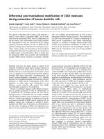
Schematic representations of unmodified and NAC-modified QDs
Schematic representations of unmodified and NAC-modified QDs. a. cysteamine-capped ("unmodified") QD (λem =
542 nm in water), b. N-acetylcysteine (NAC) c. NAC-conjugated QD (λem = 526 nm in water), d. NAC-capped QD (λem = 528
nm in water).
Page 3 of 13
(page number not for citation purposes)
Journal of Nanobiotechnology 2007, 5:1
Cytotoxicity of NAC-modified CdTe QDs in
neuroblastomas
To examine the cytotoxicity of cysteamine-QDs and NACmodified QDs, we assessed the viability of SH-SY5Y
human neuroblastoma cells by fluorescence-activated cell
scanning (FACS), and their mitochondrial metabolism
using a MTT reduction assay. Our FACS data show that
cells exposed to 5 µg/mL of cysteamine-QDs, NAC-conjugated or NAC-capped QDs yielded distinct populations of
dead cells (Figure 2a), suggesting significant toxicity
induced by these QDs. Significantly less viability was
observed in cells treated with cysteamine-QDs (52.2 ±
0.7%, p < 0.05) when compared to untreated control cells
in serum-free medium (75.9 ± 9.1%). It is noteworthy
that trophic factor deprivation, due to serum withdrawal,
contributes to cell death which explains the approximately 25% decrease in viability in the absence of QDs
(i.e. untreated control). NAC treatment can rescue cells in
this trophic factor withdrawal paradigm [26]. QDinduced cytotoxicity is prevented with cell pretreatment
with 2 mM NAC (85.5 ± 5.7%; p < 0.01; Figure 2b), confirming and complementing results from our previous
studies demonstrating the effectiveness of NAC against
both trophic factor deprivation and additional QD-insult
[14]. These multiple insults to neuroblastoma cells lead to
cell death both by apoptosis and necrosis. The latter is
characterized by mitochondrial and lysosomal swelling
and perinuclear localization of these organelles [17].
NAC-capped and NAC-conjugated QDs are still cytotoxic
(65.6 ± 5.0%, p < 0.05 and 59.1 ± 5.1%, p < 0.05 respectively) compared to control.
Results of the FACS analyses were corroborated by data
from measuring cellular MTT reduction (Figure 2c). Mitochondrial metabolic activity was most significantly
reduced in cells in the presence of cysteamine-QDs (50.1
± 5.2%; p < 0.01). NAC-conjugated QDs also significantly
reduced the cellular mitochondrial activity (62.3 ± 6.5%;
p < 0.01) compared to control. Cells treated with NACcapped QDs, on the other hand, suffered less cytotoxic
damage and showed significantly higher mitochondrial
metabolic activity (90.2 ± 2.2%; p < 0.01) compared to
cells treated with cysteamine-QDs or NAC-conjugated
QDs. Cells pretreated with NAC, prior to cysteamine-QD
addition, show significantly higher activity (106.1 ±
11.8%; p < 0.01) when compared to QD-treated cells,
again reinforcing the protective role of free NAC against
QD-induced toxicity.
Upregulation of Fas at the cell surface and internalization
of QDs by neuroblastoma cells
QD-induced cytotoxicity involves oxidative stress, specifically via the production of reactive oxygen species (ROS)
[12,15]. One cell-damaging, downstream effect of ROS
production is the upregulation of the cell surface Fas
/>
receptor. FACS analyses revealed significant upregulation
of Fas expression on the surface of SH-SY5Y cells treated
with cysteamine-QDs (net mean fluorescence intensity
(MFI) is 64.3 ± 5.5, p < 0.05) and NAC-conjugated QDs
(net MFI = 57.1 ± 3.7, p < 0.05) when compared to
untreated control cells (net MFI = 43.9 ± 1.1; Figures 3a
and 3b). No upregulation of Fas was observed in cells
treated with NAC-capped QDs (net MFI = 42.1 ± 3.7), and
Fas upregulation was completely inhibited in cells pretreated with NAC in the presence of cysteamine-QDs (net
MFI = 41.5 ± 0.4), suggesting that QD-induced Fas expression is likely due to QD-mediated oxidative stress.
In addition, free Cd2+ released from QDs and the extent of
QD uptake can contribute to the cell damage and eventually cell death. We measured intracellular and extracellular Cd2+ concentrations in SH-SY5Y cell cultures treated
with QDs. Results from this study and from our recently
published study [27] show that Cd2+ concentrations contribute to, but cannot fully explain QD-induced cytotoxicity (31.1 ± 1.7% and 58.0 ± 2.1% cytotoxicity induced by
Cd2+ and QDs respectively), suggesting that impairment
of cellular functions by QDs is multifactorial.
The extent of QD uptake was assessed by FACS analyses.
Cells treated with cysteamine-QDs, NAC-conjugated and
NAC-capped QDs show marked differences in QD uptake.
In particular, cysteamine-QD-treated cells show an evident shift in fluorescence intensity compared to the
untreated control and to both NAC-conjugated and NACcapped QD-treated cells (Figure 3c). Quantitative measurements of the mean fluorescence intensity show that
cysteamine-QDs were indeed taken up most avidly (net
MFI = 17.8 ± 0.1; Figure 3d). On the other hand, NACconjugated QDs (net MFI = 7.3 ± 0.6; p < 0.001) and NACcapped QDs (net MFI = 2.1 ± 1.2; p < 0.001) were internalized significantly less than cysteamine-QDs. The net
MFI for cells pretreated with 2 mM NAC (5.6 ± 0.8; p <
0.001) was significantly lower than in the absence of NAC
(net MFI = 17.8 ± 0.1), suggesting that NAC either reduced
QD uptake or partly quenched QD fluorescence. The latter is unlikely as spectral data show that these NAC-modified QDs have comparable, and in some cases even
higher, fluorescence intensities as the unmodified QDs
(see Additional file 1). On the other hand, measurements
of intracellular Cd2+ show reduced Cd2+ content in NAC
pretreated cells, supporting the notion that less QDs were
internalized by the cells and that extracellular Cd2+ effects
were also diminished by NAC.
QD-induced lipid peroxidation and change in membrane
potential (∆ψm) of the mitochondria
The subcellular distribution of internalized QDs has previously been reported to induce ROS production and
organelle damage [12,14]. Here we identify two intracel-
Page 4 of 13
(page number not for citation purposes)
Journal of Nanobiotechnology 2007, 5:1
/>
N
0
200
600
1000
0
NAC
1000
200
600
QD + NAC
R2
R2
R1
600
R1
R1
200
1000
R1
R2
R2
0
0
200
600
1000
0
600
1000
120
100
**
+
Q
D
AC
N
A
C
**
80
60
40
20
0
N
FSC
200
**
140
Q
D
0
+
c
A
C
R1
R2
AC
Q
D
-c
on
jQ
N
AC
D
-c
ap
Q
D
200
C
tr
l
R1
R2
Mitochondrial
metabolic activity (%)
SSC
R1
N
R2
R2
R1
600
0
AC
NAC-cap QD
20
N
NAC-conj QD
1000
40
Q
D
1000
1000
on
j
600
600
AC
-c
200
200
C
tr
l
0
0
60
D
0
*
Q
R2
*
80
-c
ap
R1
R1
R2
200
**
AC
R1
R1
100
N
R2
R2
600
Viable cells (%)
1000
120
N
b
QD
Untreated
Q
D
a
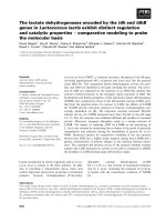
Figure
telluride and metabolic activity of human neuroblastoma (SH-SY5Y) cells treated with NAC-modified and unmodified cadmium
Viability 2QDs
Viability and metabolic activity of human neuroblastoma (SH-SY5Y) cells treated with NAC-modified and
unmodified cadmium telluride QDs. a. Quantum dot toxicity differs depending on their surface modifications by NAC.
Flow cytometry light scatter dot plots reveal two distinct cell populations corresponding to viable cells (R1), and cells in various stages of apoptotic death (R2). FSC, forward scatter (proportional to cell size); SSC, side scatter (proportional to cell complexity or granularity). b. Cell death in neuroblastomas after 24 hours of QD treatments. Graph shows percentage of dead
cells (gated on R2) for each treatment: Ctrl = cells under serum-deprivation with no drug or QD added; QD = cysteamineQDs; NAC-conj QD = NAC-conjugated QDs; NAC-cap QD = NAC-capped QDs; NAC (2 mM); NAC + QD (2 mM NAC +
5 µg/mL cysteamine-QD). All QDs were added at 5 µg/mL. Mean values and standard deviations from three independent
experiments (N = 9) are shown. (*p < 0.05; **p < 0.01). c. Mitochondrial metabolic activity was assessed using MTT and its
conversion to formazan was measured at 595 nm. All values are expressed relative to cells without any drug or QD addition
(Ctrl) taken as 100%. Note significant decrease with QD treatments and full recovery in the presence of 2 mM NAC. Mean values and standard deviations from quadruplicate measurements in two independent experiments (N = 8) are shown. (**p <
0.01).
Page 5 of 13
(page number not for citation purposes)
Journal of Nanobiotechnology 2007, 5:1
b
80
0
200
c
relative cell number
d
20
QD
net MFI
NAC-conj QD
NAC-cap QD
15
10
200
***
***
N
fluorescence intensity
***
5
0
0
Q
D
N
fluorescence intensity
+
0
AC
20
AC
0
40
A
C
200
*
N
QD-treated
400
60
Q
D
-c
on
jQ
N
AC
D
-c
ap
Q
D
untreated
*
AC
Q
D
-c
on
jQ
N
A
D
C
-c
ap
Q
N
D
A
C
+
Q
D
600
net MFI
iso ctrl
C
tr
l
800
N
1000
relative cell number
a
/>
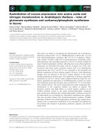
Figure 3
Fas expression and internalization of QDs
Fas expression and internalization of QDs. a. Exposure to QDs induces cell-surface Fas expression in neuroblastomas.
Fas expression was assessed by FACS in untreated cells (grey line) and in cells exposed to cysteamine-QDs for 24 hours (black
line). Dotted line shows background staining of untreated cells with isotype-matched control antibody. b. Net Fas expression
was calculated as Mean Fluorescence Intensity (MFI) of cells stained with anti-Fas antibodies subtracted by MFI of isotype control antibody-stained cells. Averages and standard deviations from three independent experiments (N = 9) are shown (*p <
0.05). c. QD uptake was assessed by flow cytometry in neuroblastoma cells treated with 5 µg/ml QDs (unmodified and NACmodified) for 24 hours. d. Net Mean Fluorescence Intensities (MFI) of cells treated with cysteamine-QDs, NAC-conjugated
and NAC-capped QDs, and cysteamine-QDs in the presence of 2 mM NAC (NAC + QD) are shown. Net MFI was calculated
as MFI of QD-treated cells subtracted by the autofluorescence of untreated cells. Note a significant decrease (p < 0.05) in the
MFI of NAC-modified QDs compared with MFI of cysteamine-QDs. Averages and standard deviations from three independent
experiments (N = 9) are shown (***p < 0.001).
Page 6 of 13
(page number not for citation purposes)
Journal of Nanobiotechnology 2007, 5:1
lular targets of this QD-induced ROS, namely membrane
lipids and mitochondria. In response to oxidative stress,
cell surface and organellar membrane lipids may undergo
peroxidation [24]. We assessed lipid peroxidation by spectrofluorometric measurements of the fluorescent BODIPY-C11 dye and by confocal microscopy (Figures 4a, b).
When compared to untreated control cells (100.0 ±
6.3%), cells treated either with cysteamine-QDs (70.2 ±
2.0%, p < 0.01) or NAC-capped QDs (76.6 ± 6.5%, p <
0.05) showed significantly reduced red (non-oxidized) to
green (oxidized) ratios. Cells treated with NAC-conjugated QDs or pretreated with free NAC, in the presence of
cysteamine-QDs, did not show significant lipid peroxidation compared to the untreated control.
Double labeling using BODIPY-C11 dye and MitoTracker
Deep Red 633 revealed lipid peroxidation of the mitochondrial membranes as shown by confocal microscopy
(Figure 4b). Co-localized oxidized BODIPY-C11(green)
and MitoTracker Deep Red 633 (red-purple) appear as
punctate yellow signals, suggesting local membrane lipid
peroxidation within the mitochondria in cells treated with
QDs.
Membrane lipid peroxidation can produce damaging
aldehydes, and at the mitochondrial level, this can impair
mitochondrial functions [28]. Confocal microscopy analyses of cells stained with JC-1 clearly show that QDtreated cells have significantly reduced mitochondrial
membrane potential (∆ψm) (Figure 4c). Compared to the
strong red fluorescence of JC-1 aggregates observed in the
untreated control, QD-treated cells show an increased
intensity in green fluorescence (JC-1 monomers) which
correlates with a decrease in ∆ψm.
Discussion
Initial reports on the potential toxicity of some types of
quantum dots (QDs) [13,14,29] prompted the development of differently modified QDs as tools in the biological sciences. Several studies describing modifications to
improve QD biocompatibility for their broad applications in the medical sciences were recently reported
[10,30,31]. At the other end of the spectrum, research
groups are also attempting to harness and apply QD toxicity in toxicotherapy. For instance, one study proposed
the application of dopamine-conjugated QDs as inducers
of cellular phototoxicity [32], while others are using QDs
in photodynamic therapy [33] and to target different
stages of cancers [11,34]. Using NAC-conjugated, NACcapped and cysteamine-capped CdTe QDs, this study
shows several cellular responses of human neuroblastoma
cells to these nanoparticles. Surface-modified nanoparticles with NAC led to reduced cell death, decreased Fas
expression and decreased mitochondrial membrane lipid
peroxidation. The negatively charged NAC-capped QDs
/>
were the most benign, followed by NAC-conjugated QDs.
Cysteamine-QDs, with a net positive surface charge,
showed significant cellular uptake, as well as increased
upregulation of Fas receptors on the cell surface and membrane lipid peroxidation, contributing to the impairment
of mitochondrial and overall cell functions. In addition to
surface charge, cytotoxicity is also affected by other physicochemical properties, including particle size, core-shell
composition and capping [14,35,36].
QD biocompatibility can be easily altered by surface modifications, such as conjugation and capping with biomolecules and polymers [31,37,38]. The Hoshino group
characterized the physicochemical properties of different
surface-modified CdSe QDs and reported that these surface modifications affect QD surface potential, QD fluorescence and QD-induced cytotoxicity [36]. In our study,
we found that QD surface conjugation and capping with
an antioxidant, N-acetylcysteine (NAC), reduced QD
uptake and cytotoxicity (Figures 2 and 3). Moreover, pretreatment of cells with free NAC fully protected cells from
QD-induced cytotoxicity (Figure 2b), as demonstrated in
our previous study in a different cell line [12]. NAC can
protect cells both from apoptosis and necrosis. Mechanisms of the cytoprotective action of NAC are well-documented, and involve NAC acting (i) as a direct thiol
antioxidant, (ii) as a glutathione precursor, (iii) as a transcription regulator for genes involved in cellular homeostasis, and (iv) as a cell survival promoter via inhibition of
apoptotic pathways including JNK and p38 [26].
Highly metabolically active mitochondria are particularly
sensitive and vulnerable targets to cellular stress [25].
Cells treated with QDs undergo a change in mitochondrial membrane potential (∆ψm) (Figure 4c). Membrane
depolarization has been widely associated with the release
of the apoptotic factor, cytochrome c, which amplifies
pro-apoptotic caspase cascades, promoting cell death
[12,25]. Among the regulators of mitochondrial membrane potential, cardiolipin, a mitochondrial membrane
specific lipoprotein, is of particular relevance in neuronal
cells [39,40]. The abundance of cardiolipin in the membranes of the mitochondria maintains the membrane
potential and regulates the release of cytochrome c. Upon
cellular stress, cardiolipin, along with other membrane
lipids, is degraded due to lipid peroxidation, and the
membrane potential is no longer stable, resulting in an
uncontrolled release of mitochondrial content [40,41]. In
addition to causing membrane instability and increasing
the vulnerability of the cell to subsequent insults [28],
lipid peroxidation can also generate harmful and relatively stable aldehyde products which add to the oxidative
stress. One of these damaging aldehydes is 4-oxo-2-nonenal (ONE), which acts by activation of the p53 signaling
Page 7 of 13
(page number not for citation purposes)
Journal of Nanobiotechnology 2007, 5:1
/>
a
120
100
80
60
40
20
0
QD-treated
BODIPY
MitoTracker
*
**
c
A
C
+
Q
D
AC
N
N
10um
N
N
AC
-c
on
j
Q
D
Q
AC
D
-c
ap
Q
D
Overlay
C
tr
l
non-oxid/oxid (%)
b
monomer
aggregate
overlay
Control
QDtreated
Figure 4
QD-induced mitochondrial lipid peroxidation and change in membrane potential
QD-induced mitochondrial lipid peroxidation and change in membrane potential. a. Spectrofluorometric assessment of lipid membrane peroxidation by ratiometric approach in untreated (Ctrl) or QD-treated cells. The ratio between the
red and green fluorescence in the control was taken as 100% and all other values with NAC or QD treatments were
expressed relative to it. All values are means from quadruplicate measurements and are obtained from three independent (N =
12) experiments (*p < 0.05; **p < 0.01). b. Confocal micrograph showing dual labeling of oxidized lipids (green fluorescence
from oxidized BODIPY-C11) within mitochondria (labeled with MitoTracker Deep Red 633). Insets show two adjacent cells
from the same field. Scale bar = 10 µm. c. Confocal micrographs of SH-SY5Y cells labeled with JC-1 reveal decrease in mitochondrial membrane potential after QD treatment. Cells were treated with 5 µg/mL QD and typical change in fluorescence
from red (Em = 590 nm) to green (Em = 530 nm) was assessed in cell cultures in serum-free medium (control) or QD (5 µg/
mL). Note an enhanced intensity of green fluorescence in QD-treated cells. The micrograph illustrates the loss in mitochondrial potential upon oxidative stress induced by QDs. Scale bar = 10 µm.
Page 8 of 13
(page number not for citation purposes)
Journal of Nanobiotechnology 2007, 5:1
pathway and induces apoptosis in SH-SY5Y neuroblastoma cells [42].
Besides intracellular targeting at the mitochondrial level,
QD treatment leads to an upregulation of cell surface Fas
expression (Figures 3a and 3b). The Fas receptor, when
activated by Fas ligand, associates with FADD which
recruits caspase-8 or caspase-10, and forms the deathinducing signaling complex (DISC). Caspases-8/10 autocatalyze their own cleavage [43-45], triggering a cascade of
caspase activation that culminates in apoptosis. This cascade may be further amplified by cleavage of the caspase8/10 substrate Bid, which then inserts into the mitochondrial membrane, resulting in loss of ∆ψm and release of
cytochrome c, further accelerating apoptosis. Fas has been
implicated as an inducer of apoptosis under conditions of
high in vivo oxidative stress [46-48], and recent studies
show that Fas expression may also be triggered upon activation of proapoptotic transcription factors, such as
FOXO3 [49].
Nanoparticles, such as the CdTe QDs investigated here,
enter cells and can get sequestered within different
organelles, changing organellar morphologies and
obstructing their functions, leading eventually to cell
death of different types [17,50,51]. For instance, our
recent study in human breast cancer cells [27] showed
that QDs induce enlargements of lysosomes and mitochondria, both of which are morphological indications of
necrotic cell death. On the other hand, intracellular accumulation of unprotected or unstable QDs can eventually
result in QD degradation and Cd2+ release from the QD
core, initiating apoptosis. The extent of apoptosis in neuroblastoma cells and Cd2+ released are, however, not
strongly correlated, suggesting additional contributors to
cell death aside from the free Cd2+.
Neuroblastoma cells that were deprived of serum-derived
trophic factors are more susceptible to additional insults
induced by QDs (i.e. ROS, Cd2+), leading to both type I
(apoptosis) and type III (necrosis) cell death [17]. Under
the circumstances in which QDs could be employed for
the detection and elimination of neuroblastoma, one
should bear in mind not only the physical properties of
QDs but also the vulnerability of healthy tissues surrounding the tumour, the rate of QD sequestration and
the rate of metal elimination from the body [51]. Collectively, earlier and current findings suggest that cell preconditioning, combined with modifications of the QD
surface with NAC and a tumour-specific ligand (e.g. Trk
mimetics to target Trk receptors) could yield an improved
nano-oncological therapeutic, sensitizing or diagnostic
agent for neuroblastomas.
/>
Conclusion
Results from this study provide new mechanistic data
(summarized in Figure 5) on the much debated issue of
QD toxicity. Cadmium telluride (CdTe) QD-induced
cytotoxicity depends on multiple QD properties including
QD core size, stability in biological media and surface
chemistry which determine the extent of cellular internalization. Mechanisms of CdTe QD-induced toxicity
include multiple organelle damage and involve increased
Fas receptor expression and cell membrane lipid peroxidation in SH-SY5Y neuroblastoma cells. These damages
bring about cell death both by apoptosis and necrosis.
Understanding the mechanisms underlying QD toxicity is
important as QDs and other nanoparticles are promising
tools in the field of nano-oncology as potential imaging
agents, photosensitizers, biosensors and nanotherapeutics.
Materials and methods
Preparation of CdTe quantum dots
Tellurium powder (200 mesh, 99.8%), sodium borohydride (99%), cadmium perchlorate hydrate, N-acetylcysteine (99%) and cysteamine hydrochloride (98%)
were purchased from Sigma-Aldrich. Milli-Q water (Millipore) was used as a solvent. Photoluminescence measurements were carried out at room temperature using a Cary
Eclipse Fluorescence spectrometer. The excitation wavelength was set at 400 nm. The excitation and emission slits
were set at 5 nm. Dialysis was performed using spectra/
por molecularporous membrane tubing (Spectrum Laboratories, Inc.) with a 6000–8000 Da molecular weight cutoff. Centrifugation was performed with Eppendorf
centrifuge 5403 (10,000 rpm) and Eppendorf centrifuge
5415 C (14,000 rpm).
Preparation of Cysteamine capped (+) CdTe
Sodium borohydride (0.8 g, 21.1 mmol) was dissolved in
water (20 mL) at 0°C under N2 atmosphere. Tellurium
powder (1.28 g, 10 mmol) was added portionwise and the
mixture was stirred at 0°C for 8 h under N2 atmosphere.
The reaction mixture was stored at 4°C in the dark and
used in the next step.
The thiol capped-QDs were prepared as described [12].
Briefly, cadmium perchlorate hydrate (500 µL, 1 M aqueous solution) and cysteamine hydrochloride (300 mg,
2.64 mmol) were dissolved in 200 mL of N2 saturated
Milli-Q water. The pH of the solution was adjusted to 5.1
with 1N NaOH aqueous solution prior to addition of an
aliquot of the previously prepared NaHTe solution (200
µL). The reaction mixture was heated to reflux for 25 min
under N2. The resulting QD solution was dialyzed against
Milli-Q water for 4 h then concentrated to 15 mL using a
rotary evaporator. QDs were precipitated using MeOH/
CHCl3 (1:1, v/v) then collected by centrifugation. The
Page 9 of 13
(page number not for citation purposes)
Journal of Nanobiotechnology 2007, 5:1
QDs were washed with MeOH/CHCl3 (1:1, v/v) two times
then dried under vacuum. The QDs were used as solutions
either in deionized water or in PBS buffer.
Preparation of NAC capped (-) CdTe
Cadmium perchlorate hydrate (500 µL, 1 M aqueous solution) and N-acetylcysteine (400 mg, 2.45 mmol) were dissolved in 200 mL of N2 saturated Milli-Q water. The pH of
the solution was adjusted to 10.5 with 1N NaOH aqueous
solution prior to addition of an aliquot of the previously
prepared NaHTe solution (200 µL). The reaction mixture
was heated to reflux for 25 min under N2. The resulting
QD solution was dialyzed against Milli-Q water for 4 h
then processed as above.
Conjugation of NAC to cysteamine capped QD
Solutions of NAC (4 mM) were freshly prepared in water
mixed with cysteamine capped (+) CdTe QD in water (2
mg/mL, λem = 542 nm) followed by 1-ethyl-3-(3'dimethylaminopropyl)carbodiimide (EDC; 12 mg, 77.3
µmol) addition. The reaction mixture was incubated for 3
h at room temperature with occasional shaking. The mixture was purified by dialysis against water for 4 h. The
emission wavelength of the resulting solution was 533
nm. The NAC-conjugated QDs were used as a solution in
water. Zeta potentials of all QD preparations were measured using Zetasizer Nano ZS (Malvern Instruments,
Worchestershire, UK).
Cell culture and treatments
The human neuroblastoma cell line SH-SY5Y was
obtained from ATCC and cultured (37°C, 5% CO2) in
DMEM medium containing phenol red and 10% FBS
(Gibco, Burlington, ON, Canada). Cells were used at 2–8
passages. For spectrofluorometric and colorimetric assays,
cells were cultured in 24-well plates (Sarstedt, Montreal,
QC, Canada) at a density of 105 cells/cm2.
One hour prior to treatments, medium containing serum
was aspirated, and cells washed with serum free medium.
Fresh serum free medium was added to all wells, including the untreated control (Ctrl). An additional set of control cells, grown in 10% FBS, was used to account for
changes in cell morphology, cell number and metabolic
activity due to the serum withdrawal.
Cells were treated with QDs (5 µg/mL) for different time
periods as specified in individual figure legends. QD solutions (5 µg/mL) were prepared from the stock (2 mg/mL)
by dilution in serum free cell culture medium. Cells were
incubated with QDs for a maximum of 24 h before biochemical analysis or live cell imaging.
NAC was dissolved in PBS (400 mM), and was added to
the culture medium 2 h before QDs. All treatments were
/>
done in triplicates or quadruplicates in three or more
independent experiments.
Flow cytometry in determining cell viability, Fas expression
and cellular uptake of QDs
SH-SY5Y cells were treated with 5 µg/ml QDs (NAC-modified and unmodified) and/or 2 mM NAC (as indicated)
for 24 h at 37°C/5% CO2 in media supplemented with
10% FBS. Adherent and non-adherent cells were harvested and pooled so as not to lose apoptotic cells which
may have detached from the plastic substrate. Cells were
resuspended at 1 × 10⁶ cells/ml in FACS buffer (PBS + 1 %
FCS). Fas expression was determined by labeling cells
with phycoerythrin (PE) conjugated anti-human Fas/
CD95 (clone DX2, BD Biosciences), and PE conjugated
isotype-matched control antibodies (mouse IgG1 kappa,
BD Biosciences) for 30 min on ice. Cells were washed
twice and resuspended in 300 µl FACS buffer. Samples
analysed for viability and/or for quantum dot-associated
fluorescence alone (FL1, PMT 488–540 nm) were not
labeled with antibodies. 10,000 events per sample were
acquired on a Becton Dickinson FACScan flow cytometer.
Data were analyzed using CellQuest software. Fas expression was determined as follows: Net Fas expression = Fas
mean fluorescence intensity (MFI) – isotype control MFI
for each individual sample, then averages and standard
deviations of three independent replicates were calculated.
MTT assay
Colorimetric MTT (3-(4,5-dimethylthiazol-2-yl)-2,5diphenyl tetrazolium bromide, Sigma) assays were performed to assess the mitochondrial activity of cells treated
as described above. After 24 h treatment, media was
removed and replaced with drug-free, serum-free media
(500 µL/well). 50 µL of stock MTT (5 mg/mL) was added
to each well and cells were then incubated for one hour at
37°C. Media were removed, cells were lysed and formazan dissolved with DMSO. Absorbance was measured at
595 nm using a Benchmark microplate reader (Bio-Rad,
Mississauga, ON, Canada). All measurements were done
in triplicates in three or more independent experiments.
Lipid peroxidation
Cells were treated with the fluorescent dye BODIPY 581/
591 C11 (BODIPY-C11, Molecular Probes), which inserts
into lipid membranes and allows for quantitative assessment of oxidized versus non-oxidized lipids by fluorescing green or red, respectively. Cells were stained for 30
min with a 10 µM solution of BODIPY-C11 prior to QD
treatment. After the QD treatment, lipids were extracted
from the cells according to the Folch method [52] by incubating twice with a mixture of chloroform and methanol
(2:1 (v/v)). After extraction, 0.2 volumes of 0.9% NaCl
solution were added and the chloroform-containing
Page 10 of 13
(page number not for citation purposes)
Journal of Nanobiotechnology 2007, 5:1
/>
QD
plasma membrane
lipid peroxidation
Fas
NAC
ROS
FADD
Caspase 8
cardiolipin
peroxidation
cytochrome c
mitochondrial
membrane lipid
peroxidation
c
caspase
cascade
n
ipi
iol
ard
NAC
∆ΨM
CELL DEATH
∆ metabolic
activity
Figure 5
Proposed mechanism of QD induced cell death involving Fas, lipid peroxidation and mitochondrial impairment
Proposed mechanism of QD induced cell death involving Fas, lipid peroxidation and mitochondrial impairment. Cells exposed to cadmium telluride quantum dots (unmodified and NAC-modified) induce ROS which causes Fas
upregulation and plasma membrane lipid peroxidation. Apoptotic cell death is induced by activation of Fas and its downstream
effectors. Lipid peroxidation also occurs at the mitochondrial membranes, degrading cardiolipin, changing the mitochondrial
membrane potential, eventually leading to the release of cytochrome c [12], and promoting apoptotic cascades. NAC bound to
the QD surface, modifies the extent of QD internalization, which is correlated with cell death, upregulation of Fas, and ROS
induced lipid peroxidation. NAC treatment (2–5 mM) abolishes oxidative stress, induces antioxidant enzymes and attenuates
mitochondrial impairment.
phase was collected. After evaporating the chloroform and
dissolving the lipids in isopropanol, spectrofluorometric
readings were taken using the SpectraMax Gemini XS
microplate spectrofluorometer (Molecular Devices Corporation, USA). Data were analyzed using the SOFTmax
Pro 4.0 program. All values are presented as normalized
means ± SEM relative to the respective serum-free control
(taken as 100%).
Confocal microscopy
Images were acquired with a Zeiss LSM 510 NLO inverted
microscope. Cells were grown in 8-well chamber slides
(Lab-Tek, Nalge Nunc International, Rochester, NY, USA).
QDs were added to designated wells and the cells were
incubated for the times indicated. Mitochondria were
stained with MitoTracker Deep Red 633 (1 µM, 1 min,
Molecular Probes; λex 644 nm, λem 665 nm) and imaged
using HeNe 633 nm excitation laser and LP 650 filter.
Lipid peroxidation was visualized using BODIPY-C11
(Molecular Probes; non-oxidized: λex 581 nm, λem 595
nm; oxidized: λex 485 nm, λem 520 nm) with the Argon
488 nm excitation laser and LP 520 nm filter, and the
HeNe 543 nm laser and LP 560 filter. Mitochondrial
depolarization was determined using JC-1 (15 µM, 30
min, Molecular Probes; monomer: λex 485 nm, λem 530
nm; aggregate: λex 535 nm, λem 590 nm). The potentialsensitive color shift was monitored using the same set of
lasers and filters as BODIPY-C11. Before imaging, cells
were washed with PBS or with serum-free medium. No
background fluorescence of cells was detected under the
settings used. Images were acquired at a resolution of 512
× 512 and 1024 × 1024. Quadruplicate samples were ana-
Page 11 of 13
(page number not for citation purposes)
Journal of Nanobiotechnology 2007, 5:1
lyzed in all the imaging experiments. Scan size was 146.2
àm ì 146.2 àm. Figures were created using Adobe Photoshop.
Statistical analysis
Data were analyzed using SYSTAT 10 (SPSS, Chicago, IL,
USA). Statistical significance was determined by Student's
t-tests with Bonferroni correction. Differences were considered significant where *p < 0.05, **p < 0.01, ***p <
0.001.
/>
5.
6.
7.
8.
9.
10.
Abbreviations
CdTe, cadmium telluride; NAC, N-acetylcysteine; QD,
quantum dot; ROS, reactive oxygen species; FACS, fluorescence-activated cell sorting; MTT, 3-(4,5-dimethylthiazol2-yl)-2,5-diphenyltetrazolium bromide; BODIPY-C11,
4,4-difluoro-5-(4-phenyl-1,3-butadienyl)-4-bora-3a,4adiaza-s-indacene-3-undecanoic acid; JC-1, 5,5',6,6'-tetrachloro-1,1',3,3'-tetraethylbenzimidazolylcarbocyanine
iodide;
11.
12.
13.
14.
Competing interests
The author(s) declare that they have no competing interests.
15.
Authors' contributions
16.
DM initiated and guided these studies. DM drafted and
AOC finalized the manuscript. AOC, SJC, JD and JL carried out the experiments. All authors read and approved
the final manuscript.
17.
Additional material
18.
19.
20.
Additional file 1
PL spectra (stability) of CdTe nanoparticles in water and PBS.
Click here for file
[ />
21.
22.
Acknowledgements
This work was supported by the Juvenile Diabetes Research Foundation
(Canada) and the Canadian Institutes of Health Research. The authors
would like to thank J. Laliberté for assistance with confocal microscopy and
U. Koppe with lipid peroxidation experiments.
23.
24.
25.
References
1.
2.
3.
4.
Schwab M, Westermann F, Hero B, Berthold F: Neuroblastoma:
biology and molecular and chromosomal pathology. Lancet
Oncol 2003, 4(8):472-480.
Brodeur GM: Neuroblastoma: biological insights into a clinical
enigma. Nat Rev Cancer 2003, 3(3):203-216.
Henry MC, Tashjian DB, Breuer CK: Neuroblastoma update. Curr
Opin Oncol 2005, 17(1):19-23.
Kushner BH: Neuroblastoma: a disease requiring a multitude
of imaging studies. J Nucl Med 2004, 45(7):1172-1188.
26.
27.
28.
Nishiyama N, Kataoka K: Current state, achievements, and
future prospects of polymeric micelles as nanocarriers for
drug and gene delivery. Pharmacol Ther 2006, 112(3):630-648.
Vicent MJ, Duncan R: Polymer conjugates: nanosized medicines
for treating cancer. Trends in biotechnology 2006, 24(1):39-47.
Portney NG, Ozkan M: Nano-oncology: drug delivery, imaging,
and sensing. Anal Bioanal Chem 2006, 384(3):620-630.
Leary SP, Liu CY, Apuzzo ML: Toward the emergence of nanoneurosurgery: part II--nanomedicine: diagnostics and imaging
at the nanoscale level. Neurosurgery 2006, 58(5):805-23; discussion 805-23.
Giepmans BN, Adams SR, Ellisman MH, Tsien RY: The fluorescent
toolbox for assessing protein location and function. Science
2006, 312(5771):217-224.
Pinaud F, Michalet X, Bentolila LA, Tsay JM, Doose S, Li JJ, Iyer G,
Weiss S: Advances in fluorescence imaging with quantum dot
bio-probes. Biomaterials 2006, 27(9):1679-1687.
Gao X, Cui Y, Levenson RM, Chung LW, Nie S: In vivo cancer targeting and imaging with semiconductor quantum dots. Nat
Biotechnol 2004, 22(8):969-976.
Lovric J, Cho SJ, Winnik FM, Maysinger D: Unmodified cadmium
telluride quantum dots induce reactive oxygen species formation leading to multiple organelle damage and cell death.
Chem Biol 2005, 12(11):1227-1234.
Hardman R: A toxicologic review of quantum dots: toxicity
depends on physicochemical and environmental factors.
Environ Health Perspect 2006, 114(2):165-172.
Lovric J, Bazzi HS, Cuie Y, Fortin GRA, Winnik FM, Maysinger D: Differences in subcellular distribution and toxicity of green and
red emitting CdTe quantum dots. Journal of Molecular MedicineJmm 2005, 83(5):377-385.
Ryman-Rasmussen JP, Riviere JE, Monteiro-Riviere NA: Surface
coatings determine cytotoxicity and irritation potential of
quantum dot nanoparticles in epidermal keratinocytes. J
Invest Dermatol 2007, 127(1):143-153.
Vogt M, Bauer MK, Ferrari D, Schulze-Osthoff K: Oxidative stress
and hypoxia/reoxygenation trigger CD95 (APO-1/Fas) ligand
expression in microglial cells. FEBS Lett 1998, 429(1):67-72.
Golstein P, Kroemer G: Cell death by necrosis: towards a
molecular definition. Trends Biochem Sci 2007, 32(1):37-43.
Nagata S, Golstein P: The Fas Death Factor. Science 1995,
267(5203):1449-1456.
Raoul C, Pettmann B, Henderson CE: Active killing of neurons
during development and following stress: a role for
p75(NTR) and Fas? Curr Opin Neurobiol 2000, 10(1):111-117.
Martin-Villalba A, Herr I, Jeremias I, Hahne M, Brandt R, Vogel J,
Schenkel J, Herdegen T, Debatin KM: CD95 ligand (Fas-L/APO1L) and tumor necrosis factor-related apoptosis-inducing ligand mediate ischemia-induced apoptosis in neurons. J Neurosci 1999, 19(10):3809-3817.
Ju ST, Panka DJ, Cui H, Ettinger R, el-Khatib M, Sherr DH, Stanger BZ,
Marshak-Rothstein A: Fas(CD95)/FasL interactions required
for programmed cell death after T-cell activation. Nature
1995, 373(6513):444-448.
Thorburn A: Death receptor-induced cell killing. Cell Signal
2004, 16(2):139-144.
Wallach D, Varfolomeev EE, Malinin NL, Goltsev YV, Kovalenko AV,
Boldin MP: Tumor necrosis factor receptor and Fas signaling
mechanisms. Annu Rev Immunol 1999, 17:331-367.
Moreira PI, Smith MA, Zhu X, Nunomura A, Castellani RJ, Perry G:
Oxidative stress and neurodegeneration. Ann N Y Acad Sci
2005, 1043:545-552.
Foster KA, Galeffi F, Gerich FJ, Turner DA, Muller M: Optical and
pharmacological tools to investigate the role of mitochondria during oxidative stress and neurodegeneration. Prog Neurobiol 2006, 79(3):136-171.
Zafarullah M, Li WQ, Sylvester J, Ahmad M: Molecular mechanisms of N-acetylcysteine actions. Cell Mol Life Sci 2003,
60(1):6-20.
Cho SJ, Maysinger D, Jain M, Roder B, Hackbarth S, Winnik FM:
Long-term exposure to CdTe quantum dots causes functional impairments in live cells. Langmuir 2007, in press:.
Lopez E, Arce C, Oset-Gasque MJ, Canadas S, Gonzalez MP: Cadmium induces reactive oxygen species generation and lipid
peroxidation in cortical neurons in culture. Free radical biology
& medicine 2006, 40(6):940-951.
Page 12 of 13
(page number not for citation purposes)
Journal of Nanobiotechnology 2007, 5:1
29.
30.
31.
32.
33.
34.
35.
36.
37.
38.
39.
40.
41.
42.
43.
44.
45.
46.
47.
48.
49.
50.
51.
Derfus AM, Chan WCW, Bhatia SN: Probing the cytotoxicity of
semiconductor quantum dots. Nano Letters 2004, 4(1):11-18.
Michalet X, Pinaud FF, Bentolila LA, Tsay JM, Doose S, Li JJ, Sundaresan G, Wu AM, Gambhir SS, Weiss S: Quantum dots for live cells,
in vivo imaging, and diagnostics.
Science 2005,
307(5709):538-544.
Zhelev Z, Ohba H, Bakalova R: Single quantum dot-micelles
coated with silica shell as potentially non-cytotoxic fluorescent cell tracers. J Am Chem Soc 2006, 128(19):6324-6325.
Clarke SJ, Hollmann CA, Zhang Z, Suffern D, Bradforth SE, Dimitrijevic NM, Minarik WG, Nadeau JL: Photophysics of dopaminemodified quantum dots and effects on biological systems.
Nat Mater 2006, 5(5):409-417.
Samia AC, Dayal S, Burda C: Quantum dot-based energy transfer: perspectives and potential for applications in photodynamic therapy.
Photochemistry and photobiology 2006,
82(3):617-625.
Voura EB, Jaiswal JK, Mattoussi H, Simon SM: Tracking metastatic
tumor cell extravasation with quantum dot nanocrystals and
fluorescence emission-scanning microscopy. Nat Med 2004,
10(9):993-998.
Kirchner C, Liedl T, Kudera S, Pellegrino T, Munoz Javier A, Gaub HE,
Stolzle S, Fertig N, Parak WJ: Cytotoxicity of colloidal CdSe and
CdSe/ZnS nanoparticles. Nano Lett 2005, 5(2):331-338.
Hoshino A, Fujioka K, Oku T, Suga M, Sasaki YF, Ohta T, Yasuhara M,
Suzuki K, Yamamoto K: Physicochemical Properties and Cellular Toxicity of Nanocrystal Quantum Dots Depend on Their
Surface Modification . Nano Lett 2004, 4(11):2163-2169.
Sheng W, Kim S, Lee J, Kim SW, Jensen K, Bawendi MG: In-situ
encapsulation of quantum dots into polymer microspheres.
Langmuir 2006, 22(8):3782-3790.
van Vlerken LE, Amiji MM: Multi-functional polymeric nanoparticles for tumour-targeted drug delivery. Expert Opin Drug Deliv
2006, 3(2):205-216.
Iverson SL, Orrenius S: The cardiolipin-cytochrome c interaction and the mitochondrial regulation of apoptosis. Arch Biochem Biophys 2004, 423(1):37-46.
Sen T, Sen N, Tripathi G, Chatterjee U, Chakrabarti S: Lipid peroxidation associated cardiolipin loss and membrane depolarization in rat brain mitochondria.
Neurochem Int 2006,
49(1):20-27.
Kroemer G, Galluzzi L, Brenner C: Mitochondrial membrane
permeabilization in cell death. Physiological reviews 2007,
87(1):99-163.
Shibata T, Iio K, Kawai Y, Shibata N, Kawaguchi M, Toi S, Kobayashi
M, Kobayashi M, Yamamoto K, Uchida K: Identification of a lipid
peroxidation product as a potential trigger of the p53 pathway. J Biol Chem 2006, 281(2):1196-1204.
Beyaert R, Van Loo G, Heyninck K, Vandenabeele P: Signaling to
gene activation and cell death by tumor necrosis factor
receptors and Fas. Int Rev Cytol 2002, 214:225-272.
Curtin JF, Cotter TG: Live and let die: regulatory mechanisms
in Fas-mediated apoptosis. Cell Signal 2003, 15(11):983-992.
Peter ME, Krammer PH: The CD95(APO-1/Fas) DISC and
beyond. Cell Death Differ 2003, 10(1):26-35.
Matsushita K, Wu Y, Qiu J, Lang-Lazdunski L, Hirt L, Waeber C,
Hyman BT, Yuan J, Moskowitz MA: Fas receptor and neuronal
cell death after spinal cord ischemia. J Neurosci 2000,
20(18):6879-6887.
Martin LJ, Chen K, Liu Z: Adult motor neuron apoptosis is mediated by nitric oxide and Fas death receptor linked by DNA
damage and p53 activation. J Neurosci 2005, 25(27):6449-6459.
Raoul C, Estevez AG, Nishimune H, Cleveland DW, deLapeyriere O,
Henderson CE, Haase G, Pettmann B: Motoneuron death triggered by a specific pathway downstream of Fas. potentiation
by ALS-linked SOD1 mutations. Neuron 2002, 35(6):1067-1083.
Yang JY, Xia W, Hu MC: Ionizing radiation activates expression
of FOXO3a, Fas ligand, and Bim, and induces cell apoptosis.
Int J Oncol 2006, 29(3):643-648.
Maysinger D, Lovric J, Eisenberg A, Savic R: Fate of micelles and
quantum dots in cells. Eur J Pharm Biopharm 2006 in press.
Fischer HC, Liu L, Pang KS, Chan WC: Pharmacokinetics of nanoscale quantum dots: in vivo distribution, sequestration, and
clearance in the rat. Adv Funct Mater 2006, 16(10):1299-1305.
/>
52.
Folch J, Lees M, Sloane Stanley GH: A simple method for the isolation and purification of total lipides from animal tissues. J
Biol Chem 1957, 226(1):497-509.
Publish with Bio Med Central and every
scientist can read your work free of charge
"BioMed Central will be the most significant development for
disseminating the results of biomedical researc h in our lifetime."
Sir Paul Nurse, Cancer Research UK
Your research papers will be:
available free of charge to the entire biomedical community
peer reviewed and published immediately upon acceptance
cited in PubMed and archived on PubMed Central
yours — you keep the copyright
BioMedcentral
Submit your manuscript here:
/>
Page 13 of 13
(page number not for citation purposes)