báo cáo khoa học: "Optical characterization of colloidal CdSe quantum dots in endothelial progenitor cells" pptx
Bạn đang xem bản rút gọn của tài liệu. Xem và tải ngay bản đầy đủ của tài liệu tại đây (1.14 MB, 8 trang )
RESEA R C H Open Access
Optical characterization of colloidal CdSe
quantum dots in endothelial progenitor cells
Mátyás Molnár
1,2,3
, Ying Fu
1*
, Peter Friberg
2,3
, Yun Chen
2,3*
Abstract
We have quantitatively analyzed the confocal spectra of colloidal quantum dots (QDs) in rat endothelial progenitor
cells (EPCs) by using Leica TCS SP5 Confocal Microscopy System. Comparison of the confocal spectra of QDs
located inside and outside EPCs revealed that the interaction between the QDs and EPCs effectively reduces the
radius of the exciton confinement inside the QDs so that the excitonic energy increases and the QD fluorescence
peak blueshifts. Furthermore, the EPC environment surrounding the QDs shields the QDs so that the excitation of
the QDs inside the cells is relatively weak, whereas the QDs outside the cells can be highly excited. At high excita-
tions, the occupation of the ground excitonic state in the QD outside the cells becomes saturated and high-energy
states excited, resulting in a large relaxation energy and a broad fluorescence peak. This permits, in concept, to use
QD biomarkers to monitor EPCs by characterizing QD fluorescence spectra.
Background
The use of collo idal quantum dots (QDs) is one of the
most exciting developments in nanobiotechnology.
Because of their high durability and unique optical
properties QDs are widely used as fluorescent labelling
agents for in vitro and in vivo bioimagings, such as cel-
lular labeling, deep tissue imaging, and fluoresce nt reso-
nance energy transfer donors [1]. Surf ace modified and
water-soluble QDs open a new era in cell imaging and
bio targeting as transport vehicles f or therapeutic drug
delivery to different diseases s uch as cancer an d athero-
sclerosis [2-5].
Endothelial progenitor cells (EPCs) are h eterogeneous
groups of endothelial cell pr ecurso rs which are circulat-
ing in the blood vessel. These cells play an important
role in atherogenesis and cardiovascular regeneration
[6-9]. One of the important challenges in cardiovascular
research is to develop a sensitive tool that allows non-
invasive in vivo tracking of EPCs, which can provide
important information about site specific EPCs incor-
poration throughout the vasculature and whether the
stage of disease alters the way EPCs are targeted.
In this work we carefully characterized confocal
microscopic spectra of QDs after uptaken by EPCs. The
main aim of this work is to find quanti tative indicators
about the interaction between the QDs and the EPCs so
that we can rely on these indicators to characterize che-
mical and physical interactions between QDs and EPCs
for in vivo tracking of EPCs.
Materials and methods
Rat peripheral blood derived EPCs were obtained by
isolating peripheral blood monocytes and incubating
monocytes in endothelial cell basal medium supplied
with SingleQuots (Lonza, Denmark), 10% fetal bovine
serum, penicillin/streptomycin/glutamine, and 0.25
μg/mL amphotericin B (Invitrogen, Sweden). After 7
days incubation, the EPCs were identified by their
endothelial cell-like cobblestone morphology [Fig. 1
(a)] and their ability to form capillary-like structure
[Fig. 1(b)] on Matrigel (BD Bioscience, Sweden). The
EPCs were then detached with trypsin, plated on a
glass-bottom dish (MatTe k Corporation , USA) and
incubated in the cell culture medium for two days
before QD labeling.
Colloidal CdSe QDs with one monolayer CdS shell
(the external CdS shell was introduced for COOH deri-
vatization), with a nominal diameter 5.5 nm and an
emission wavelength of 625 nm (denoted as QD625),
were chemically synthesised following the common
* Correspondence: ;
1
Department of Theoretical Chemistry, School of Biotechnology, Royal
Institute of Technology, S-106 91 Stockholm, Sweden
2
Department of Molecular and Clinical Medicine/Clinical Physiology,
Wallenberg Laboratory, The Sahlgrenska Academy, Gothenburg, Sweden
Molnár et al. Journal of Nanobiotechnology 2010, 8:2
/>© 2010 Molnár et al; licens ee BioMed Central Ltd. This is an Open Acces s article distri buted under the terms of the Creative Commons
Attribution License ( which permits unrestricted use, distribution, and reproduction in
any medium , provided the original work is properly cited.
standard method (see detailed description in Ref. [10]
and references therein), which were octadecylamine
coated so that they were not water soluble. They were
dissolved in chloroform and a same volume of a water
solution containing 3-mercaptopropionic acid (3-MPA)
(1 m ol/L = M) was then added under v igorous stirring
for 2 hours after which QDs become wate r soluble.
After resting the mixture for a while, chloroform and
water were separated and the aqueous layer, which con-
tained mercapto-coate d QDs, was extracted. After cen-
trifugation and decantation with water twice, an
aqueous Na
2
CO
3
solution was added to form a clear
solution which was washed to remove residual 3-MPA
ligands. Successive re-dispersion of QDs into water at
pH 10.8 yielded a clear solution containing w ater-solu-
ble QDs coated with carboxyl groups. Similar QDs wer e
purchased from Invitrogen. Same optical characteriza-
tions w ere obtained using our QDs and the ones from
Invitrogen so that in the f ollowing presentation we do
not make further distinctions between them. QD625
were diluted in the cell culture medium to a final con-
centration of 16 nano-M (nM) and w ere added to the
EPCs. Here unlike c onventional organic and inorganic
chemicals, the concentration of colloidal QDs is difficult
to determine by gravimetric methods. It is usually
expressed as molar concentration determined via molar
extinction coefficient measurement [11]. T he cells were
incubated with QD625 for 30 hours. After QD incuba-
tion the cells were washed with phosphate-buffered sal-
ine (PBS, pH 7.2), fixed with 4% paraformaldehyde for
10 minutes and then stored in PBS at 5°C for confocal
microscopy measurements (note that the confocal
microscopy measurements were performed at room
temperature).
Leica TCS SP5 Confocal Microscopy System was used
to characterize the optical properties of these samples.
Images were captured with a scanning speed of 400 Hz
and image resolution of 512 × 512 pixels, and then ana-
lysed using Leica Application Suite 2.02.
Results and discussion
Fig. 2 shows typical confocal slice images using the exci-
tation laser source at 458 nm.
We analyzed optical spectra of QD clusters located
inside EPCs [QD cluster 1 as marked in Fig. 2(b)] and
one aggregated QD cluster located outside the EP Cs
(not shown). Their confocal spectra are shown in Fig. 3.
The d iameter of the areas measured were 24 μminall
cases. Two major effects can be observed in Fig. 3. The
first one is the strong reflection of the excitation radia-
tion from the EPCs (458 nm) from the area of QD clus-
ter 1 [see Fig. 2(b)] as compared with the areas outside
EPCs. It can be more clearly observed in Fig. 2(a) which
was obtained at 450 nm. Note that the central wave-
length of the excitation laser 458 nm is 458 nm and its
full width at half maximum (FWHM) is about 12 nm
[obtained from the 10% excitation-power spectrum, see
Fig. 4(a) below]. The signals at 458 nm were already
saturated when 20% excitation power was used so that
Fig. 2(a) is shown at 450 nm (i.e., at the edge of the
excitation peak which is centred at 458 nm with a
FWHM of about 12 nm) in order to be able to show
the spatial structure of the sample. Strong reflection
indicates less transmission and thus less excitation so
that the ratios between the excitation radiation signal
and the QD fluorescence are different for QD cluster 1
and QD clusters outside EPCs. Another important effect
is the blue-shift of the fluorescence from QD cluster 1
inside the EPC (616 nm with respect to 625 nm outside
EPCs).
Note that the cells were washed after QD incubation
so that QDs are not expected to remain outsid e cells in
the sample. However, we occasionally observed QD
clusters stuck to small particles. These particles
Figure 1 After 7 days incubation, rat EPCs display endothelial
cell-like cobblestone morphology (a) and form capillary-like
structure on Matrigel (b).
Molnár et al. Journal of Nanobiotechnology 2010, 8:2
/>Page 2 of 8
remained loosely attached to the bottom of the dish
after washing steps. The optical spectrum of one of such
QD cluster outside EPCs is shown in Fig. 3 measured by
using a wavelength scanning step of 12 nm. It was diff i-
cult to measure confoca l spectra a t smaller scanning
steps since these loosely attached QDs were moving. In
order to be able to do precise quantitative comparison,
we prepared reference QD samples (QD cluster 3 and
cluster 4) simply by drying one drop of 8 μM carboxyl-
coated QD625 solution on a glass-bottomed dish so that
QDs are not mobile.
Fig. 4(a-b) show the confocal spectra of clusters 1 and
2 in Fig. 2, together with those of clusters 3 and 4, mea-
sured using ten di fferent excitation power settings (low-
est = 10% and highest = 100%). To ensure the
intracellular localization of QDs, a series of 163 sequen-
tial images that covers the whole cell volume (from the
top down to the bottom, total thickness 19.6 μm) were
acquired by the confocal microscope from which three-
dimensional image (Fig. 5) was re-constructed showing
that QDs (red) did locate in the middle of the cell sur-
rounded by the cell membrane. Note furthermore that
EPCs under investigation were disk like with a breadth
of about 20 μm and a thickness of about 20 μm, see Fig.
5. Fig. 6 shows the fluorescence spectra similar to Fig. 4
but obtai ned by using the built-in excitation laser at 514
nm.
Note th e difference in fluorescent emission peaks (616
nm and 613 nm) when 458 nm and 514 nm LASER
wavelengths are used for c luster 1 and 2 in Figs. 4(a-b)
and 6(a-b). The most probable reason is the merging of
the laser signal with the QD fluorescent signal when the
514 nm laser is used, especially at high excitation
powers. The fitted wavelength of the peak at about 613
nm in Fi gs. 6(a-b) actually blue shifts from 615 to 613
nm following the increase of the 514-nm laser power.
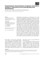
The energy band structure of the CdSe QD is schema-
ticallyshowninFig.7,where CB denotes the conduc-
tion ban d edge and VB the valence band edge. Referring
to the vacuum level as potential energy zero, the CB of
CdSe is -4.95 eV (electro n affinity of CdSe), the band
gap E
g
(energy difference between CB and VB) is 1.74
eV, and the quantum confinement energy for the
valencebandholeis1.5eV[12].ForourCdSeQDs
with a diameter of 5.5 nm (including the one monolayer
CdS shell), the energy separation between the ground
electron state, i.e., E
c0
in Fig. 7, in the conduction band
and the ground hole state (E
v0
)inthevalencebandis
1.988 eV, corresponding to the emission wavelength of
625 nm. Because of the quantum confinement effects in
QDs, electron states in the conduction band (hole states
in the valence band) become quantized as E
c0
, E
c1
etc
(E
v0
, E
v1
etc), where E
c0
and E
v0
denote the ground elec-
tron and hole state, respectively. QD fluorescence due
Figure 2 Confocal imaging of QD-uptaken EPCs at 450 nm (a)
and at 616 nm (b). (c) is the confocal imaging at 616 nm merged
with a differential interference contrast image. Built-in excitation
laser source at 458 nm was used. Excitation power control was 20%.
Molnár et al. Journal of Nanobiotechnology 2010, 8:2
/>Page 3 of 8
Figure 4 Fluorescence spectra of QDs inside and outside cells. Ten fluorescence spectra for each QD cluster were obtained using ten
microscopy excitation powers (lowest = 10% and highest = 100%). Excitation wavelength is 458 nm. (a) QD cluster 1 inside cell, (b) QD cluster 2
inside cell, see Fig. 2; (c) QD cluster 3 outside cells; (d) QD cluster 4 outside cells.
Figure 3 Confocal spectra of intracellular QD1 (hollow stars) and an aggregated QD cluster located outside EPCs (solid stars).The
wavelength of the excitation laser source is 458 nm. Excitation power control is 20%. The wavelength scanning step is 12 nm.
Molnár et al. Journal of Nanobiotechnology 2010, 8:2
/>Page 4 of 8
to the recombination of electron at E
c0
and hole at E
v0
is described by a Lorentzian peak [13]
y
A
()
()
2
0
22
(1)
where ħ ω is the photon energy, ħω
0
= E
c0
- E
v0
is the
excitonic energy in t he QD, Γ is the relaxation energy,
A the fluorescence intensity. The values of these fitting
parameters for spectra in Figs. 4 and 6 are shown in
Figs. 8 and 9.
In the course of this work two major effects were
observed. First is the blue shift of QD fluorescence peak
following their uptake by the EPCs. It has been shown
that QDs with carboxylic acid surface coatings were
recognized by lipid rafts in human epide rmal keratino-
cytes and internalised into early endosomes then trans-
ferred to late endosomes or lysosomes [14]. For our
Figure 5 Three-dimensional confocal imaging at 616 nm.A
cross section of an endothelial progenitor cell is shown in the
upper left corner. QDs (red) are located in the middle of the EPC
surrounded by the cell membrane.
Figure 6 Same as Fig. 4 except the excitation wavelength is
514 nm.
Figure 7 (a) Geometric structure of the CdSe QD with one monolayer CdS shell. (b) Schematic energy band structure of the CdSe QD.
Molnár et al. Journal of Nanobiotechnology 2010, 8:2
/>Page 5 of 8
QDs inside EPCs shown in Fig. 5, the most probably
modifications to the quantum confinement of electrons
and holes in the CdSe QD are interactions between sur-
face atoms and lipids and proteins (mo stly interacting
with Cd atoms) as well as ions such as K
+
(mostly inter-
acting with S atoms) inside the cell so that the covalent
electrons of the surface Cd and S atoms are no longer
in the energy band structure of Fig. 7. The effective
radius of quantum confinement is reduced for the exci-
ton inside t he QD and the excitonic energy becomes
increased. As was shown [15]
E
EE
g
r
r2( )
(2)
where E = ħω
0
is the excitonic energy, r is the QD
radius, E
g
is the energy bandgap of the QD material, δr
and δE are modifications in radius and excitonic energy.
For our CdSe QD625, the nominal diameter is 5.5 nm.
Assuming one monolayer modification (about 0.3 nm
Figure 8 Confocal spectral characterizations of QDs. Excitation wavelength is 458 nm. (a) Fluorescence intensity; (b) Relaxation energy. Solid
stars: QDs inside cells; hollow stars: QDs outside cells (curves are grouped by circle).
Figure 9 Same as Figs. 8 but excitation wavelength is 514 nm.
Molnár et al. Journal of Nanobiotechnology 2010, 8:2
/>Page 6 of 8
[12]) in the radius, Eq. (2) gives us δE =30meV,which
agrees very well with Figs. 8 and 9. Note that the fitted
fluorescence peak position for 458 nm excitation is dif-
ferent from the 514 nm excitation, 616 nm vs 613 nm
in Figs. 4 and 6, which we believe is due to the mixtures
between the excitation signal a nd the QD fluorescence.
For 514 nm excitation, the mixture is stronger so that
the blue shift appeared to be larger.
Zhang et al. reported similar blue shift of fluorescence
peak of thiol-capped CdTe QDs within less than 10 min of
QD uptaking in living cells caused by surface photooxida-
tion [16]. The reported blue shift in CdTe QDs is much
larger than our cases. Furthermor e, the peak width of
CdTe QDs is largely increased, while it remains basically
unchanged for our CdSe QDs. The major differences
between CdTe QDs and our CdSe QDs are probably due
to the fact that the oxidation of Te atoms are relatively
easy, therefore CdTe QDs are less chemically stable.
The other important finding is that the relaxation
energy in the QDs inside cells is relatively small and
independent of the excitation power, while it increases
quickly in the QDs outside of cells then saturates as a
function of the excitation power, see Figs. 8(b) and 9
(b). The large relaxation energy is actually an indica-
tion of the saturation of the ground excitonic state
occupation and the occupations o f high-energy exci-
tonic states due to the large optical pumping by the
excitation radiation.
The same effects (blue shift and the relaxation energy
behavior) were obtained for QD625 (emission wavelength
625 nm) under the excitations of 458 and 514 nm wave-
lengths. The insensitivity to the excitation w avelength
can be theoretically expected when the excitation energy
is not too high compared with the excitonic energy of
QDs (i.e., in the range of one-photon and multiphoton
excitations) [17]. High energy radiation (larger than twice
the excitonic energy) was shown t o induce multicarrier
excitation [18] so that it may induce different charac teri-
zations in the QD fluorescence spectrum.
Similar m easurements were repeated two and four
months late on randomly chosen QD clusters, a nd we
found that both the samples and measurement results
were very stable when the same measurement setups
were used. We noticed th at as long as measurement per-
formances are careful, there are no significant change s in
the confocal spectral characteristics (i.e., the fluorescence
intensity, excitonic energy and relaxation energy).
Conclusions
We have shown that the uptaking of colloidal QDs by
EPCs effectively reduces the radius of the exciton con-
finement inside the QDs so that the excitonic energy
increases and the peak of the QD fluorescence blue
shifts. Furthermore, the cell environment surrounding
the QDs shields the QDs so that the excitation of the
QDs inside the cells is usually weaker. QDs outside the
cells are excited to higher degree, which leads to the
saturation of the ground exciton ic state. The excitation
of high-energy states results in a broader fluorescence
peak.
Our study shown that intracellular environment can
affect optical characteristics of QDs and that such
changes are quantifiable. Therefore, changes of QD
fluorescence spectra should allow one to characterize
the interaction between colloidal QDs and EPCs. This
should facil itat e the developm ent of QD biomarkers for
monitoring EPCs at sub-cellular level.
Acknowledgements
Swedish Vinnova support to project “Molecular study of early atherosclerosis
with quantum dots” (Pro-jektnummer P35914-1) and computing resources
from the Swedish National Infrastructure for Computing (SNIC 001-09-52) are
acknowledged.
Author details
1
Department of Theoretical Chemistry, School of Biotechnology, Royal
Institute of Technology, S-106 91 Stockholm, Sweden.
2
Department of
Molecular and Clinical Medicine/Clinical Physiology, Wallenberg Laboratory,
The Sahlgrenska Academy, Gothenburg, Sweden.
3
University Hospital,
University of Gothenburg, SE 41345 Gothenburg, Sweden.
Authors’ contributions
All authors contributed equally, read and approved the final manuscript.
Competing interests
The authors declare that they have no competing interests.
Received: 16 September 2009
Accepted: 4 February 2010 Published: 4 February 2010
References
1. Medintz IL, Uyeda HT, Goldman ER, Mattoussi H: Quantum dot
bioconjugates for imaging, labelling and sensing. Nature Materials 2005,
4:435-446.
2. Vashist SK, Tewari R, Bajpai RP, Bharadwaj LM, Raiteri R: Review of
Quantum Dot Technologies for Cancer Detection and Treatment
AZojono J Nanotechnology Online 2006, 2:1-14.
3. Park JH, von Maltzahn G, Ruoslahti E, Bhatia SN, Sailor MJ: Micellar Hybrid
Nanoparticles for Simultaneous Magnetofluorescent Imaging and Drug
Delivery. Angew Chem Int Ed 2008, 47:7284-7288.
4. Yezhelyev MV, Qi L, O’Regan RM, Nie S, Gao X: Proton-sponge coated
quantum dots for siRNA delivery and intracellular imaging. J Am Chem
Soc 2008, 130:9006-9012.
5. Derfus AM, Chen AA, Min DH, Ruoslahti E, Bhatia SN: Targeted Quantum
Dot Conjugates for siRNA Delivery. Bioconjugate Chem 2007, 18:1391-1396.
6. Hirschi KK, Ingram DA, Yoder MC: Assessing identity, phenotype, and fate
of endothelial progenitor cells. Arterioscler Thromb Vasc Biol. 2008,
28(9):1584-1595.
7. Fadini GP, Baesso I, Albiero M, Sartore S, Agostini C, Avogaro A: Technical
notes on endothelial progenitor cells: ways to escape from the
knowledge plateau Atherosclerosis 2008, 197:496-503.
8. Kawamoto A, Losordo DW: Endothelial progenitor cells for cardiovascular
regeneration. Trends Cardiovasc Med 2008, 18:33-37.
9. Brunt KR, Hall SRR, Ward CA, Melo LG: Endothelial Progenitor Cell and
Mesenchymal Stem Cell Isolation, Characterization, Viral Transduction.
Methods in Molecular Medicine, Vascular Biology Protocols Sreejayan N, Ren J
139:197-210.
10. Li JJ, Wang YA, Guo W, Keay JC, Mishima TD, Johnson MB, Peng X: Large-
scale synthesis of nearly monodisperse CdSe/CdS core/shell nanocrystals
Molnár et al. Journal of Nanobiotechnology 2010, 8:2
/>Page 7 of 8
using air-stable reagents via successive ion layer adsorption and
reaction J Am Chem Soc 2003, 125:12567-75.
11. Yu WW, Qu L, Guo W, Peng X: Experimental Determination of the
Extinction Coefficient of CdTe, CdSe, and CdS Nanocrystals. Chem Mater
2003, 15:2854-2860.
12. Data in Science and Technology: Semiconductors other than Group IV
Elements and III-V Compounds. Springer, BostonMadelung O 1992.
13. Fu Y, Han TT, Luo Y, Ågren H: Dynamic analysis of multiple-photon
optical processes in semiconductor quantum dots. J Phys Condens Matter
2006, 18:9071-82.
14. Zhang LW, Monteiro-Riviere NA: Mechanisms of quantum dot
nanoparticle cellular uptake. Toxicological Sciences 2009, 110:138-55.
15. Fu Y, Han TT, Ågren H, Lin L, Chen P, Liu Y, Tang GO, Wu J, Yue Y, Dai N:
Design of semiconductor CdSe core ZnS/CdS multishell quantum dots
for multiphoton applications. Appl Phys Lett 2007, 90(3):173102.
16. Zhang Y, He J, Wang PN, Chen JY, Lu ZY, Lu DR, Guo J, Wang CC, Yang WL:
Time-dependent photoluminescence blue shift of the quantum dots in
living cells: effect of oxidation by singlet oxygen. J Am Chem Soc 2006,
128:13396-13401.
17. Fu Y, Han TT, Luo Y, Ågren H: Multiphoton excitation of quantum dots by
ultrashort and ultraintense laser pulses. Appl Phys Lett 2006, 88(3):221114.
18. Schaller RD, Klimov VI: High efficiency carrier multiplication in PbSe
nanocrystals: implications for solar energy conversion. Phys Rev Lett 2004,
92(4):186601.
doi:10.1186/1477-3155-8-2
Cite this article as: Molnár et al.: Optical characteriza tion of colloidal
CdSe quantum dots in endothelial progenitor cells. Journal of
Nanobiotechnology 2010 8:2.
Submit your next manuscript to BioMed Central
and take full advantage of:
• Convenient online submission
• Thorough peer review
• No space constraints or color figure charges
• Immediate publication on acceptance
• Inclusion in PubMed, CAS, Scopus and Google Scholar
• Research which is freely available for redistribution
Submit your manuscript at
www.biomedcentral.com/submit
Molnár et al. Journal of Nanobiotechnology 2010, 8:2
/>Page 8 of 8