báo cáo khoa học: "Laser ablation-based one-step generation and bio-functionalization of gold nanoparticles conjugated with aptamers" potx
Bạn đang xem bản rút gọn của tài liệu. Xem và tải ngay bản đầy đủ của tài liệu tại đây (1.71 MB, 11 trang )
RESEARC H Open Access
Laser ablation-based one-step generation and
bio-functionalization of gold nanoparticles
conjugated with aptamers
Johanna G Walter
1
, Svea Petersen
2
, Frank Stahl
1
, Thomas Scheper
1
, Stephan Barcikowski
2*
Abstract
Background: Bio-conjugated nanoparticles are important analytical tools with emerging biological and medical
applications. In this context, in situ conjugation of nanoparticles with biomolecules via laser ablation in an aqueous
media is a highly promising one-step method for the production of functional nanoparticles resulting in highly
efficient conju gation. Increased yields are required, particularly considering the conjugation of cost-intensive
biomolecules like RNA aptamers.
Results: Using a DNA aptamer directed against streptavidin, in situ conjugation results in nanoparticles with
diameters of approximately 9 nm exhibiting a high aptamer surface density (98 aptamers per nanoparticle) and a
maximal conjugation efficiency of 40.3%. We have demonstrated the functionality of the aptamer-conju gated
nanoparticles using three independent analytical methods, including an agglomeration-based colorimetric assay,
and solid-phase assays proving high aptamer activity. To demonstrate the general applicability of the in situ
conjugation of gold nanoparticles with aptamers, we have transferred the method to an RNA aptamer directed
against prostate-specific memb rane antigen (PSMA). Successful detection of PSMA in human prostate cancer tissue
was achieved utilizing tissue microarrays.
Conclusions: In comparison to the conventional generation of bio-conjugated gold nano particles using chemical
synthesis and subsequent bio-functionalization, the laser-ablation-based in situ conjugation is a rapid, one-step
production method. Due to high conjugation efficiency and productivity, in situ conjugation can be easily used for
high throughput generation of gold nanoparticles conjugated with valuable biomolecules like aptamers.
Background
Gold nanoparticles (AuNPs) feature unique optical
properties, including high surface plasmon resonance
(SPR), enhanced absorbance and s cattering with high
quantum efficiency. In addition to their resistance
against p hotobleaching, AuNPs perfectly fulfill require-
ments for use a s colorimetric sensors and markers.
For sensing or labeling of DNA targets, AuNPs can
fairly easily be functionalized with DNA via thiol l in-
kers, resulting in a highly ordered, self-assembled
monolayer (SAM) [1,2]. Numerous c olorimetric appli-
cations of DNA-conjugat ed AuNPs have already been
developed[3].
More recently, several applications of aptamer-conju-
gated AuNPs have been reported[4,5]. Aptamers are
short, single-stranded DNA or RNA molecules that
exhibit high specificity and affinity towards their corre-
sponding target. Thus, aptamers can be thought of as
nucleic acid analogues to antibodies that can be selected
in vitro via SELEX (systematic evolution of ligands by
exponential enrichment) against virtually any molecule,
including proteins as well as small molecules like metal
ions [6-8]. Aptamer-conjugated AuNPs have already
bee n successfu lly used for the detection of proteins in a
dry-reagent strip biosensor, [9] for detection of throm-
bin o n surfaces, [4] for colorimetric detection of plate-
let-derived growth factor, [5] f or detection of adenosine
and potassium ions in an agglomeration-based approach,
[10] for detection of t hrombin in a dot blot assay [11]
and for targeting and therapy of cancerous cells [12,13].
* Correspondence:
2
Laser Zentrum Hannover, Hollerithallee 8, 30419 Hannover, Germany
Full list of author information is available at the end of the article
Walter et al. Journal of Nanobiotechnology 2010, 8:21
/>© 2010 Walter et al; licensee BioMed Central Ltd. This is an Open Access article distributed under the terms of the Creativ e Commons
Attribution License ( which permits unr estricted use, distribution, and reprod uction in
any medium, provided the original work is properly cited.
All applications of aptamer-conjugated AuNPs pub-
lished s o far have be en based on c hemical synthesis of
AuNPs in the presence of reducing and stabilizing
agents, and subsequent (ex situ) ligand exchange with
aptamers. This ligand exchange might require heating
and buffering in order to achieve satisfactory yields and
surface coverage. The latter might be limited by interfer-
ence from remaining reducing agents with the aptamer
during the replacement p rocess. Additionally, remaining
precursors and/or reducing agents might result in a pos-
sible restriction of AuNPs use in biomedical applications
[14,15].
Recently, laser ablation of gold in a liquid environ-
ment has been used for the production of AuNPs
[16,17], using surfactants for growth quenching, result-
ing in narrow nanoparticle size distributions[18]. The
advantages of laser-generated AuNPs include high purity
in combination with uniq ue surface characteristics. The
Au surface of laser-generated AuNPs is part ially oxi-
dized, resulting in electrostatic stabilization of the col-
loid without the need for chemical additives. These
partially positively-charged AuNPs, acting as electron
acceptors, can interact directly with electron donors like
amino or thiol groups in the ablation medium[19,20].
During laser ablation, the DNA acts as a capping agent,
allowing precise size control of the resulting AuNPs, as
it has been previously reported for the addition of cyclo-
dextrines, biopolymers, etc[21]. Recently, a direct
comparison of conventional ex situ conjugation of laser-
generated AuNPs and laser-ablation-b ased in situ conju-
gation of AuNPs with DNA has revealed a four times
higher conjugation efficiency when using the laser-abla-
tion-based procedure. In comparison to AuNPs pro-
duced by chemical synthesis and subsequent ex situ
conjugation, AuNPs generated using laser-ablation-
based in situ conjugation exhibit up to five times higher
surface coverage[22]. Hence, bio-conjugation during
laser ablation presents a rapid and efficient preparation
method, especially for the conjugation of valuable bio-
molecules like aptamers or vectors. The high surface
coverage of DNA-modified AuNPs produced by in situ
conjugation may be especially advantageous for applica-
tions including cellular uptake of AuNPs. In this con-
text, Giljohann et al. have found that the extent of
cellular uptake of DNA-modified AuNPs can be
increased by enhancing the DNA loading[23]. Moreo ver,
high DNA densities can also facilitate cooperative bind-
ing, re sulting in increased association constants with a
given target, e.g. in intracellular gene regulation[24].
Another imp ortant parameter that can be modulated via
surface coverage is the immune response induced by
DNA-modified AuNPs. Higher DNA densities efficiently
limit the immune response as measured by Interferon-b
expression in mouse macrophages[25].
In spite of these benefits, t he use of laser-ablation-
based in situ conjugation for the generation of aptamer-
conjugated AuNPs has not yet been reported.
We show the functionalization of nanoparticles with
aptamers during femtosecond-pulsed, laser-induced
gold nano particle formation in a n aqueous media using
a DNA aptamer directed against streptavidin as a
model system. In order to demo nstrate the a pplicabil-
ity of aptamer-conjugated AuNPs generated via laser
ablation in complex biomedical applications, we have
used an RNA aptamer directed against prostate-speci-
ficmembraneantigen(PSMA)forthedetectionof
PSMA in human prostate cancer tissue utilizing tissue
microarrays.
Results and Discussion
Choice of aptamer orientation and spacer design
In order to ensure aptamer activity, several factors con-
cerning the ability of the aptamer to fold into the cor-
rect three-dimensional structure have been considered.
We have previously reported the application of an apta-
mer directed against streptavidin (referred to as miniS-
trep) in a protein microarray format[26,27]. Using this
approach, we have found that the miniStrep aptamer
requires an additional spacer placed between the apta-
mer and the substrate to show activity that is slightly
higher when immobilized via its 3’ terminus. Thus, we
decided to use 3’ orientation. An additional oligothymi-
dine (T10) spacer was placed between the disulfide
group and the aptamer sequence. Tymidine was chosen,
sincethisnucleotidehasthelowestaffinitytowardsthe
gold surface[28]. Thus, nonspecific binding of the spacer
bases to gold is minimized, which should increase the
surface loading and improve elevation of the aptamer
away from the nanoparticle surface. Taking these con-
siderations into account, the miniStrep aptamer con-
struct used in this work was the following: TCT GTG
AGA CGA CGC ACC GGT CGC AGG TTT TGT
CTC ACA G -T
10
-(CH
2
)
3
-S-S-(CH
2
)
6
OH.
We decided to immobilize the anti-PSMA aptamer via
the 3’ terminus. According to Lupold et al., the aptamer
can be subjected to 3’ truncation of up to 15 nucleotides
without losing its affinity to PSMA[29]. Since the 3’
terminal bases are not necessary for target recognition,
we decided to omit the use of an additional oligonucleo-
tide spacer. Instead, hexaethylenglycol was chosen as a
spacer, because it does not exhibit intermolecular repul-
sion, which is one cause of low DNA loadin g on AuNPs
[30]. Furthermore, it only occu pies a small surface area,
which a llows high packing d ensities,[30] and is known
to minimize nonspecific protein binding[31]. Therefore,
the aptamer construct used was the following: GGG
AGG ACG AUG CGG AUC AGC CAU GUU UAC
GUC ACU CCU UGU CAA UCC UCA UCG GCA
Walter et al. Journal of Nanobiotechnology 2010, 8:21
/>Page 2 of 11
GAC GAC UCG CCC GA-(CH
2
CH
2
O)
6
-(CH
2
)
6
-S-S-
(CH
2
)
6
OH.
The aptamers were directly used in the laser ablation
process without prior dithiothreitol (DTT) treatment
(Figure 1A). According to Dougan et al., this does not
affect surface coverage[32]. Moreover, the mercaptohex-
anol (MCH) of the mixed disulfide (aptamer-S-S-(CH
2
)
6
OH) may serve as a co-adsorb ent, eliminating unspeci-
fic binding to the g old surface, by occupying free bind-
ing sites[33]. Due to the formation of a mixed
monolayer consisting of aptamers and short organic
residues, the available space for optimal aptamer folding
is enhanced (Figure 1B).
In situ conjugation
Due to rapid, one-step processing, laser-ablation-based
in situ conjugation enables fast screening of different
conjugation conditions. Utilizing this high throughput
potential, we have determined optimal conjugat ion con-
ditions by using different concentrations of miniStrep
aptamer i n a Tris(hydroxymethyl)-aminomethan (Tris)
buffer during laser ablation. Per investigated concentra-
tion, the laser ablation p rocess took less than two min-
utes. A UV/VIS spectrum of AuNPs produced via laser
ablation in the presence of 5 μM aptamer can be found
in Figure 2.
DLS measurements demonstrate that the hydrody-
namic diameter (d
h
) of the AuNPs increases with
increasing aptamer concentrations (Figure 3A). While d
h
after ablation in Tris buffer (without aptamer) is 7 nm,
d
h
increases with increasing aptamer concentrations up
to 5 μM, and finally reaches a plateau of approximately
60 - 70 nm (Figure 3A). We assume that this d
h
increase
is a result of cumulative aptamer loading on the gold
surface. At low surface coverage, the aptamer lays flat
on the surface, due to non-specific bind ing via the lone
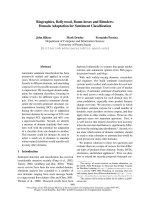
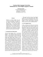
Figure 1 Generation of aptamer-conjugated AuNPs via in situ conjugation. (A) Schematic illustratio n of in situ con jugation of AuNPs with
aptamers during laser ablation in an aqueous aptamer solution. (B) Spacer design and resulting mixed monolayer conjugated nanoparticles.
Mixed monolayer formation and careful spacer design contribute to correct aptamer folding.
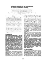
Figure 2 UV/VIS spectrum of aptamer-conjugated AuNPs.The
spectrum was obtained with an as prepared AuNP solution after in
situ conjugation with 5 μM anti-streptavidin aptamer.
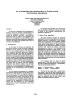
Figure 3 Characterization of aptamer-conjugated AuNPs.(A)
Hydrodynamic diameter and Feret diameter of the aptamer AuNP
conjugates as a function of the aptamer concentration used during
laser ablation. (B) Suggested mechanism of the observed increase of
the size of the conjugates. At low surface coverage, the negatively
charged aptamer lays flat on the positively charged AuNP surface.
With increasing surface coverage, the aptamers straightened up on
the surface, resulting in an increased hydrodynamic diameter. Scale
bars illustrate the proportions of linearized aptamer and AuNP.
Walter et al. Journal of Nanobiotechnology 2010, 8:21
/>Page 3 of 11
nitrogen electron pairs of the nucleotides. As the surface
coverage increases, the aptamers are forced to adopt a
more perpendicular conformation, due to electrostatic
repulsion of the aptamers’ negatively charged phosphate
backbones, resulting in a d
h
increase (Figure 3B). We
estimated the length o f the aptamer (including T
10
spacer) to be 21.5 nm, using a base to b ase distance for
ssDNA o f 0.43 nm[34]. For an aptamer-conjugated
AuNP of 9 nm core size, this results in a diameter of
approximately 52 nm, which is close to the observed
plateau of d
h
, and supports our assumption (Figure 3B).
On first sight, the AuNPs hydrodynamic diameter
increase seems to be contradictory to our previous find-
ing of a growth quenching effect induced by increasing
DNA concentrations[19]. But in contrast to our previous
work, here the ablation was perfo rmed in Tris buffer.
Tris interacts with the surface of the embryonic AuNPs,
resulting in prevention of further post-ablation nanopar-
ticle agglomeration . Consequently, the AuNPs produced
in Tris buffer are already stabilized by the buffer mole-
cule, r esulting in reduced diame ters (d
h
=7.1±0.8nm
in Tris buffer versus 54.2 ± 0.6 nm in ddH
2
O(Figure
3A)) and a diminished influence of the oligonucleotide
concentration on the nanoparticle size. This assumption
is supported by TEM analysis data. The Feret diameter
(d
Feret
) of AuNPs produced in Tris buffer slightly
decreases from 12.1 to 7 .3 nm with increasing aptamer
concentration (Figure 3A). In comparison to the Tris
molecule, the thiolated aptamer exhibits a higher affinity
towards the gold surface, resulting in better stabilization
of embryonic particles, and thus smaller AuNPs, as
detected via TEM analysis. Although there may be some
portion of Tris-aptamer ligand exchange after nanoparti-
cle generation, the size quenching effect observed by
TEM analysis confirms successful in situ bio-conjuga-
tion during laser ablation.
In addition to the AuNP size, we have determined
aptamer loading (Table 1). For AuNPs produced by
laser ablation in a 5 μ M aptamer solution (in situ), we
found a loading of 9 8 aptamers per nanoparticle, corre-
sponding to 65 pmol/cm
2
. This aptamer loading is
higher than the results achievedbypostproduction(ex
situ) modification of chemically synthesized AuNPs with
short oligonucleotides (Demers et al.: 34 pmol/cm
2
),[35]
and the aptamer loading to chemically synthesized
AuNPs reported by Huang et al. (13 pmol/cm
2
)[5]. The
high aptamer loading achieved by in situ conjugation
confirms high availability of laser-generated AuNPs for
bio-conjugation. The conjugation efficiency was calcu-
lated as the portion of provided aptamer bound to the
nanoparticle surface (Table 1). At a 1.25 μM aptamer
concentration, 40.3% of the available aptamer binds to
the AuNPs, which demonstrates the suitability of the
method for efficient conjugation of valuable biomole-
cules.Sincewehaveobservednodenaturationofthe
aptamer during laser ablation (as discussed in the next
section), the remaining aptamer can be reused.
After the ablation process, conjugates were slowly
transferred into the aptamer selection buffer by adding
NaCl and MgCl
2
. During this salting process, we
observed precipitation of AuNPs produced at aptamer
concent rations lower than 5 μM. The higher stability of
AuNPs c onjugated at aptamer c oncentrations of 5 μM
(or h igher) coincides with the plateau in the hydrody-
namic diameter (Figure 3B), and indicates better stabili-
zation due to higher surface coverage.
All further experiments were performed with AuNPs
produced in a 5 μM apt amer solution, which was a
compromise between maximal aptamer density and
minimal aptamer consumption (Table 1). Under these
conditions, we could produce 75 μg(150μg/ml ) miniS-
trep-conjugated AuNPs in less than two minutes. The
free aptamer was removed by centrifugation. In order to
maintain conjugate activity, centrifugation was per-
formed under rather mild conditions (16600 × g). This
procedure results in a slightly increased average Feret
diameter (14.6 nm) due to loss of small nanoparticles
(Figure 4).
It should be noted that the high immobilization effi-
ciency, and thus high aptamer consumption, results in
decreasing aptamer concentrations during the laser abla-
tion process. As we suppo se that the aptamer conjuga-
tion takes place in the millisecond to second regime
after the collapse of the cavitation bubble, the aptamer
loading of NPs will also decrease over this period of
time. If more homogeneo us aptamer lo adings are
Table 1 Characterization of aptamer-conjugated gold nanoparticles
c
(miniStrep)
[μM]
d
h
(DLS)
a
[nm]
d
Feret
b
[nm]
Aptamer/AuNP Aptamer/A
AuNP
c
[pmol/cm
2
]
E
con
d
[%]
0 7.1 ± 0.8 12.1 ± 9.8 - - -
1.25 37.5 ± 2.3 11.2 ± 4.2 80 ± 2 33.93 ± 1.04 40.3 ± 0.3
5 61.1 ± 1.9 9.0 ± 5.0 98 ± 4 64.58 ± 1.82 19.8 ± 0.7
25 71.0 ± 2.0 7.3 ± 2.5 74 ± 11 73.90 ± 7.22 6.1 ± 0.9
Summary of data obtained for AuNPs conjugated with miniStrep aptamer in Tris buffer (
a
Hydrodynamic diameter,
b
Feret diameter,
c
Aptamers per AuNP
surface,
d
Conjugation efficiency).
Walter et al. Journal of Nanobiotechnology 2010, 8:21
/>Page 4 of 11
required, this can be achieved by applying higher apta-
mer concentrations and/or by shortening ablation time.
Functionality of miniStrep-conjugated AuNPs
Functionality of the immobilized miniStrep aptamer was
confirmed by using three independent methods. First, a
classical, agglomeration-based m ethod was applied. A
fixed amount of AuNPs (0.69 nM) conjugated with min-
iStrep aptamer was incubated with different amounts of
streptavidin (0 - 15.9 nM), and UV/VIS was detected.
Since streptavidin is a tetrameric pro tein, agglomeration
can be observed as a red shift of SPR
Max
(Figure 5A).
The shift in SPR
Max
increases with increasing concentra-
tions of streptavidin, and reaches a maximum at a strep-
tavidin concentration of 2 nM. In addition to the
SPR
Max
shift, we observed the formation of a red film
on the wall of the reaction vessel at streptavidin concen-
trations from 1 nM to 4 nM. Simultaneously, we
observed a loss of AuNPs in the solution. Based on
absorbance at 380 nm, the loss of particles was calcu-
lated to be 86.5%. We assume that the red film is com-
posed of large agglomerates, while small agglomerates
stay in the solution and ca n be detected via the shift of
SPR
Max
and TEM analysis. TEM micrographs of the
agglomerates indicated a defined composit ion and tetra-
hedral structure of these agglomerates (Figure 6). Based
on TEM analysis, we determined an agglomerate size of
35 nm (edge-to-edge length). In order to verify the pro-
posed tetrahedral structure, we calculated the size of the
agglomerates based on the observed shift of SPR
Max
, uti-
lizing the “plasmon ruler equation":[36]
Δ
0
018
023
≈×
−
⎛
⎝
⎜
⎞
⎠
⎟
⎛
⎝
⎜
⎜
⎜
⎜
⎞
⎠
⎟
⎟
⎟
⎟
, exp
,
s
D
This approximation describes the dependency between
the observed shift of SPR
Max
( Δl) on t he interparticle
gap (s) and na noparticle size (D). Using our experim en-
tal results (Δl = 6 nm; l0 = 523.5 nm; D = d
Feret
= 14.6
nm), we calculated an interparticle distance of 9.3 nm,
and an edge-to-edge length of the proposed tetrahedron
of 38.5 nm. Taking into account that equation (1) is an
empirical approximation established for a pair of inter-
acting nanoparticles rather than for a tetramer, and con-
sidering that the geometry of streptavidin is not
perfectly tetrahedral, the deviation of 10% between the
agglomerate sizes measured by TEM analysis and calcu-
lated f rom the shift of SPR
Max
seems to be acceptable.
Good agreement between the agglomeration sizes
obtained using two independent methods supports the
proposed tetrahedral structure of the agglomerates.
At streptavidin concentrations above 2 nM, the
SPR
Max
shift decreases, due to saturation of aptamers
immobilized on the AuNPs surface with streptavidin
(Figure 5B). This saturatio n effect is in accordance with
the observations of Huang et al., who used an aptamer
against a dimeric pr otein (pl atelet-derived growth fac-
tor)[5].
In order to gain quantitat ive insight into streptavidin
binding and thus aptamer activity, the AuNPs were
incubated with an excess of Cy3 labeled streptavidin.
The ag glomerates of aptamer-coated nanoparticles and
attached streptavidin were removed using ultracentrifu-
gation, and the amount of bound streptavidin was deter-
mined by measuring the remaining streptavidin
concentration in the supernatant.
Since aptamer loading was determined for the whole
AuNP population generated by laser ablation (d
Feret
=
9.0 nm), and the binding of Cy3 labeled streptavidin was
performed with the AuNP subpopulation resulting from
Figure 4 TEM analysis of aptamer-conjugated AuNPs.TEM
micrographs and AuNP size distributions (lognormal fit) of AuNPs
produced by laser ablation in 5 μM miniStrep solution before (A),
and after (B) removal of free aptamer using centrifugation.
Walter et al. Journal of Nanobiotechnology 2010, 8:21
/>Page 5 of 11
removal of the free aptamer by centrifugation (d
Feret
=
14.6 nm), it was not possible to compare aptamer load-
ing directly to the amount of bound streptavidin. In
order to estimate the aptamer activity, it was assumed
that aptamer density (pmol/cm
2
) is not significantly
affected by the nanoparticle diameter. Based on this
assumption, the aptamer loading was calculated to be
64.58 ± 1.82 pmol/cm
2
, and the amount of streptavidin
bound to the AuNP surface was 67.23 ± 0.76 pmol/cm
2
,
resulting in approximately 100% aptamer activity. This
indicates that the aptamer is not degraded d uring the
laser ablation process, and optimal aptamer folding was
achieved by careful design of the spacer.
To examine the applicability of the aptamer-conju-
gated AuNPs in solid phase assays, we performed a sim-
ple dot blot assay (Figure 7A). Within this assay, BSA
was used as a negative control. BSA was chosen becaus e
it exhi bits a mildly acidic isoelectric point (pI) similar to
the pI of streptavidin (pI 5-6) resulting in a comparable
Figure 5 Verification of the activity of aptamer-conjugated AuNPs. (A) The shift of the SPR maximum clearly indicates the formation of
agglomerates in the presence of streptavidin. (B) Schematic illustration of the formation of agglomerates as a function of streptavidin
concentration: At low streptavidin concentrations, relatively small agglomerates occur (i). At medium streptavidin concentrations, the tetrameric
protein induces the formation of large agglomerates (ii). An excess of streptavidin inhibits the formation of agglomerates by saturation of the
aptamers bound to the nanoparticle surface (iii).
Figure 6 TEM analysis of AuNPs conjugated with aptamers
against streptavidin. TEM micrographs of AuNPs without
streptavidin (i), and after incubation with streptavidin (ii). The insert
displays a scheme of the proposed composition of the
agglomerates. Please note that the agglomerates are displayed in a
simplified manner in the scheme, de facto streptavidin is not planar
but tetrahedral.
Walter et al. Journal of Nanobiotechnology 2010, 8:21
/>Page 6 of 11
neg ative net charge of the two proteins under the given
conditions (pH 7.4). In order to exclude the possibility
of electrostatic interactions between the negatively
charged proteins and the positively charged AuNPs, the
experiment was repeated w ith unconjugated nanoparti-
cles. The nanoparticle aptamer conjugates bind only to
the immobilized streptavidin, and no binding can be
observed to BSA. Using unconjugated nanoparticles, no
binding of AuNPs to the immobilized proteins occurred
(data not shown). This clearly demonstrates the specific
binding of AuNPs to streptavidin via the aptamer conju-
gated to the nanoparticle surface.
The AuNPs bound to streptavidin during the dot blot
assay were further analyzed via ESEM. The ESEM
micrograph affirm s the Feret diameter of the nanoparti-
cles determined by TEM (Figure 7B).
Functionality of anti-PSMA-conjugated AuNPs
Encouraged by the positive performance of the dot blot
assay, our next aim was to prove the applicability of
aptamer-conjugated AuNPs in more co mplex and
demanding solid-phase assays. Therefore, we used
AuNPs conjugated with an aptamer directed against
PSMA for detection of PSMA in prostate cancer (adeno-
carcinoma) tissue sections.
AuNPs conjugated with anti-PSMA aptamer show a
staining pattern similar to anti-PSMA antib ody. In bo th
cases, a positive staining of acinar epithelial cells was
observed (Figure 8). In tissue sections treated with anti-
PSMA aptamer-conjugated AuNPs, an additional staining
of muscle cells was observed that was not detected in the
positive control. To ensure that the binding to PSMA is
based on the affinity o f the anti-PSMA aptamer r ather
than on electrostatic interaction between the target pro-
tein and the highly negative ly charged aptamers, AuNPs
conjugated with anti-streptavidin aptamers were used.
Since the negative control does not show positive binding
to epithelial cells or false positive binding to muscle cells,
we assume the binding of anti-PSMA conjugates to mus-
cle cells to be induced by the specific three-dimensional
structure of the anti-PSMA aptamer. A positive staining
of smooth muscle cells in prostate cancer has also been
reported for one monoclonal PSMA antibody (7E11),[37]
and some authors assume that there may be a “PSMA-
like” tar get in smooth muscle cells[38,39]. Follow ing this
consideration, the binding of AuNPs conjugated with
anti-PSMA aptamer to muscle cells may be the result of
cross-reactivity of the aptamer with this unknown
“PSMA-like” target. In summary, our results demonstrate
that the anti-PSMA aptamer AuNP conjugates can detect
PSMA in acinar epithelial cells of human prostate cancer.
This exemplifies the broad applicability of aptamer-
Figure 7 Dot blot. Dot blot detection of streptavidin, using
aptamer-conjugated gold nanoparticles (A). ESEM image of
nanoparticles bound to streptavidin immobilized on the
nitrocellulose membrane (B).
Figure 8 DetectionofPSMAinhumanprostate cancer tissue.
Detection of PSMA positive structures in prostate cancer tissue
sections by immunohistochemical staining using anti-PSMA aptamer
(PSMA apt)-conjugated AuNPs. As a negative control, AuNPs
conjugated with miniStrep aptamer (miniStrep apt) were used. A
polyclonal antibody directed against PSMA (PSMA pAb) was used as
a positive control. Positive control was additionally stained with
Haematoxylin and Eosin. Black arrows indicate specific staining,
while white arrows flag unspecific binding.
Walter et al. Journal of Nanobiotechnology 2010, 8:21
/>Page 7 of 11
conjugated AuNPs, even in highly complex biological
matrices and bio-imaging applications.
Conclusions
We have demonstrated the suitability of laser-ablation-
based in situ bio-conjugation for the production of func-
tional, aptamer-conjugated gold nanoparticles. Exploiting
the potential of this rapid, one-step method for high
throughput screening, we have optimized the conjugation
regarding aptamer loading and conjugati on efficiency. To
address the general applicability of the method, we have
utilized two different aptamers composed of DNA and
RNA. The high degree of aptamer activity determined on
AuNP surface verifies that there is no heat-induced dena-
turation of the aptamer during laser ablation. We have
proven the functionality of conjugates using three differ-
ent methods (agglomeration-based assay, dot blot assay,
tissue microarray), indicating the broad applicability of
aptamer-conjugated gold nanopar ticles for bio-analytical
applications, even in highly demanding assays. Moreover,
in situ conjugation avoids possible contamination by
toxic educts, residual reducing agents or preservatives.
Thus, this method could also be esp ecially advantageous
for use in medical applications.
Since in situ conjugation is a fast and simple one-step
approach to generate pure conjugated AuNPs with h igh
conjugation efficiency and productivity, it can be easily
used for the high throughput production of large
amounts of different conjugated nanopartic les. The
higher conjugation efficiencies are beneficial for high-
priced biomolecules, and the comparably high surface
coverage is desirable for cellular uptake, which depends
on the DNA density on the AuNPs surface. Moreover,
suc h high surface densities may assist cooperative bind-
ingandmaydecreasetheimmuneresponseagainst
AuNPs.
Methods
Materials
All chemicals were purchased from Sigma-Aldrich
(Steinheim, Germany) or Fluka Chemie AG (Tauf-
kirchen, Germany), and used as received. The aptamer
against streptavidin (TCT GTG AGA CGA CGC ACC
GGT CGC AGG TTT TGT CTC ACA G -T
10
-(CH
2
)
3
-
S-S-(CH
2
)
6
OH, referred to as miniStrep) [26] and anti-
PSMA aptamer (GGG AGG ACG AUG CGG AUC
AGC CAU GUU UAC GUC ACU CCU UGU CAA
UCC UCA UCG GCA GAC GAC UCG CCC GA-
(CH
2
CH
2
O)
6
-(CH
2
)
6
-S-S-(CH
2
)
6
OH) [29] were pur-
chased from Biospring GmbH (Frankfurt, Germany).
The gold foil was 0.1 mm thick and had >99.99% purity,
and was obtained from Goodfellow GmbH (Bad Nau-
heim, Germany).
Generation of aptamer-conjugated AuNPs
Laser ablation was performed in the same buffer system
the aptamer was originally selected in. For the miniStrep
aptamer, 50 mM Tris(hydroxymethyl)-aminomethan
(Tris) pH 8.0 was used, and the anti-PSMA aptamer was
conjugated in 20 mM N’-2-Hydroxyethylpiperazine-N’-2
ethanesulphonic acid (HEPES) pH 7.4. Laser generation
of AuNPs was performed utilizing a Spitfire Pro femto-
second laser system (Spectra-Physics) providing 120 fs
laser pulses at a wavelength of 800 nm. 5×5 mm gold
foils were placed in the wells of a 24 well plate filled
with 500 μl of aptamer solution in the respectiv e buffer.
Ablation was performed while moving the plate at a
constant speed of 60 mm×min-1 in a spiral (outer
radius: 3 mm, inner radius: 1.5 mm), using an axis sys-
tem. Recently, we have optimized the laser parameters
for laser-ablation-based generation of DNA-conjugated
AuNPs[19]. Here, laser f luence was optimiz ed in regard
to maximal productivity, while avoiding degradation of
the oligonucleotide. In the present study, t he optimized
parameters were chosen, the pulseenergywasfixedat
100 μJ, and the repetition rate was 5 kHz. In order to
avoid heat-induced degradation of the aptamer, the
focus position was adjusted t o be 2 mm beneath the
focus position determined in air[19].
Post-generation processing of the aptamer-conjugated
AuNPs
After laser ablation, the conjugates were allowed to age
overnight at 4°C before NaCl was added in increments
of 25 mM by addition of 2 M NaCl in Tris-Cl or
HEPES respectively. After each NaCl addition, the col-
loidal solution was mixed and incubated for 1 h at room
temperature. The addition of MgCl
2
and CaCl
2
was per-
formed after another overnight incubation at 4°C, by
addition of 1 M MgCl
2
and 1 M CaCl
2
. Final buffer
compositions were the following: miniStrep: 150 mM
NaCl, 10 mM MgCl
2
, 50 mM Tris-Cl pH 8.0; anti-
PSMA:150mMNaCl,1mMMgCl
2
, 1 mM CaCl
2
,
0.05% Tween 20, 20 mM HEPES pH 7.4.
To remove the free aptamer, the ab lation medium was
centrifuged for 15 m in at 15000 rpm. The supernatant
was transferred into a new centrifugal tube and centri-
fuged for another 30 min. The superna tant was dis-
carded, and the pellets were pooled and resuspended in
the respective buffer. This process was repeated 4 times.
Characterization methods
UV/VIS spectra of the AuNP solutions were recorded
using a Shimadzu 1650 spectrophotometer. In order to
determine the AuNP concentration, the absorption at
380 nm (mainly corresponding to the interband transi-
tion of gold) was measured. Intensities were converted
Walter et al. Journal of Nanobiotechnology 2010, 8:21
/>Page 8 of 11
to AuNP mass concentrations by interpolation from a
linear standard calibration curve (R
2
=0.99).Standard
curves were prepared with known concentrations of
AuNP produced by weighing a gold target three times
before and after ablation.
Transmission electron micrographs (T EM) were com-
missioned at Stiftung Tierärztliche Hochschule, Institut
für Pathologie (Prof. Dr. W. Baumgärtner, Kerstin
Rohn), and were obtained by utilizing a TEM Philip
CM30witha0.23nmresolution.Onedropofthecol-
loidal solution was placed on a carbon-coated, formvar-
covered copper grid, and then dried at room tempera-
ture. Given diameters were averaged for at least 200
AuNPs. Dynamic light scattering (DLS) measurements
were performed, using a Zetasizer ZS (Malvern). Three
consecutive measur ements were carried out and average
values are presented.
The a mount of aptamer bound per nanoparticle was
determined by measuring the concentration of the
unbound aptamer. Aptamer-conjugated AuNPs were
removed by ultracentrifugation (Beckman Coulter
Optima Max, 30000 × g), and the adsorption of the
supernatant was measured at 260 nm against a serial
dilution of aptamer in Tris buffer. Mean values of three
measurements are presented.
Determination of miniStrep aptamer functionality
The agglomeration-based streptavidin assay was per-
formed by incubating a fi xed amount of miniStrep -con-
jugated AuNPs (0.69 nM) with varying concentrations
of streptavidin (0 - 15.9 nM) for 16 h at room tempera-
ture. UV/VIS was measured to monitor the shift of
SPR
Max
.
Furthermore, the aptamer activity was determined in a
“ golden blot” [40] format similar to the method pub-
lished by Wang et al[11]. In brief, streptavidin (0.5 μl, 1
mg/ml in PBS) was spotted in 10 replicates onto a nitro-
cellulose membrane (Sartorius, Goett ingen, Germany).
After1hincubationatroomtemperature, blocking of
the membrane was performed with 1% BSA in miniS-
trep selection buffer. The membrane was washed in the
same buffer and incubated with a solution of AuNPs
(20 μg/ml) for 2 h. Finally, t he membrane was washed
with miniStrep selection buffer. As a negative control,
BSA (0.5 μ l, 1 mg/ml in PBS) was spotted on the mem-
brane. Furthermore, the experiment was repeated with
“bare” AuNP produced in Tris buffer in the absence of
aptamer. In this experiment, t he miniStrep selectio n
buffer was replaced by 50 mM Tris-Cl pH 8.0, in order
to maintain colloidal stability of the non-stabilized nano-
particles. Environmental scanning electron microscopy
(ESEM) of the membrane after incubation with AuNPs
was performed with a Quanta 400 F (FEI, Eindhoven,
Netherlands) in low vacuum conditions. A piece of
membrane was placed on an aluminum holder and
visualized without previous sputtering.
In order to determine the activity of the miniStrep
aptamer bound to the AuNP surface, the conjugate
(28.5 μg/ml, 0.15 nM) was incubated with Cy3-labeled
streptavidin (166.7 μg/ml, 2.8 μM) for 16 h at room
temperature, in the dark. The conjugates and bound
streptavidin were removed by ultracentrifugation. The
amount of streptavidin bound to the nanoparticles was
deter mined by measuring the streptav idin concentratio n
remaining in the supernatant, utilizing a Fluoroskan
ascent fluorescence plate reader (Ex: 544 nm, Em: 590
nm). Mean values of 4 measurements are presented.
Determination of anti-PSMA aptamer functionality
The activity of anti-PSMA aptamers conjugated to
AuNPs was investigated, using a tissue microarray con-
sisting of paraffin-embedded prostate cancer tissues (US
Biomax, Rockville, MD, USA). After baking the slides at
60°C for 30 min, paraffin was removed using two wash-
ing steps in xylene (10 min each). The tissue arrays
were rehydrated by consecutive washes in 100%, 95%
and 70% ethanol, followed by a washing step in ddH
2
O
(5 min each). Antigen retrieval was performed by pla-
cing the slides in 0.01 M sodium citrate pH 6.0 for
15 min at 95°C. Consequently slides were was hed with
ant i-PSMA aptamer selection buffer, and blocked in 5%
goat serum (Millipore) in the same buffer. The anti-
PSMA selection buffer was used for all consequent assay
steps. Incubation with the aptamer-modified AuNPs
(20 μg/ml) was performed for 2 h at 20°C and 300 rpm
in an Eppendorf shaker equipped with a slide adaptor,
after placing a secure seal incubation chamber (Grace
Biolabs, Bend, OR, USA) filled with 800 μlofthe
respective AuNP solution on the slide. Slides were
washed two times for 5 min with 1% goat serum, and
fixed for 15 min with 2.5% glutaraldehyde solution. Sil-
ver enhancement was performed using a silver enhancer
kit (Sigma), according to the instructions provided by
the manufacturer.
AuNPs conjugated with miniStrep Aptamer in HEPES
buffer were chosen as a negative control. All washing and
incubation steps were performed as described above. As
a positive control, a rabbit anti-PSMA antibody directed
against the C-terminal domain of human PSMA (Milli-
pore) was used[41]. Here, all washing and incubation
steps were performed using PBS. After incubation with
2.5 μg/ml rabbit anti-PSMA for 2 h, the slides were
washed two times for 5 min with 1% goat serum, and
consequently incubated with a 1:20 dilution of 12 nm
colloi dal gold conjugated with goat ant i-rabbit IgG (Jack-
son Immuno Research; OD at 520 n m of stock solution:
2) for 1.5 h. High background of developed tissue arrays
was removed as described by Springall et al[42].
Walter et al. Journal of Nanobiotechnology 2010, 8:21
/>Page 9 of 11
Acknowledgements
This work was funded by the German Research Foundation Society DFG
within the Excellence Cluster REBIRTH (From Regenerative Biology to
Reconstructive Therapy). The authors thank Prof. C. Urbanke, PD U. Curth
and Frank Hartmann (Medizinische Hochschule Hannover) for the possibility
to use the ultracentrifugation facilities.
Author details
1
Institut für Technische Chemie, Leibniz Universität Hannover, Callinstrasse 3,
30167 Hannover, Germany.
2
Laser Zentrum Hannover, Hollerithallee 8, 30419
Hannover, Germany.
Authors’ contributions
JGW and SP carried out the in situ conjugations and partial drafting of the
manuscript. JGW carried out the determination of aptamer functionality.
JGW and FS carried out the tissue microarray experiments. SB carried out
the principal study design, manuscript drafting and supervision of
nanoparticle generation. TS participated in the conception design and
supervised aptamer-related work. All authors read and approved the final
manuscript.
Competing interests
The authors declare that they have no competing interests.
Received: 30 March 2010 Accepted: 23 August 2010
Published: 23 August 2010
References
1. Alivisatos AP, Johnsson KP, Peng X, Wilson TE, Loweth CJ, Bruchez MP Jr,
Schultz PG: Organization of ‘nanocrystal molecules’ using DNA. Nature
1996, 382:609-611.
2. Mirkin CA, Letsinger RL, Mucic RC, Storhoff JJ: A DNA-based method for
rationally assembling nanoparticles into macroscopic materials. Nature
1996, 382:607-609.
3. Sato K, Hosokawa K, Maeda M: Colorimetric biosensors based on DNA-
nanoparticle conjugates. Anal Sci 2007, 23:17-20.
4. Pavlov V, Xiao Y, Shlyahovsky B, Willner I: Aptamer-functionalized Au
nanoparticles for the amplified optical detection of thrombin. JAm
Chem Soc 2004, 126:11768-11769.
5. Huang CC, Huang YF, Cao Z, Tan W, Chang HT: Aptamer-modified gold
nanoparticles for colorimetric determination of platelet-derived growth
factors and their receptors. Anal Chem 2005, 77:5735-5741.
6. Tuerk C, Gold L: Systematic evolution of ligands by exponential
enrichment: RNA ligands to bacteriophage T4 DNA polymerase. Science
1990, 249:505-510.
7. Ellington AD, Szostak JW: In vitro selection of RNA molecules that bind
specific ligands. Nature 1990, 346:818-822.
8. Robertson DL, Joyce GF: Selection in vitro of an RNA enzyme that
specifically cleaves single-stranded DNA. Nature 1990, 344:467-468.
9. Xu H, Mao X, Zeng Q, Wang S, Kawde AN, Liu G: Aptamer-functionalized
gold nanoparticles as probes in a dry-reagent strip biosensor for protein
analysis. Anal Chem 2009, 81:669-675.
10. Zhao W, Chiuman W, Lam JC, McManus SA, Chen W, Cui Y, Pelton R,
Brook MA, Li Y: DNA aptamer folding on gold nanoparticles: from colloid
chemistry to biosensors. J Am Chem Soc 2008, 130:3610-3618.
11. Wang Y, Li D, Ren W, Liu Z, Dong S, Wang E: Ultrasensitive colorimetric
detection of protein by aptamer-Au nanoparticles conjugates based on
a dot-blot assay. Chem Commun (Camb) 2008, 2520-2522.
12. Medley CD, Smith JE, Tang Z, Wu Y, Bamrungsap S, Tan W: Gold
nanoparticle-based colorimetric assay for the direct detection of
cancerous cells. Anal Chem 2008, 80:1067-1072.
13. Huang YF, Sefah K, Bamrungsap S, Chang HT, Tan W: Selective
photothermal therapy for mixed cancer cells using aptamer-conjugated
nanorods. Langmuir 2008, 24:11860-11865.
14. Besner S, Kabashin A, Winnik F, Meunier M: Ultrafast laser based “
green”
synthesis of non-toxic nanoparticles in aqueous solutions. Applied Physics
A 2008, 93:955-959.
15. Connor EE, Mwamuka J, Gole A, Murphy CJ, Wyatt MD: Gold nanoparticles
are taken up by human cells but do not cause acute cytotoxicity. Small
2005, 1:325-327.
16. Sylvestre J, Kabashin A, Sacher E, Meunier M: Femtosecond laser ablation
of gold in water: influence of the laser-produced plasma on the
nanoparticle size distribution. Applied Physics A 2005, 80:753-758.
17. Barcikowski S, Hahn A, Kabashin A, Chichkov B: Properties of nanoparticles
generated during femtosecond laser machining in air and water. Applied
Physics A 2007, 87:47-55.
18. Mafune F, Kohno J, Takeda Y, Kondow T, Sawabe H: Formation of gold
nanoparticles by laser ablation in aqueous solution of surfactant. JPHYS
CHEM B 2001, 105:5114-5120.
19. Petersen S, Barcikowski S: In Situ Bioconjugation: Single Step Approach to
Tailored Nanoparticle-Bioconjugates by Ultrashort Pulsed Laser Ablation.
ADV FUNCT MATER 2009, 19:1167-1172.
20. Petersen S, Jakobi J, Barcikowski S: In situ bioconjugation-Novel laser
based approach to pure nanoparticle-conjugates. APPL SURF SCI 2009,
255:5435-5438.
21. Sylvestre JP, Kabashin AV, Sacher E, Meunier M, Luong JH: Stabilization and
size control of gold nanoparticles during laser ablation in aqueous
cyclodextrins. J Am Chem Soc 2004, 126:7176-7177.
22. Petersen S, Barcikowski S: Conjugation Efficiency of Laser-Based
Bioconjugation of Gold Nanoparticles with Nucleic Acids. J Phys Chem C
2009, 113:19830-19835.
23. Giljohann DA, Seferos DS, Patel PC, Millstone JE, Rosi NL, Mirkin CA:
Oligonucleotide loading determines cellular uptake of DNA-modified
gold nanoparticles. Nano Lett 2007, 7:3818-3821.
24. Rosi NL, Giljohann DA, Thaxton CS, Lytton-Jean AK, Han MS, Mirkin CA:
Oligonucleotide-modified gold nanoparticles for intracellular gene
regulation. Science 2006, 312:1027-1030.
25. Massich MD, Giljohann DA, Seferos DS, Ludlow LE, Horvath CM, Mirkin CA:
Regulating immune response using polyvalent nucleic acid-gold
nanoparticle conjugates. Mol Pharm 2009, 6:1934-1940.
26. Bittker JA, Le BV, Liu DR: Nucleic acid evolution and minimization by
nonhomologous random recombination. Nat Biotechnol 2002,
20:1024-1029.
27. Walter JG, Kokpinar O, Friehs K, Stahl F, Scheper T: Systematic investigation
of optimal aptamer immobilization for protein-microarray applications.
Anal Chem 2008, 80:7372-7378.
28. Kimura-Suda H, Petrovykh DY, Tarlov MJ, Whitman LJ:
Base-dependent
competitive adsorption of single-stranded DNA on gold. J Am Chem Soc
2003, 125:9014-9015.
29. Lupold SE, Hicke BJ, Lin Y, Coffey DS: Identification and characterization of
nuclease-stabilized RNA molecules that bind human prostate cancer
cells via the prostate-specific membrane antigen. Cancer Res 2002,
62:4029-4033.
30. Hurst SJ, Lytton-Jean AK, Mirkin CA: Maximizing DNA loading on a range
of gold nanoparticle sizes. Anal Chem 2006, 78:8313-8318.
31. Zhang F, Skoda MW, Jacobs RM, Zorn S, Martin RA, Martin CM, Clark GF,
Goerigk G, Schreiber F: Gold nanoparticles decorated with oligo(ethylene
glycol) thiols: protein resistance and colloidal stability. J Phys Chem A
2007, 111:12229-12237.
32. Dougan JA, Karlsson C, Smith WE, Graham D: Enhanced oligonucleotide-
nanoparticle conjugate stability using thioctic acid modified
oligonucleotides. Nucleic Acids Res 2007, 35:3668-3675.
33. Balamurugan S, Obubuafo A, Soper SA, Spivak DA: Surface immobilization
methods for aptamer diagnostic applications. Anal Bioanal Chem 2008,
390:1009-1021.
34. Park S, Brown K, Hamad-Schifferli K: Changes in oligonucleotide
conformation on nanoparticle surfaces by modification with
mercaptohexanol. NANO LETT 2004, 4:1925-1929.
35. Demers LM, Mirkin CA, Mucic RC, Reynolds RA, Letsinger RL, Elghanian R,
Viswanadham G: A fluorescence-based method for determining the
surface coverage and hybridization efficiency of thiol-capped
oligonucleotides bound to gold thin films and nanoparticles. Anal Chem
2000, 72:5535-5541.
36. Jain P, Huang W, El-Sayed M: On the universal scaling behavior of the
distance decay of plasmon coupling in metal nanoparticle pairs: A
plasmon ruler equation. NANO LETT 2007, 7:2080-2088.
37. Kinoshita Y, Kuratsukuri K, Landas S, Imaida K, Rovito PM Jr, Wang CY,
Haas GP: Expression of prostate-specific membrane antigen in normal
and malignant human tissues. World J Surg 2006, 30:628-636.
38. Chang SS, Reuter VE, Heston WD, Bander NH, Grauer LS, Gaudin PB: Five
different anti-prostate-specific membrane antigen (PSMA) antibodies
Walter et al. Journal of Nanobiotechnology 2010, 8:21
/>Page 10 of 11
confirm PSMA expression in tumor-associated neovasculature. Cancer Res
1999, 59:3192-3198.
39. Gong MC, Chang SS, Sadelain M, Bander NH, Heston WD: Prostate-specific
membrane antigen (PSMA)-specific monoclonal antibodies in the
treatment of prostate and other cancers. Cancer Metastasis Rev 1999,
18:483-490.
40. Brada D, Roth J: “Golden blot"–detection of polyclonal and monoclonal
antibodies bound to antigens on nitrocellulose by protein A-gold
complexes. Anal Biochem 1984, 142:79-83.
41. Murphy GP, Greene TG, Tino WT, Boynton AL, Holmes EH: Isolation and
characterization of monoclonal antibodies specific for the extracellular
domain of prostate specific membrane antigen. J Urol 1998,
160:2396-2401.
42. Springall DR, Hacker GW, Grimelius L, Polak JM: The potential of the
immunogold-silver staining method for paraffin sections. Histochemistry
1984, 81:603-608.
doi:10.1186/1477-3155-8-21
Cite this article as: Walter et al.: Laser ablation-based one-step
generation and bio-functionalization of gold nanoparticles conjugated
with aptamers. Journal of Nanobiotechnology 2010 8:21.
Submit your next manuscript to BioMed Central
and take full advantage of:
• Convenient online submission
• Thorough peer review
• No space constraints or color figure charges
• Immediate publication on acceptance
• Inclusion in PubMed, CAS, Scopus and Google Scholar
• Research which is freely available for redistribution
Submit your manuscript at
www.biomedcentral.com/submit
Walter et al. Journal of Nanobiotechnology 2010, 8:21
/>Page 11 of 11