Báo cáo y học: " Treatment of stasis dermatitis using aminaphtone: Giant lipoma of the adrenal gland: a case report." pot
Bạn đang xem bản rút gọn của tài liệu. Xem và tải ngay bản đầy đủ của tài liệu tại đây (743.59 KB, 2 trang )
CAS E REP O R T Open Access
Giant lipoma of the adrenal gland: a case report
Rashmi D Patel
1*
, Aruna V Vanikar
1
, Pranjal R Modi
2
Abstract
Introduction: Lipoma of the adrenal gland is rare with a reported incidence of between 2% to 4%. Improved
imaging techniques have helped in the diagnosis of these lesions.
Case presentation: We report an incidentally detected giant adrenal lipoma in a 43-year-old Asian man with a six
year history of hypertension. He had a myocardial infarction one year earlier, for which he was taking an
antiplatelet agent in addition to antihypertensive medication.
The tumor was detected by computed tomography and magnetic resonance imaging, and was a large, well-defined,
altered signal intensity lesion 12 cm in size in the right suprarenal region. The tumor was resected laparoscopically and
sent for histopathologic evaluation. It measured 15 cm × 11.5 cm × 6.5 cm on gross examination, weighed 810 g and
had a homogenous yellow cut surface. The postoperative course was smooth. Microscopy revealed mature adipose
tissue with myxoid degeneration. Over the course of a four month follow-up the patient recovered.
Conclusion: Giant lipoma of the adrenal gland, a benign tumor, is rare compared with myelolipoma. Improved
radiologic modalities have led to increased reporting of these benign tumors. Laparoscopic removal of the tumor
has helped in early recovery and in reinstating patients to normal lives.
Introduction
Lipomatous tumors of the adrenal gland are rare.
Improved diagnostic moda lities, including hig h-resolu-
tion ultrasonography, computed tomography (CT) and
magnetic resonance imaging (MRI), have led to
increased reporting and management of such tumors.
We report one such case of an incidentally detected
adrenal lipoma.
Case presentation
A 43-year-old Asian man was incidentally detected to
have a loose, well-defined, homogenously enhancing soft
tissue density lesion in the right adrenal gland. This
lesion was found while the patient was undergoing
investigations for hypertension of six years’ duration
being controlled with a-andb-blockers and a diuretic.
The patient had a history of myocardia l infarction ni ne
earlier for which he underwent coronary angioplasty
and he was taking an antiplatelet agent.
During this admission, he had undergone ultrasonogra-
phy of the abdomen, which revealed an adrenal mass. No
other significant history or findings were noted.
On examination, the patient’s blood pre ssure was 150/90
mm Hg. A chest radiograph revealed prominent vascular
markings in bilateral lung fields with cardiomegaly. MRI
suggested adrenal myolipoma with a large, well-defined,
altered signal intensity lesion in the right suprarenal region
measuring 119 × 105 mm in size. Other organs were unre-
markable. No lymph node enlargement was noted. CT
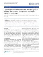
scan revealed a large, well-defined, soft tissue density lesion
with intrinsic fat density areas in the right adrenal region
measuring 116 × 100 mm in size (Figure 1). On laboratory
investigations, the patient’s random blood sugar was 82.6
mg/dL and his renal and liver functions and electrolytes
were within normal range. The patient’s hemoglobin was
10.6 g/dL, and counts were within normal range and were
nonreactive to HIV, hepatitis B surface antigen and hepati-
tis C virus on enzyme-linked immunosorbent assay. Urine
analysis and microscopic findings were unremarkable. His
24-hour urinary vanillylmandelic acid level was 4.7 mg
(reference range, 2-8 mg/24 h). The patient was subjected
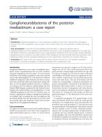
to transperitoneal laparoscopic adrenalectomy. On gros s
examination, a well-encapsulated tumor mass weighing
810 g measured 15 × 11.5 × 6.5 cm in size (Figure 2A).
* Correspondence:
1
Department of Pathology, Laboratory Medicine, Transfusion Services and
Immunohematology, G.R. Doshi and K.M. Mehta Institute of Kidney Diseases
and Research Centre and Dr. H.L. Trivedi Institute of Transplantation
Sciences, Ahmedabad, India
Full list of author information is available at the end of the article
Patel et al. Journal of Medical Case Reports 2011, 5:78
/>JOURNAL OF MEDICAL
CASE REPORTS
© 2011 Pa tel et al; licensee Bio Med Central Ltd. This is an Open Access article distributed under t he terms of the Creative Commons
Attribution License (http://creativec ommons.o rg/ licenses/by/2.0), which permits unrestricted use, distributio n, and reproduction in
any medium, provided the original work is properly cited.
The external surface was smooth and grey-yellow colored,
and th e adjacent adr enal gland m easuring 6 × 1.5 × 0.5 cm
in size was unremarkable. The cut surface was homoge-
nous, yellow and fatty with myxomatous areas or a jelly-
like appearance and soft to firm in consistency. Microscopy
revealed a well-encapsulated tumor composed of mature
adipose tissue with myxoid degenerative changes and nor-
mal adrenal parenchyma (Figure 2B).
The patient had an uneventful postoperative course
and is stable with the same medications continued for
one month. The patient is now taking a beta-blocker
and antiplatelet agents, and he has stopped taking the
alpha-blocker and diuretic that he needed previou sly for
blood pressure control.
Discussion
Adrenal lipomas are benign tumors with a reported inci-
dence of 2% to 4% of all adrenal tumors [1-4]. The
differential diagnoses include myelolipoma, angiomyoli-
poma, liposarcoma and teratoma. The histogenesis of
these mesenchymal tumors is still little understood. Lipo-
mas are known to occur on the right side with male pre-
dominance as opposed to myelolipomas, which have no
gender or site predilection. The s ize of these tumors is
usually smaller than 4 cm; however, the term giant
lipoma is preferred when the size exceeds 8 cm. Our
patient had a tumor of more than 11 cm in diameter,
thus falling in the category of “giant adrenal lipoma.” No
calcification was noted despite the lipoma’s large size.
Conclusion
Giant adrenal lipomas are rare but are being reported
more frequently because of improved modern imaging
technologies. This tumor was removed with the rarely
reported technique of transperitoneal laparoscopic adre-
nalectomy and was confirmed histologically.
Consent
Written informed consent was obtained from the patient
for publication o f this case report and accompanying
images. A copy of the written consent is available for
review by the Editor-in-Chief of this journal.
Author details
1
Department of Pathology, Laboratory Medicine, Transfusion Services and
Immunohematology, G.R. Doshi and K.M. Mehta Institute of Kidney Diseases
and Research Centre and Dr. H.L. Trivedi Institute of Transplantation
Sciences, Ahmedabad, India.
2
Department of Urology and Transplantation, G.
R. Doshi and K.M. Mehta Institute of Kidney Diseases and Research Centre
and Dr. H.L. Trivedi Institute of Transplantation Sciences, Ahmedabad, India.
Authors’ contributions
RDP was the major contributor in writing the manuscript and analysis and
interpretation of patient data regarding the histopathologic disease. AVV
performed the histologic examination and helped in preparation of the final
manuscript. PRM did the transperitoneal laparoscopic adrenalectomy and
postoperative follow-up of the patient. All authors read and approved the
final manuscript.
Competing interests
The authors declare that they have no competing interests.
Received: 21 October 2009 Accepted: 24 February 2011
Published: 24 February 2011
References
1. Lam KY, Lo CY: Adrenal lipomatous tumors: a 30 year clinicopathological
experience at a single institution. J Clin Pathol 2001, 54(9):707-712.
2. Milathianakis KN, Farfarelos CD, Mpogdanos IM, Karamanolakis DK: Giant
lipoma of the adrenal gland. J Urol 2002, 167(4):1777.
3. Buettner A: Lipoma of the adrenal gland. Pathol Int 1999,
49(11):1007-1009.
4. Reinig JW, Doppman JL, Dwyer AJ, Johnson AR, Knop RH: Adrenal masses
differentiated by MR. Radiology 1986, 158(1):81-4.
doi:10.1186/1752-1947-5-78
Cite this article as: Patel et al.: Giant lipoma of the adrenal gland: a case
report. Journal of Medical Case Reports 2011 5:78.
Figure 1 Computed tomography scan showing a large, well-
defined, soft tissue density mass in the right suprarenal region
(bidirectional arrow).
Figure 2 A) Cut surface of the giant adrenal lipoma with
golden yellow-tan color with an arrow showing the uninvolved
adrenal gland. B) Photomicrograph (hematoxylin and eosin stain)
showing the tumor made up of mature adipose tissue intermingled
with normal adrenal cortical parenchyma.
Patel et al. Journal of Medical Case Reports 2011, 5:78
/>Page 2 of 2