Báo cáo y học: ": Gastrointestinal relapse of multiple myeloma and sustained response to lenalidomide: a case repo" pps
Bạn đang xem bản rút gọn của tài liệu. Xem và tải ngay bản đầy đủ của tài liệu tại đây (1.04 MB, 3 trang )
CAS E REP O R T Open Access
Gastrointestinal relapse of multiple myeloma and
sustained response to lenalidomide: a case report
Patrick R Benusiglio
1*
, Thomas A McKee
2
, Xavier Montet
3
, Jean-Marc Dumonceau
4
, Laurence Favet
1
,
Anne-Claude George
1
and Pierre-Yves Dietrich
1
Abstract
Introduction: Gastrointestinal relapse in patients with multiple myeloma is very rare and, when reported, always
associated with a poor prognosis.
Case presentation: We describe the case of a 71-year-old Caucasian man who presented with life-threatening
hematemesis and melena due to a digestive relapse of his multiple myeloma. Despite the active hemorrhage, we
initiated a third-line treatment with lenalidomide. The response was spectacular and long-lasting.
Conclusions: Clinicians must consider digestive tract involvement in myeloma patients presenting with a
gastrointestinal hemorrhage. Furthermore, myeloma patients do benefit from novel oral drugs, even when they are
critically ill.
Introduction
The involvement of the gastrointestinal tract years after
an ini tial diagnosis of multiple my eloma (MM) is excep-
tional and, when reported, always associated with a poor
prognosis [1-4]. We report the case of a 71-year-old
man with MM who had been heavily pre-treated and
who presented with hematemesis and melena due to a
gastrointestinal relapse of his disease. The bleeding
lasted for over two weeks and soon became life-threa-
tening. Despite the active hemorrage, we initiated a
third-line treatment with lenalidomide. The response
was spectacular.
Case presentation
We report the case of a 71-year-old Caucasian diabetic
man with severe diabetic neuropathy who was diagnosed
with stage IIIA IgG l MM in 2004. He was initially trea-
ted with three cycles of vincristin e, doxorubicin and
dexamethasone, followed by high-dose melphalan and
autologous st em-cell transplantation, which resulted in a
partial response. His monoc lonal IgG had dropped from
65 g/L before treatment to 11 g/L after transplantation.
In 2007, a second-line chemotherapy treatment
(melphalan and prednisone, six cycles ) for a relapse
characterized by diffuse spinal involvement and an
increase in monoclonal IgG (22 g/L) stabilized the
disease. His neuropathy had precluded treatment with
thalidomide or bortezomib. Eight months after the sec-
ond-line chemotherapy, an irradiation to the T10-L1
vertebrae (30 Gy) was undertaken for symptomatic,
localized bone involvement. Ten months later, an
increase in his level of IgG (34 g/L), combined with
widespread bone pain and a worsening of his general
condition, led to the introduction of high-dose steroids.
After a week of steroid treatment, he was admitted to
our ho spital for the first time with chest pain and dys-
pnea. He was febrile (38.4°C) and his inflammatory para-
meters were increased (C-reactive protein 91 mg/L).
A urinary test for the Legionella pneumophila antigen
was positive and a computed tomography (CT) scan
showed trilobar consolidation and a bilateral pleural
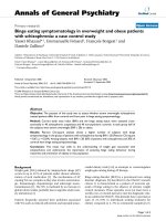
effusion. A heterogeneous solid mass extending from
the retroperitoneal to the peritoneal spaces (Figure 1A,
B) provided evidence for the progression of the MM. At
the time, priority was given to the treatment of the pul-
monary infection and he recovered after three weeks of
oral levofloxacin.
Shortlyaftertheantibiotictherapywasdiscontinued,
he presented with sudden hematemesis and melena,
requiring fifteen 500 ml units of packed red cells, in
* Correspondence:
1
Centre for Oncology, Geneva University Hospital, 4 rue Gabrielle Perret-
Gentil, 1211 Geneva 14, Switzerland
Full list of author information is available at the end of the article
Benusiglio et al. Journal of Medical Case Reports 2011, 5:110
/>JOURNAL OF MEDICAL
CASE REPORTS
© 2011 Benusiglio et al; licensee BioMed Central Ltd. This is an Open Access article distributed under the terms of the Creative
Commons Attribution License (http://creativeco mmons.org /licenses/by/2.0), which permits unrestricted use, distribution, and
reproduction in any medium, provided the original work is properly cited.
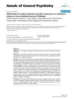
total, over a period of twenty days. His platelet count
and coagulation parameters were normal. A bleeding,
ulcerat ed jejunal mass was reveal ed by an upper gastro-
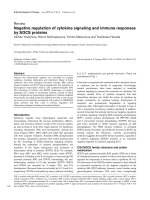
intestinal endoscopy (Figure 2) and biopsies showed a n
infiltration of the intestinal mucosa by neoplastic plasma
cells producing monoclonal l light chains (Figure 3A,B).
Despite the active bleeding, a third-line therapy with
lenalidomide (25 mg daily) and dexamethasone (40 mg
once-weekly) was initiated; the lenalidomide was given
in three-week cycles followed by a one-week break [5,6].
An excellent resp onse was achieved after the first cycle:
his paraprotein levels dropped to 10 g/L and there was
no recurrence of the hematemesis or melena. His gen-
eral condition improved rapidly and he was discharged
after the second cycle had commenced. A re peat CT
four months later showed a dramatic shrinkage of the
retroperitoneal mass (Figure 1C,D). This response lasted
for a total of 10 months and resulted in an excellent
Figure 1 CT of the abdomen before (A, B) and after (C, D) four months of treatment with lenalidomide.
Figure 2 Jejunal mass as seen on the upper gastroin testinal
endoscopy.
Figure 3 Jejunal mucosa infil trate d by multiple myeloma.A:
magnification ×200, hematoxylin and eosin. B: magnification ×400,
l chains.
Benusiglio et al. Journal of Medical Case Reports 2011, 5:110
/>Page 2 of 3
quality of life for the patient during the whole period.
He declined further treatment at retroperitoneal pro-
gression and died a few weeks later.
Discussion
Gastrointestinal involvement in MM is very rare. It most
often occurs in the context of an isolated, primary, extra-
medullary plasmacytoma [7]. Patients with newly-
diagnosed MM rarely present with symptoms related to
gastrointestinal involvement [8]. A gastrointestinal
relapse in patients with long-term MM, such as that
observed in the case of our patient, is exceptional.
Amongst a total of 553 patients with MM included in
two large European studies, 87 experience d an extrame-
dullary relapse but none of these involved the gastroin-
testinal tract [1,2]. Only one out of six extramedullary
relapses reported by a North American Institution
involved the gastrointestinal tract [3]. All of these cases
had a poor prognosis, with a maximal survival rate of 106
days from diagnosis. Finally, Dawson et al. reported the
case of a 60-year-old patient with MM with hematemesis,
melena and gastroduodenal mucosal lesions [4]. The
patient died two weeks after presentation. The gastroin-
testinal lesions were not biopsied, but their myelomatous
nature was likely, as a biopsy of a right breast mass pro-
vided pathological evidence of an extramedullary relapse.
Conclusion
Our case report is well documented and highly informa-
tive. It reminds us th at, in addition to much more com-
mon causes (for example, ulcers), clinicians must
consider digestive tract involvement in patien ts with
MM presenting with a gastrointestina l hemorrhage. It
also shows that patie nts with MM who have been heav-
ily pre-treated can benefit from novel drugs, even when
they are critically ill. We suggest that the major clinical
improvement has to be linked to lenalidomide, since
high-dose steroids had been ineffective in this case. Our
patient’ s recovery and the drop i n his monoclonal IgG
were very rapid. This effect of lenalidomide has already
been observed in other life-threatening situations asso-
ciated with MM, such as severe renal impairment or
high-output heart failure secondary to intramedullary
arteriovenous fistulas [9,10]. Finally, it should be empha-
sized that a response to this oral drug was obtained
despite active bleeding in the upper digestive tract.
Consent
Written informed consent was obtained from the
patient’s next-of-kin for publication of this case report
and any accompanying images. A copy of the written
consent is available for review b y the Editor-in-Chief of
this journal.
Author details
1
Centre for Oncology, Geneva University Hospital, 4 rue Gabrielle Perret-
Gentil, 1211 Geneva 14, Switzerland.
2
Department of Pathology, Geneva
University Hospital, 4 rue Gabrielle Perret-Gentil, 1211 Geneva 14,
Switzerland.
3
Department of Radiology, Geneva University Hospital, 4 rue
Gabrielle Perret-Gentil, 1211 Geneva 14, Switzerland.
4
Department of
Gastroenterology, Geneva University Hospital, 4 rue Gabrielle Perret-Gentil,
1211 Geneva 14, Switzerland.
Authors’ contributions
PRB, JMD, LF, ACG and PYD were directly involved in the management of
the patient. PRB wrote the manuscript wit h support from TAM and PYD.
TAM and XM reviewed and interpreted the pathology slides and CT scan
images, respectively. All authors read and approved the final manuscript.
Competing interests
The authors declare that they have no competing interests.
Received: 5 August 2010 Accepted: 19 March 2011
Published: 19 March 2011
References
1. Damaj G, Mohty M, Vey N, Dincan E, Bouabdallah R, Faucher C, Stoppa AM,
Gastaut JA: Features of extramedullary and extraosseous multiple
myeloma: a report of 19 patients from a single center. Eur J Haematol
2004, 73:402-406.
2. Zeiser R, Deschler B, Bertz H, Engelhardt M: Extramedullary vs medullary
relapse after autologous or allogeneic hematopoietic stem cell
transplantation (HSCT) in multiple myeloma (MM) and its correlation to
clinical outcome. Bone Marrow Transplant 2004, 34:1057-1065.
3. Cerny J, Fadare O, Hutchinson L, Wang SA: Clinicopathological features of
extramedullary recurrence/relapse of multiple myeloma. Eur J Haematol
2008, 81:65-69.
4. Dawson MA, Polizzotto MN, Gordon A, Roberts SK, Spencer A:
Extramedullary relapse of multiple myeloma presenting as hematemesis
and melena. Nat Clin Pract Oncol 2006, 3:223-226.
5. Weber DM, Chen C, Niesvizky R, Wang M, Belch A, Stadtmauer EA, Siegel D,
Borrello I, Rajkumar SV, Chanan-Khan AA, Lonial S, Yu Z, Patin J,
Olesnyckyj M, Zeldis JB, Knight RD, Multiple Myeloma (009) Study
Investigators: Lenalidomide plus dexamethasone for relapsed multiple
myeloma in North America. N Engl J Med 2007, 357:2133-2142.
6. Rajkumar SV, Jacobus S, Callander NS, Fonseca R, Vesole DH, Williams ME,
Abonour R, Siegel DS, Katz M, Greipp PR, Eastern Cooperative Oncology
Group: Lenalidomide plus high-dose dexamethasone versus
lenalidomide plus low-dose dexamethasone as initial therapy for newly
diagnosed multiple myeloma: an open-label randomised controlled trial.
Lancet Oncol 2010, 11(1):29-37.
7. Alexiou C, Kau RJ, Dietzfelbinger H, Kremer M, Spiess JC, Schratzenstaller B,
Arnold W: Extramedullary plasmacytoma: tumor occurrence and
therapeutic concepts. Cancer 1999, 85:2305-2314.
8. Herbst A, Renner SW, Ringenberg QS, Fass R, Krouse RS: Multiple myeloma
presenting with a colonic obstruction and bony lesions: a clinical
dilemma. J Clin Oncol 2008, 26:5645-5647.
9. Ludwig H, Zojer N: Renal recovery with lenalidomide in a patient with
bortezomib-resistant multiple myeloma. Nat Rev Clin Oncol 2010,
7(5):289-294.
10. Robin J, Fintel B, Pikovskaya O, Davidson C, Cilley J, Flaherty J: Multiple
myeloma presenting with high-output heart failure and improving with
anti-angiogenesis therapy: two case reports and a review of the
literature. J Med Case Reports 2008, 2:229.
doi:10.1186/1752-1947-5-110
Cite this article as: Benusiglio et al.: Gastrointestinal relapse of multiple
myeloma and sustained response to lenalidomide: a case report. Journal
of Medical Case Reports 2011 5:110.
Benusiglio et al. Journal of Medical Case Reports 2011, 5:110
/>Page 3 of 3