Báo cáo y học: " Papillary renal cell carcinoma with metastatic laparoscopic port site and vaginal involvement: a case report" pot
Bạn đang xem bản rút gọn của tài liệu. Xem và tải ngay bản đầy đủ của tài liệu tại đây (952.96 KB, 3 trang )
CAS E REP O R T Open Access
Papillary renal cell carcinoma with metastatic
laparoscopic port site and vaginal involvement:
a case report
Xue En Chuang
1
, Hwai Liang Loh
2
, Hong Gee Sim
3
, Kah Leng Fong
4
and Min-Han Tan
1*
Abstract
Introduction: Laparoscopic port-site metastasis is a rare but well recognized outcome following surgery in
urological cancers, with its etiology not clearly understood. Additionally, vaginal metastasis in clear cell renal cell
carcinoma is rare, and has not been previously reported in the setting of papill ary renal cell carcinoma.
Case presentation: We present the case of a 71-year-old Chinese woman with metastatic type II papillary renal
cell carcinoma with histologically verified vaginal involvement and a concurrent laparoscopic port-site metastasis.
This was also associated with a unique constellation of widely disseminated metastatic sites, which include a local
relapse, the peritoneum and the urethra.
Conclusion: Laparoscopic port-site metastases are associated with the presence of advanced cancer with multiple
sites of metastasis. We hypo thesize from the findings of our report and background data that this phenomenon is
more likely to be related to tumo r factors rather than operative factors. We also present what is, to the best of our
knowledge, the first reported case in the literature of vaginal and urethral metastasis and the second reported case
of laparoscopic port-site recurrence.
Background
Renal cell carcinoma is well known for its ability to metas-
tasize widely to nearly every organ in the body. While
vaginal metastases are very rare, with the mode of spread
still currently obscure, it is critical to differentiate these
metastases from primary vaginal carcinomas, which are
rare and constitute approximately 2% of all malignant neo-
plasms of the female genital tract [1]. To date, all renal cell
carcinoma (RCC) metastases to the vagina have been
reported to be of the clear cell subtype. Additionally, up to
September2007,therewereonly28casesofport-site
metastases involving urological malignancies reported.
The etiology of port-site metasta ses has not been clearly
established, though it appears to be multi-factorial [2].
Case report
A 71-year-ol d Chinese woma n, with ischemic heart dis-
ease and a metallic stent and on prophylactic warfarin
anticoagulation, presented to our institution with inter-
mittent gross hematuria. A computed tomography (CT)
scan showed an 8 cm mass in the upper pole of the
right kidney, with no evidence of metastasis. Subse-
quently, a laparoscopic radical nephrectomy was per-
formed, with the specimen bagged and removed
through a lower abdominal incision. Histology results
showed a type II papillary RCC, pT3A, nuclear grade 3,
without sarcomatoid differentiation, with focal invasion
of adjacent perirenal fat but with sparing of Gerota’s fas-
cia (Figure 1). Our patient relapsed six months after sur-
gery, with local recurrence and multiple lesions in the
lungs, liver, peritoneum, mesentery, iliac, and abdominal
wall, as well as a laparoscopic port site metastasis
(Figure 1). She was started on sunitinib 37.5 mg daily,
and one week later, she presented with vaginal bleeding.
Her international normalized ratio (INR) was 1.46.
Colposcopy revealed a urethral mass as well as a hard nod-
ular bleeding mass on the right vaginal wall (Figure 1).
A vaginal biopsy yielded a papillary carcinoma, histolo-
gically consistent with the earlier diagnosis of RCC.
She underwent palliative radiotherapy (30 to 36Gy in
* Correspondence:
1
Department of Medical Oncology, National Cancer Centre Singapore,
Singapore
Full list of author information is available at the end of the article
Chuang et al. Journal of Medical Case Reports 2011, 5:131
/>JOURNAL OF MEDICAL
CASE REPORTS
© 2011 Chuang et al; li cense e BioMed Central Ltd. This is an Open Access article distributed under the terms of the Creative Commons
Attribution License ( which permits unre stricted use, distribution, and reproduction in
any medium, provided the original work is properly cited.
10 to 12 fractions) and the bleeding was halted. She
declined further systemic treatment, and died six
months later, approximately one year after initial
nephrectomy. Informed consent for this publication
was obtained from her family.
Discussion
Approximately 80 cases of vaginal metastasis fr om renal
cell carcinoma have been reported to date, with indeter-
minate prognostic implications from conflicting case
reports. After undergoing treatment, mostly in the form
of nephrect omy and excision of the vagina lesion, some
patients continue to live with no evidence of the disease,
whereas others show rapid deterioration. Our report
represents the first case of papillary RCC metastasizing
to the vagina, with the second such report of a concur-
rent laparoscopic port-site metastasis. This case is high-
lighted because the patterns of metastasis for clear cell
renal cell carcinoma and papillary renal cell carcinoma
are recognized as being different [3]. Rare and unex-
pected sites of metastases in RCC are usually associated
with the clear cell subtype.
Although immunohistochemical studies have suggested
a common cellular origin for clear cell RCC and papillary
RCC, there are distinct underlying genetic differences.
Inactivation of the von Hippel-Lindau (VHL) gene occurs
in patients with clear cell renal cell carcinoma in both
the germline and somatic settings [4], whereas the under-
lying pathways that drive papillary RCC, particularly in
the somatic setting, are less established. Reports indicat-
ing the identification of a familial cancer syndrome
including type II papillary RCC from an underlying
germline mutation in the fumarate hydratase (FH)gene
and different activation patterns of cell cycle pathways
between type I and type II papillary RCC have led to the
role of metabolic signaling to be examined [5].
An anatomical explanation for vaginal metastasis has
been advanced, supporting a predominant left-sided
renal origin [6]. Consistent with the concept of retro-
grade venous spread as a mechanism of vaginal metasta-
sis from renal cell carcinoma, retrograde flow of
contrast medium from the left renal vein to the left
ovarian vein, followed by filling of the ovarian and vagi-
nal plexus has bee n demonstrated in patients with renal
cell carcinoma [7]. Our case had a right-sided renal ori-
gin, but there was naturally widespread involvement of
the systemic circulation including the lungs, which may
account for this metastasis pattern.
Several hypotheses have been advanced to account for
port-site metastasis, which is a recognized phenomenon
[8-10], including contamination during laparoscopic sur-
gery via surgical apparatus stained with exfoliated tumor
cells, pneumoperitoneum or preferential growth of
tumor cells at sites of high cellular proliferation during
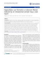
Figure 1 Images of the metastatic papillary renal cell carcinoma (RCC). (A) Laparoscopic port-site metastasis (arrow); (B) local recurrence at
the right renal bed (black arrow) and ring enhancing liver metastasis (white arrow); (C) a superficial mass representing tumor metastasis at the
urethral orifice; (D) a similar tumor located at the right vaginal wall imaged on colposcopy, from which a biopsy was taken; (E) a hematoxylin
and eosin stained histological section of the primary papillary RCC (20 × magnification); (F) a hematoxylin and eosin stained histological section
of the metastatic vaginal lesion (20 × magnification).
Chuang et al. Journal of Medical Case Reports 2011, 5:131
/>Page 2 of 3
wound healing at the por t site [11]. It is recogn ized that
although port-site metastasis are rare, they normally
occur in the presence of advanced disease [12]. Given
that the single previous report of a port-site metastasis
in type 2 papillary RCC had a similar profile of meta-
static sites involving the peritoneum and liver i n addi-
tion to the port site [13], our case repo rt provides
minor support for the hypothesis that port-site metasta-
sis is related to tumor factors rather than operative
factors.
Conclusion
In summary, we report the first case of papillary renal
cell carcinoma with metastasis to the vagina, with the
second such report of a laparoscopic port-side metasta-
sis. Our case report documenting a second port-site
metastasis in a rare tumor provides support for the
hypothesis that port-site metastases are related to tumor
factors, and not operative factors.
Consent
Written informed consent was obtained from the
patient’s next-of-kin for publication of this case report
and any accompanying images. A copy of the written
consent is available for review by the Editor-in-Chief of
this journal.
Acknowledgements
None.
Author details
1
Department of Medical Oncology, National Cancer Centre Singapore,
Singapore.
2
Department of Pathology, Singapore General Hospital,
Singapore.
3
Department of Urology, Singapore General Hospital, Singapore.
4
Department of Obstetrics and Gynaecology, Singapore General Hospital,
Singapore.
Authors’ contributions
XEC and MHT wrote the report; HGS, KLF and MHT participated in the care
of our patient; HLL provided an independent pathological review. All
authors read and approved the final manuscript.
Competing interests
The authors declare that they have no competing interests.
Received: 21 December 2009 Accepted: 1 April 2011
Published: 1 April 2011
References
1. Marchal F, Leroux A, Hoffstetter S, Granger P: Vaginal metastasis revealing
colon adenocarcinoma. Int J Colorect Dis 2006, 21:861-862.
2. Eng MK, Katz MH, Bernstein AJ, Shikanov S, Shalhav AL, Zorn KC:
Laparoscopic port-site metastasis in urologic surgery. J Endourol 2008,
22:1581-1586.
3. Mai KT, Landry DC, Robertson SJ, Commons AS, Burns BF, Thijssen A,
Collins J: A comparative study of metastatic renal cell carcinoma with
correlation to subtype and primary tumor. Pathol Res Pract 2001,
197:671-675.
4. Maher ER: Von Hippel-Lindau disease. Curr Mol Med 2004, 4:833-842.
5. Yang XJ, Tan MH, Kim HL, Ditlev JA, Betten MW, Png CE, Kort EJ, Futami K,
Furge KA, Takahashi M, Kanayama HO, Tan PH, Teh BS, Luan C, Wang K,
Pins M, Tretiakova M, Anema J, Kahnoski R, Nicol T, Stadler W,
Vogelzang NG, Amato R, Seligson D, Figlin R, Belldegrun A, Rogers CG,
Teh BT: A molecular classification of papillary renal cell carcinoma.
Cancer Res 2005, 65:5628-5637.
6. Milathianakis CN, Karamanolakis DK, Massoud WA, Roumier X, Bogdanos IM,
Perrin P: Vaginal metastases from renal cell carcinoma [in French]. Prog
Urol 2005, 15:319-321.
7. Mulcahy JJ, Furlow WL: Vaginal metastasis from renal cell carcinoma:
radiographic evidence of possible route of spread. J Urol 1970, 104 :50-52.
8. Dorrance HR, Oien K, O’Dwyer PJ: Effects of laparoscopy on
intraperitoneal tumor growth and distant metastases in an animal
model. Surgery 1999, 126:35-40.
9. Tan BJ: Is carbon dioxide insufflation safe for laparoscopic surgery? A
model to assess the effects of carbon dioxide on transitional-cell
carcinoma growth, apoptosis, and necrosis. J Endourol 2006, 20:965-969.
10. Neuhaus SJ, Watson DI, Ellis T, Rofe AM, Jamieson GG: Influence of
cytotoxic agents on intraperitoneal tumor implantation after
laparoscopy. Dis Colon Rectum 1999, 42:10-15.
11. Patton MS, Park KG: Laparoscopic port site recurrence in the absence of
intra-abdominal disease. JR Coll Surg Edinb 2001, 46:184-185.
12. Zivanovic O, Sonoda Y, Diaz JP, Levine DA, Brown CL, Chi DS, Barakat RR,
Abu-Rustum NR: The rate of port-site metastases after 2251 laparoscopic
procedures in underlying malignant disease. Gynecol Oncol 2008,
111:431-437.
13. Masterson TA, Russo P: A case of port-site recurrence and locoregional
metastasis after laparoscopic partial nephrectomy. Nat Clin Pract Urol
2008, 5:345-349.
doi:10.1186/1752-1947-5-131
Cite this article as: Chuang et al.: Papillary renal cell carcinoma with
metastatic laparoscopic port site and vaginal involvement: a case
report. Journal of Medical Case Reports 2011 5:131.
Submit your next manuscript to BioMed Central
and take full advantage of:
• Convenient online submission
• Thorough peer review
• No space constraints or color figure charges
• Immediate publication on acceptance
• Inclusion in PubMed, CAS, Scopus and Google Scholar
• Research which is freely available for redistribution
Submit your manuscript at
www.biomedcentral.com/submit
Chuang et al. Journal of Medical Case Reports 2011, 5:131
/>Page 3 of 3