Báo cáo y học: " Ocular coherence tomography of symptomatic phototoxic retinopathy after cataract surgery: a case report" ppsx
Bạn đang xem bản rút gọn của tài liệu. Xem và tải ngay bản đầy đủ của tài liệu tại đây (2.23 MB, 4 trang )
CAS E REP O R T Open Access
Ocular coherence tomography of symptomatic
phototoxic retinopathy after cataract surgery: a
case report
Ahmad M Mansour
1,2*
, Muhammad H Yunis
1,2
and Walid A Medawar
3
Abstract
Introduction: High-resolution ocular coherence computed tomography enables unprecedented visualization of the
retinal microarchitecture. To the best of our knowledge, this is the first report of high-resolution ocular coherence
tomography findings in the healed form of photic post-cataract retinopathy.
Case presentation: A 76-year-old Caucasian man complained of paracentral scotoma, persisting for six weeks after
cataract surgery.
Conclusion: Ocular coherence tomography demonstrated a localized juxta-foveal area of retinal atrophy involving
the photoreceptor layer, and the retinal pigment epithelium layer.
Introduction
Operating microscope light-induced foveal damage is a
well recognized occurrence following ocular surgery
including complicated or lengthy cataract extraction and
complex anterior segment procedures [1-5]. While the
majority of injuries produce minimal symptoms, sco-
toma and permanent central vision loss have occurred
in some patients. Retinal edema is typically discernable
a few days after exposure, while prominent pigmentary
changes of the fundus are not apparent prior to two to
three weeks after exposure. The recent advent of high-
definition ocular coherence computed tomography can
help clinicians in analyzing the level and degree of ret-
inal damage after photic damage induced by surgical
microscope.
Case presentation
A healthy 76-year-old Caucasian man underwent pha-
coemulsification under retrobulbar anesthesia in his
right eye with torn posterior capsule at the completion
of cortex aspiration. Anterior vitrectomy was performed
and a 5 × 6 mm intra-ocular lens was implanted in the
sulcus. A coaxial illuminated microscope (OPMI CS-XY;
Zeiss, Oberkochen, Germany) was used for surgery that
last ed 45 minutes. At six weeks postoperatively, his best
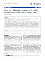
corrected visual acuity was 0.5. His main complaint
was a para central scotoma confirmed by perimetry
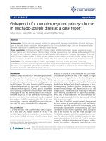
(Figure 1). Fundoscopy reve aled a well circumscribed flat
yellowish retinal lesion, approximately double the disc
diameter in size, inferotemporal to the fovea (Figure 2).
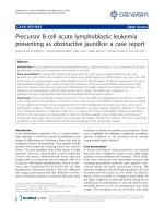
The retinal lesion was prominent by autofluorescence
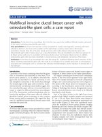
(Figure 3) stained with fluorescein dye with speckled
blockage of fluorescence (Figure 4). Spectral domain ocu-
lar coherence tomography (OCT) confirmed thinning of
the retinal lesion with loss of the inner/outer photorecep-
tor layer and retinal pigment epithelium (Figure 5). At
nine months after surgery, repeat OCT revealed cystoid
macular edema induced by topical travoprost initiated
over the past month to control ocular hypertension
(Figure 6). There was persistent disruption of the inner/
outer photoreceptor layer and retinal pigment epithelium.
Discussion
Most mild phototoxic retinal injuries probably remain
undiagnosed in routine postoperative examination [1-5].
Retinal phototoxic lesions first appear a few days after
exposure as well circumscribed outer retinal whitening
with mild disturbances of the retinal pigment epithe-
lium, often with a light border. After the first week,
lesions are characterized by coarse alterations of the
* Correspondence:
1
Department of Ophthalmology, American University of Beirut, Beirut,
Lebanon
Full list of author information is available at the end of the article
Mansour et al. Journal of Medical Case Reports 2011, 5:133
/>JOURNAL OF MEDICAL
CASE REPORTS
© 2011 Mansour et al; licensee BioMed Central Ltd. This is an Open Access article distributed under the terms of the Creative
Commons Attribution License ( nses/by/ 2.0), which permits unrest ricted use, distribution , and
reproduction in any me dium , provided the original work is properly cited.
retinal pigment epithelium layer with fluores cein angio-
graphy demonstrating shar ply demar cated characteristic
early discrete mottled hyperfluorescence with late stain-
ing. Historically, these lesions are typical ly located infer-
iortothefoveaasaresultoftheslightdowngaze
during extracapsular cataract surgery. The shape of the
lesion often matches the shape of the illuminating
source of the particular operating microscope used.
Such lesions were noted in 3% of the most recent catar-
act surgery series [4], even in phacoemulsification of
short duration. While the majority of injuries produce
minimal symptoms, scotoma and permanent central
vision loss have occurred in some patients [3,5]. Risk
factors for retinal photi c injuries have included angle of
light incidence, light intensity, exposure time, and inten-
sity of the blue light component [1-5]. It is recom-
mended to use the minimal light intensity needed to
perform surgery, use oblique light or filters or pupil
shields. Implantation of an intra-ocular lens, including
multi-focal lenses, is an importa nt factor in the produc-
tion of maculopathy [2], on account of its light-focusing
effect on the retina.
Figure 1 Automated central 22 field testing of the right eye
shows the right blind spot corresponding to the optic disc and
the superotemporal paracentral scotoma.
Figure 2 The fundus photograph shows a well demarcated,
elliptical, yellowish, mottled retinal pigment epithelium
alteration approximately twice the size of the optic disk, and
encroaching on the fovea.
Figure 3 Autofluorescence image of the posterior pole of the
right eye shows a dendritiform pattern of autofluorescence at
the inferotemporal macula (BluePeak Blue Laser
Autofluorescence, Heidelberg Engineering GmbH, Heidelberg,
Germany).
Figure 4 Fluorescein angiography shows an irregular
fluorescein transmission pattern without leakage.
Mansour et al. Journal of Medical Case Reports 2011, 5:133
/>Page 2 of 4
Acute histological changes in photic injuries have
included localized necrosis of the retinal pigment
epithelium, extensive disruption of the outer lamellae of
the photoreceptors, and edema of the inner segments
[1]. Rodriguez-Marco et al. [5] presented late OCT find-
ings in a 39-year-old patient who underwent two conse-
cutive pterygium surgeries lasting 1.5 hours. Visual
acuity was 0.4 with metamorphopsia. The fundus exhib-
ited a hypo-pigmented rounded lesion in the macular
area with earl y hyperfluorescent foveal area on fluores-
cein angiography. OCT revealed a detachment of the
retinal pigment epithelium.
Conclusion
We present, to the best of our knowledge, the first
report of high-definition OCT findings in the healing
stage (six week s and nine months after surgery) of pho-
tic post-cataract retinopathy, showing atrophy of the
photoreceptor and retinal pigment epithelium layers.
Consent
Written informed consent was obtained from the patient
for publication of this case report and any accompany-
ing images. A copy of the written consent is available
for review by the Editor-in-Chief of this journal.
Author details
1
Department of Ophthalmology, American University of Beirut, Beirut,
Lebanon.
2
Department of Ophthalmology, Rafic Hariri University Hospital,
Beirut, Lebanon.
3
Department of Internal Medicine, American University of
Beirut, Beirut, Lebanon.
Authors’ contributions
AM analyzed and interpreted our patient’s fluorescein angiography and OCT
data. MY performed the surgery and eye examinations of our patient. WM
was a major contributor in writing the manuscript. All authors read and
approved the final manuscript.
Competing interests
The authors declare that they have no competing interests.
Received: 1 August 2010 Accepted: 1 April 2011 Published: 1 April 2011
Figure 5 At six weeks after surgery, a spectral domain ocular coherence tomography (OCT) scan (scan angle 45°, 0.25 mm spacing, 6
mm scan length) showed thinning of the juxta-foveal temporal retinal lesion with effacement of the photoreceptor layer, and retinal
pigment epithelium layer (Cirrus, Carl Zeiss Meditec, Oberkochen, Germany).
Figure 6 At nine months after surgery, there is disruption of the photoreceptor layer and retinal pigment epithelium layer with intact
Bruch’s membrane, as seen on spectral domain ocular coherence tomography (OCT) imaging of the temporal macula (Spectralis,
Heidelberg Engineering GmbH, Heidelberg, Germany).
Mansour et al. Journal of Medical Case Reports 2011, 5:133
/>Page 3 of 4
References
1. Michels M, Sternberg P: Operating microscope-induced retinal
phototoxicity: pathophysiology, clinical manifestations and prevention.
Surv Ophthalmol 1990, 34:237-252.
2. Kleinmann G, Hoffman P, Schechtman E, Pollack A: Microscope-induced
retinal phototoxicity in cataract surgery of short duration. Ophthalmology
2002, 109:334-338.
3. Byrnes GA, Chang B, Loose I, Miller SA, Benson WE: Prospective incidence
of photic maculopathy after cataract surgery. Am J Ophthalmol 1995,
119:92-93.
4. Gomolin JE, Koenekoop RK: Presumed photic retinopathy after cataract
surgery: an angiographic study. Can J Ophthalmol 1993, 28:221-224.
5. Rodríguez-Marco NA, Andonegui-Navarro J, Compains-Silva E, Rebollo-
Aguayo A, Aliseda-Pérez-de-Madrid D, Aranguren-Laflin M: Optical
coherence tomography and macular phototoxicity [in Spanish]. Arch Soc
Esp Oftalmol 2008, 83:267-271.
doi:10.1186/1752-1947-5-133
Cite this article as: Mansour et al.: Ocular coherence tomography of
symptomatic phototoxic retinopathy after cataract surgery: a case
report. Journal of Medical Case Reports 2011 5:133.
Submit your next manuscript to BioMed Central
and take full advantage of:
• Convenient online submission
• Thorough peer review
• No space constraints or color figure charges
• Immediate publication on acceptance
• Inclusion in PubMed, CAS, Scopus and Google Scholar
• Research which is freely available for redistribution
Submit your manuscript at
www.biomedcentral.com/submit
Mansour et al. Journal of Medical Case Reports 2011, 5:133
/>Page 4 of 4