Vaginal Surgery for Incontinence and Prolapse - part 4 pot
Bạn đang xem bản rút gọn của tài liệu. Xem và tải ngay bản đầy đủ của tài liệu tại đây (871.03 KB, 30 trang )
Outcome Measures for Assessing Efficacy of Incontinence Procedures 81
Good test-retest reliability with 7-day void-
ing diaries has been shown with regard to the
frequency of micturition, episodes of inconti-
nence, incontinence-associated symptoms (urge-
or stress-related), and urinary urgency (48).
Three- and 4-day diaries have similar reliability
with regard to these parameters. Investigators
have reported that 4- and 7-day diaries record-
ing voiding frequency and volumes are statisti-
cally indistinguishable (49).
A question distinct from test-retest reliability
relates to the length of time required to detect
any effect of intervention with a voiding diary.
Utilizing mathematical modeling, 7-day voiding
diaries are long enough to reasonably detect
changes in either stress- or urge-related inconti-
nent episodes (50). This time course is depen-
dent on the initial frequency of the outcome
variable being evaluated, with a lower number of
episodes requiring a longer voiding diary period
in order to detect signifi cant changes owing to
intervention.
One confounding aspect regarding utilization
of voiding diaries for outcomes assessment is
that the very act of keeping track of voiding
symptomatology can affect those symptoms.
This behavioral modifi cation owing to increased
focusing on and awareness of urinary issues
is referred to as “self-monitoring” (51). This
effect has been shown to occur early, though it
can dissipate after 3 days. The impact of self-
monitoring is most strongly felt on shorter-
term diaries and should be considered when
interpreting these data.
Voiding diaries have been established as the
standard outcome tool in evaluating new medical
therapy for incontinence, particularly for mea-
suring effectiveness in treating symptoms of
overactive bladder (OAB). Nearly all studies of
new pharmaceutical agents for OAB incorporate
a voiding diary, since the main outcome mea-
sures (i.e., voiding frequency, urge-leakage epi-
sodes) can be readily obtained from a micturition
diary (52,53). The role of diaries in evaluating
treatment for SUI has also been addressed (54),
but without some assessment of bother associ-
ated with leakage, the results of leakage fre-
quency that can be gleaned from a voiding diary
might tell only part of the story. The largest role
for voiding diaries in outcome assessment may
be in combination with other methods of evalu-
ation. In this regard, scoring systems that in-
corporate results from voiding diaries with symp-
tom questionnaires and pad tests have been
proposed and used to evaluate surgical interven-
tions for incontinence (55).
Physiologic Assessment:
Urodynamic Testing
Uses of Urodynamic Testing
Urodynamic testing is ideally suited to assess
objective outcome following incontinence sur-
gery because it can assess success as well as
failure. Specifi cally, urodynamic testing can
evaluate for the fi nding that prompted the inter-
vention (i.e., by testing leak point pressures or
urethral pressure profi les for SUI), but it can
also evaluate for possible (sometimes asymp-
tomatic) sequelae of the intervention, such as
detrusor overactivity, altered voiding dynam-
ics, and elevated postvoid residuals. However,
because urodynamic testing is more expensive
than other noninvasive measures, it may not be
a practical method. Since many referral centers
have standardized urodynamic protocols and
therefore generate comparable data, it may be
most useful as a research tool to assess both the
etiology of successes and sources of failures in
treating SUI.
Accuracy of Urodynamic Testing
It is important to stress that urodynamic mea-
surements of voiding parameters are evaluating
a different entity than the normal daily voiding
experience. The lack of correlation between
some reported symptom scales and urody-
namic fi ndings (56) is not entirely surprising, as
it may be explained by the differences between
a patient’s normal spontaneous voids and those
performed in the milieu of the urodynamic
laboratory. Similarly, these changes may eluci-
date the poor correlation between validated
quality of life instruments and urodynamic
fi ndings (57). Deviation from an individual’s
normal voiding experience to that which is rep-
resented on the urodynamic tracing may be due
to the instrumentation required by the study,
the laboratory environment, the presence of
observers, nonphysiologic fi lling velocities, or
other alterations. This does not mean that these
objective measurements are any less valuable,
but that it is critical that they are considered in
82 Vaginal Surgery for Incontinence and Prolapse
their appropriate context. For example, since
alterations have been shown to exist between
free and intubated fl ow rates (58), comparison
of postoperative free fl ow rates to preoperative
intubated fl ow rates should not be considered a
reliable or meaningful outcomes assessment.
Assessing Treatment Success
Urodynamics have proven quite useful in under-
standing mechanisms of success. In a nonran-
domized study of 327 women who underwent
surgery for incontinence, Tamussino and col-
leagues (59) performed urodynamics before and
5 years after either anterior colporrhaphy (AC),
AC with needle suspension, or Burch colposus-
pension. They found that the effi cacy of surgical
therapy depends not only on the procedure
chosen and urodynamic changes effected, but
also on the preoperative severity of the inconti-
nence. For example, colposuspension was sig-
nifi cantly more likely to increase the pressure
transmission ratio (PTR) across the urethra
than an anterior colporrhaphy and much more
likely to cure incontinence. Klutke and col-
leagues (60) noted that patients cured of incon-
tinence following colposuspension were more
likely to have a higher mean urethral resistance
following surgery (0.099) than those who failed
(0.041), which suggests that enhanced resistance
rather than anatomic restoration is the key to
surgical success. Similarly, Bump and colleagues
(61) noted that patients with low PTRs (<90%)
were much more likely, 6 weeks and 6 months
after bladder neck surgery for incontinence
(Burch or vaginal suspensions), to remain incon-
tinent than those with appropriate (90–110%) or
high (>110%) PTR. This alteration in PTR has
been shown to be durable over long-term follow-
up (62). In contrast, urethral axis angle did not
correlate with success of the operation. Elevated
levels of urethral resistance have been found in
patients following tension-free vaginal tape pro-
cedures (63). These data support the hypothesis
that an element of dynamic urethral obstruction
may be the most important factor in the success
of anti-incontinence operations.
Assessing Adverse Outcomes
But when is urethral obstruction excessive? Uro-
dynamics taken after a procedure also allow
surgeons to determine when obstruction may
alter both voiding dynamics and resting bladder
function. Bump et al’s (61) study demonstrated
that when the PTR was too high (>110%), patients
were signifi cantly more likely to have detrusor
overactivity and voiding dysfunction. Nearly all
studies that investigate pressure fl ow relation-
ships during voiding after anti-incontinence
operations note increased detrusor pressure at
maximum fl ow. In fact, increased voiding pres-
sures have been noted after modifi ed Pereyra
bladder neck suspension (64), Stamey proce-
dures (65), Burch colposuspension (66), pubo-
vaginal sling (67), and tension-free vaginal tape
(68), though in most cases, clinical symptoms
were not apparent. Urodynamics can also assess
for development and resolution of detrusor
overactivity following anti-incontinence proce-
dures (69,70), which is a factor that must be con-
sidered given that the likelihood of new-onset
urgency and the resolution of urge symptoms
are important aspects of informed consent.
As with the other methods of outcomes assess-
ment discussed, the information generated by
multichannel urodynamics should not be viewed
in a vacuum. Evaluation of these studies in light
of the patient’s symptom complex must be per-
formed to ensure correct interpretation. One
outcome assessment study found that of patients
with urodynamically demonstrated sphincteric
incontinence following surgery, only 50% were
symptomatic (71). Given that anti-incontinence
surgery is undertaken to correct urinary incon-
tinence, it would not seem accurate to classify
these patients as failures in the absence of
reported incontinent episodes solely because of
low leak point pressures. It is important to keep
this in mind when interpreting study results that
only report urodynamic data on “failures,” as
alterations or persistent abnormalities in voiding
parameters may exist in “successfully” treated
populations.
Objective Measures to Assess
Anatomic Changes Following
Treatment
Radiologic Studies
There are several reasons to consider anatomic
assessment of the bladder and/or urethra as an
outcome measure after an intervention for
Outcome Measures for Assessing Efficacy of Incontinence Procedures 83
incontinence. Historically, surgical correction
of incontinence was based on the principle of
restoring the urethra to a more anatomically
correct position, as this was thought to restore
physiologic pressure transmission to the urethra
during times of increased intraabdominal pres-
sure. Whether this is the mechanism by which
some surgical approaches produce continence
is debatable, but restoring the urethra to an
intrapelvic position and reducing urethral
hypermobility (UH) remains a goal of some
approaches. Additionally, development of sec-
ondary cystoceles has been noted following
certain suspension procedures in which no
support of the more proximal anterior vaginal
wall was applied.
Although physical examination alone offers
some insight into urethral mobility and anterior
vaginal wall descent, its accuracy and interexam-
iner reliability remain uncertain. For this reason,
radiologic studies may offer a more objective
assessment. The standing cystogram has been
utilized to assess for anterior vaginal descent
following suspension procedures (72). It is also
a reliable means to assess urethral mobility
with increases in intraabdominal pressure and
changes in urethral angle following anti-
incontinence procedures (73). Vaginal ultraso-
nography has been used to assess for anatomic
changes in the position of the bladder neck
following colposuspension (74).
Q-Tip Test
During a Q-tip test, the urethral angle is
assessed by placing a cotton swab in the urethra
to the level of the bladder neck and then mea-
suring defl ection from the horizontal at rest
and with straining. This simple test can be con-
ducted in the offi ce and without more inva-
sive or costly testing, and it seems a fairly
reliable indicator of urethral mobility associ-
ated with straining maneuvers (75). However,
some investigators question its accuracy and
overall value. For example, like any assessment
of urethral position, the presence of hypermo-
bility alone does not necessarily indicate SUI.
That is, the specifi city of this test for predicting
SUI is quite low (76), and using specifi c cutoff
values (such as 30- to 35-degree defl ection with
straining) to differentiate incontinent women
is unreliable (77). Thus, although a properly
performed Q-tip test gives reasonably accurate
assessment of urethral position, its current role
in evaluating incontinent patients and the
outcome of procedures aimed at treating incon-
tinence is questionable.
Stress Test
Several authors have reported on the use of a
provocative stress test to assess for urinary
incontinence during pelvic examination. The
technique for performing the stress test has dif-
fered, generally based on the degree of bladder
fi lling, which has varied from empty (inconti-
nence assessed 20 minutes after catheteriza-
tion) (78) to 200 cc (79) or higher (80). In general,
the observation of leakage at the urethral meatus
during performance of either a Valsalva maneu-
ver or cough is considered a positive test. Most
studies have shown excellent correlation with
urodynamic parameters used to measure ure-
thral function (such as low Valsalva leak point
pressures) indicating intrinsic sphincteric dys-
function (ISD). Positive predictive values (for
predicting ISD) of greater than 95% have been
reported, though the fi nding of stress-induced
leakage does not rule out the possibility of
coexisting detrusor overactivity. Of potentially
greater interest is the fi nding of negative pre-
dictive values of 80% to 90%, indicating that
women without leakage during stress testing
are unlikely to have ISD in most instances (81).
The supine stress test is a useful clinical tool in
patients with severe ISD without features of
mixed incontinence.
Recent Trends
A multidimensional approach to defi ning cure
for incontinence has been advocated by most
professional organizations dealing with this
issue, including the ICS (82), the Society for Uro-
dynamics and Female Urology (83), and the
World Health Organization International
Consultation on Incontinence (84). Recent
large-scale, multiinstitutional randomized
trials comparing incontinence treatments have
adopted this approach, including both the
National Institutes of Health (NIH) Urinary
Incontinence Network trial comparing Burch
colposuspension versus pubovaginal sling (22)
and the United Kingdom–based trial of TVT
versus Burch (21). In the former, a strict
84 Vaginal Surgery for Incontinence and Prolapse
defi nition of cure incorporating both subjective
and objective outcome measures is being uti-
lized, whereas in the latter, the primary outcome
was a 1-hour pad test, though secondary out-
comes included subjective measures of success.
Others have utilized a similar approach to
defi ning treatment success. Groutz and col-
leagues (85) combined a questionnaire as a sub-
jective assessment with a 24-hour pad test and
voiding diary to assess success of a pubovaginal
sling operation, and, predictably, the cure rate
using this rigid approach was lower than histori-
cally quoted values. It seems clear that a rigid
system such as this may provide more realistic
outcome data for most women, though it may
underestimate success in women who have dra-
matic improvement albeit with some persistent
leakage. Although overall, it is true that most
patients may perceive questionnaire results as
the most important outcome following inconti-
nence procedures (86), surveying multiple
domains, including both subjective and objec-
tive measures, seems to be the most reliable
means of assuring continued improvement in
the therapies we offer patients.
Conclusion
To improve treatment for women with inconti-
nence, it is imperative to do further studies to
enhance current assessment practices. Outcome
assessment has evolved over the past few
decades and must continue to do so. This is
particularly true for a disease entity such as
incontinence, for which the desired goal is
enhanced lifestyle rather than enhanced sur-
vival. For this reason, worsening a patient’s
symptoms or creating new, unexpected symp-
toms are not acceptable outcomes. Therefore,
assessing other domains potentially affected,
such as sexual function, and also investigating
for the development of new urinary symptoms
must be incorporated into standard outcome
assessment in addition to an analysis of the cure
of urinary leakage. Existing tools can be modi-
fi ed, and new ones should emerge to evaluate
these other domains.
References
1. Govier FE, Gibbons RP, Correa RJ, Weissman RM,
Pritchett TR, Hefty TR. Pubovaginal slings using fascia
lata for the treatment of intrinsic sphincter defi ciency.
J Urol 1997;157:117–121.
2. Sirls LT, Keoleian CM, Korman HJ, Kirkemo AK. The
effect of study methodology on reported success rates
of the modifi ed Pereyra bladder neck suspension.
J Urol 1995;154:1732–1735.
3. Rodriguez LV, Blander DS, Dorey F, Raz S, Zimmern P.
Discrepancy in patient and physician perception of
patient’s quality of life related to urinary symptoms.
Urology 2003;62:49–53.
4. Lee PS, Reid DW. Psychosocial vs. physical impact of
urinary incontinence: corroboration of patient with
physician perceptions. Can J Aging 1998;11:400–411.
5. Shumaker SA, Wyman JF, Uebersax JS, McClish D,
Fantl JA. Health-related quality of life measures for
women with urinary incontinence: the Incontinence
Impact Questionnaire and the Urogenital Distress
Inventory. Continence Program in Women (CPW)
Research Group. Qual Life Res 1994;3:291–306.
6. Uebersax JS, Wyman JF, Shumaker SA, McClish DK,
Fantl JA. Short forms to assess life quality and symptom
distress for urinary incontinence in women: the Incon-
tinence Impact Questionnaire and the Urogenital Dis-
tress Inventory. Continence Program for Women
Research Group. Neurourol Urodyn 1995;14:131–139.
7. Hagen S, Hanley J, Capewell A. Test-retest reliability,
validity, and sensitivity to change of the urogenital dis-
tress inventory and the incontinence impact question-
naire. Neurourol Urodyn 2002;21:534–539.
8. van der Vaart CH, de Leeuw JR, Roovers JP, Heintz AP.
Measuring health-related quality of life in women with
urogenital dysfunction: the urogenital distress inven-
tory and incontinence impact questionnaire revisited.
Neurourol Urodyn 2003;22:97–104.
9. Lemack GE, Zimmern PE. Predictability of urodynamic
fi ndings based on the Urogenital Distress Inventory-6
questionnaire. Urology 1999;54:461–466.
10. Raz S, Erickson D. SEAPI QMM Incontinence Classifi -
cation System. Neurourology Urodynamics 1992;11:
187–199.
11. Stothers L. Reliability, validity, and gender differences
in the quality of life index of the SEAPI-QMM inconti-
nence classifi cation system. Neurourol Urodyn 2004;
23:223–228.
12. Jackson S, Donovan J, Brookes S, Eckford S,
Swithinbank L, Abrams P. The Bristol Female Lower
Urinary Tract Symptoms questionnaire: development
and psychometric testing. Br J Urol 1996;77:805–812.
13. Swithinbank LV, Donovan JL, Rogers CA, Abrams P.
Nocturnal incontinence in women: a hidden problem.
J Urol 2000;164:764–766.
14. Temml C, Haidinger G, Schmidbauer J, Schatzl G,
Madersbacher S. Urinary incontinence in both sexes:
prevalence rates and impact on quality of life and
sexual life. Neurourol Urodyn 2000;19:259–271.
15. Kelleher CJ, Cardozo LD, Khullar V, Salvatore S. A new
questionnaire to assess the quality of life of urinary
incontinent women. Br J Obstet Gynaecol 1997;104:
1374–1379.
16. Avery K, Donovan J, Peters TJ, Shaw C, Gotoh M,
Abrams P. ICIQ: a brief and robust measure for evalu-
ating the symptoms and impact of urinary inconti-
nence. Neurourol Urodyn 2004;23:322–330.
17. Herzog AR, Diokno AC, Brown MB, Normolle DP,
Brock BM. Two-year incidence, remission, and change
Outcome Measures for Assessing Efficacy of Incontinence Procedures 85
patterns of urinary incontinence in noninstitutional-
ized older adults. J Gerontol 1999;45:M67–M74.
18. Patrick DL, Martin ML, Bushnell DM, Yalcin I, Wagner
TH, Buesching DP. Quality of life of women with
urinary incontinence: further development of the
Incontinence Quality of Life Instrument (I-QOL).
Urology 1999;53:71–76.
19. Shaw C, Matthews RJ, Perry SI, et al. Validity and reli-
ability of a questionnaire to measure the impact of
lower urinary tract symptoms on quality of life: the
Leicester Impact Scale. Neurourol Urodyn 2004;23:
229–236.
20. Rogers RG, Kammerer-Doak D, Villarreal A, Coates K,
Qualls C. A new instrument to measure sexual
function in women with urinary incontinence or
pelvic organ prolapse. Am J Obstet Gynecol 2001;184:
552–558.
21. Ward K, Hilton P. Prospective multicentre randomised
trial of tension-free vaginal tape and colposuspension
as primary treatment for stress incontinence. BMJ
2002;325:67.
22. UITN. Design of the SISTER Study A Randomized Sor-
gical Trial comparing the Burch Colposuspension
and the Autologous Fascial Sling. Urology 2005;66:
1213–1217.
23. Aslan E, Beji NK, Coskun A, Yalcin O. An assessment
of the importance of pad testing in stress urinary
incontinence and the effects of incontinence on the
life quality of women. Int Urogynecol J Pelvic Floor
Dysfunct 2003;14:316–319.
24. Karantanis E, Fynes M, Moore KH, Stanton SL. Com-
parison of the ICIQ-SF and 24-hour pad test with other
measures for evaluating the severity of urodynamic
stress incontinence. Int Urogynecol J Pelvic Floor
Dysfunct 2004;15:111–116.
25. Fleischmann N, Flisser AJ, Blaivas JG, Panagopoulos G.
Sphincteric urinary incontinence: relationship of
vesical leak point pressure, urethral mobility and sever-
ity of incontinence. J Urol 2003;169:999–1002.
26. Matharu GS, Assassa RP, Williams KS, et al. Objective
assessment of urinary incontinence in women: com-
parison of the one-hour and 24-hour pad tests. Eur
Urol 2004;45:208–212.
27. Caldwell KP. Proceedings: clinical use of recording
nappy. Urol Int 1974;29:172–173.
28. Sutherst J, Brown M, Shawer M. Assessing the severity
of urinary incontinence in women by weighing perineal
pads. Lancet 1981;1:1128–1130.
29. Klarskov P, Hald T. Reproducibility and reliability of
urinary incontinence assessment with a 60 min test.
Scand J Urol Nephrol 1984;18:293–298.
30. Kinn AC, Larsson B. Pad test with fi xed bladder volume
in urinary stress incontinence. Acta Obstet Gynecol
Scand 1987;66:369–371.
31. Lose G, Rosenkilde P, Gammelgaard J, Schroeder T.
Pad-weighing test performed with standardized bladder
volume. Urology 1988;32:78–80.
32. Jakobsen H, Kromann-Andersen B, Nielsen KK,
Maegaard E. Pad weighing tests with 50% or 75%
bladder fi lling. Does it matter? Acta Obstet Gynecol
Scand 1993;72:377–381.
33. Abrams P, Blaivas JG, Stanton S, Anderson JT. The
standardization of terminology of lower urinary tract
function recommended by the International Conti-
nence Society. Int Urogyn J 1990;1:45–48.
34. Soroka D, Drutz HP, Glazener CM, Hay-Smith EJ, Ross
S. Perineal pad test in evaluating outcome of treatments
for female incontinence: a systematic review. Int Uro-
gynecol J Pelvic Floor Dysfunct 2002;13:165–175.
35. Simons AM, Yoong WC, Buckland S, Moore KH. Inad-
equate repeatability of the one-hour pad test: the need
for a new incontinence outcome measure. Br J Obstet
Gynaecol 2001;108:315–319.
36. Versi E, Orrego G, Hardy E, Seddon G, Smith P, Anand
D. Evaluation of the home pad test in the investigation
of female urinary incontinence. Br J Obstet Gynaecol
1996;103:162–167.
37. Groutz A, Blaivas JG, Chaikin DC, et al. Noninvasive
outcome measures of urinary incontinence and lower
urinary tract symptoms: a multicenter study of mictu-
rition diary and pad tests. J Urol 2000;164:698–701.
38. Singh M, Bushman W, Clemens JQ. Do pad tests and
voiding diaries affect patient willingness to participate
in studies of incontinence treatment outcomes? J Urol
2004;171:316–318.
39. Nygaard I, Zmolek G. Exercise pad testing in continent
exercisers: reproducibility and correlation with voided
volume, pyridium staining, and type of exercise.
Neurourol Urodyn 1995;14:125–129.
40. Wall LL, Wang K, Robson I, Stanton SL. The pyridium
pad test for diagnosing urinary incontinence. A com-
parative study of asymptomatic and incontinent
women. J Reprod Med 1990;35:682–684.
41. Karantanis E, O’Sullivan R, Moore KH. The 24-hour
pad test in continent women and men: normal values
and cyclical alterations. Br J Obstet Gynaecol 2003;110:
567–571.
42. Ryhammer AM, Laurberg S, Djurhuus JC, Hermann
AP. No relationship between subjective assessment of
urinary incontinence and pad test weight gain in a
random population sample of menopausal women. J
Urol 1998;159:800–803.
43. Burgio KL, Goode PS, Locher JL, et al. Predictors
of outcome in the behavioral treatment of urinary
incontinence in women. Obstet Gynecol 2003;102:
940–947.
44. Subak LL, Quesenberry CP, Posner SF, Cattolica E,
Soghikian K. The effect of behavioral therapy on
urinary incontinence: a randomized controlled trial.
Obstet Gynecol 2002;100:72–78.
45. Quinn P, Goka J, Richardson H. Assessment of an elec-
tronic daily diary in patients with overactive bladder.
BJU Int 2003;91:647–652.
46. Wyman JF, Choi SC, Harkins SW, Wilson MS, Fantl JA.
The urinary diary in evaluation of incontinent women:
a test-retest analysis. Obstet Gynecol 1988;71:812–817.
47. Robb SS. Urinary incontinence verifi cation in elderly
men. Nurs Res 1985;34:278–282.
48. Brown JS, McNaughton KS, Wyman JF, et al. Measure-
ment characteristics of a voiding diary for use by men
and women with overactive bladder. Urology 2003;61:
802–809.
49. Schick E, Jolivet-Tremblay M, Dupont C, Bertrand PE,
Tessier J. Frequency-volume chart: the minimum
number of days required to obtain reliable results.
Neurourol Urodyn 2003;22:92–96.
50. Homma Y, Ando T, Yoshida M, et al. Voiding and
incontinence frequencies: variability of diary data and
required diary length. Neurourol Urodyn 2002;21:
204–209.
86 Vaginal Surgery for Incontinence and Prolapse
51. Locher JL, Goode PS, Roth DL, Worrell RL, Burgio KL.
Reliability assessment of the bladder diary for urinary
incontinence in older women. J Gerontol [A] Biol Sci
Med Sci 2001;56:M32–M35.
52. Anderson RU, Mobley D, Blank B, Saltzstein D, Susset
J, Brown JS. Once daily controlled versus immediate
release oxybutynin chloride for urge urinary inconti-
nence. OROS Oxybutynin Study Group. J Urol 1999;161:
1809–1812.
53. Mattiasson A, Blaakaer J, Hoye K, Wein AJ. Simplifi ed
bladder training augments the effectiveness of toltero-
dine in patients with an overactive bladder. BJU Int
2003;91:54–60.
54. Culligan PJ, Goldberg RP, Sand PK. A randomized con-
trolled trial comparing a modifi ed Burch procedure
and a suburethral sling: long-term follow-up. Int Uro-
gynecol J Pelvic Floor Dysfunct 2003;14:229–233.
55. Chou EC, Flisser AJ, Panagopoulos G, Blaivas JG. Effec-
tive treatment for mixed urinary incontinence with a
pubovaginal sling. J Urol 2003;170:494–497.
56. FitzGerald MP, Brubaker L. Urinary incontinence
symptom scores and urodynamic diagnoses. Neurou-
rol Urodyn 2002;21:30–35.
57. Stach-Lempinen B, Kirkinen P, Laippala P, Metsanoja
R, Kujansuu E. Do objective urodynamic or clinical
fi ndings determine impact of urinary incontinence or
its treatment on quality of life? Urology 2004;63:67–71.
58. Baseman AG, Baseman JG, Zimmern PE, Lemack GE.
Effect of 6F urethral catheterization on urinary fl ow
rates during repeated pressure-fl ow studies in healthy
female volunteers. Urology 2002;59:843–846.
59. Tamussino KF, Zivkovic F, Pieber D, Moser F, Haas J,
Ralph G. Five-year results after anti-incontinence oper-
ations. Am J Obstet Gynecol 1999;181:1347–1352.
60. Klutke JJ, Klutke CG, Bergman J, Elia G. Bladder neck
suspension for stress urinary incontinence: how does it
work? Neurourol Urodyn 1999;18:623–627.
61. Bump RC, Hurt WG, Elser DM, et al. Understanding
lower urinary tract function in women soon after
bladder neck surgery. Continence Program for Women
Research Group. Neurourol Urodyn 1999;18:629–637.
62. Langer R, Lipshitz Y, Halperin R, Pansky M, Bukovsky
I, Sherman D. Long-term (10–15 years) follow-up after
Burch colposuspension for urinary stress incontinence.
Int Urogynecol J Pelvic Floor Dysfunct 2001;12:323–
326.
63. Sander P, Moller LM, Rudnicki PM, Lose G. Does the
tension-free vaginal tape procedure affect the voiding
phase? Pressure-fl ow studies before and 1 year after
surgery. BJU Int 2002;89:694–698.
64. Leach GE, Yip CM, Donovan BJ. Mechanism of conti-
nence after modifi ed Pereyra bladder neck suspen-
sion. Prospective urodynamic study. Urology 1987;29:
328–331.
65. Pope AJ, Shaw PJR, Coptcoat MJ, Worth PHL. Changes
in bladder function following a surgical alteration in
outfl ow resistance. Neurourology and Urodynamics
1990;9:503–508.
66. Belair G, Tessier J, Bertrand PE, Schick E. Retropubic
cystourethropexy: is it an obstructive procedure? J Urol
1997;158:533–538.
67. Fulford SC, Flynn R, Barrington J, Appanna T,
Stephenson TP. An assessment of the surgical outcome
and urodynamic effects of the pubovaginal sling for
stress incontinence and the associated urge syndrome.
J Urol 1999;162:135–137.
68. Gateau T, Faramarzi-Roques R, Le NL, Glemain P,
Buzelin JM, Ballanger P. Clinical and urodynamic
repercussions after TVT procedure and how to dimin-
ish patient complaints. Eur Urol 2003;44:372–376.
69. Cardozo LD, Stanton SL, Williams JE. Detrusor insta-
bility following surgery for genuine stress incontinence.
Br J Urol 1979;51:204–207.
70. Langer R, Ron-el R, Newman M, Herman A, Caspi E.
Detrusor instability following colposuspension for
urinary stress incontinence. Br J Obstet Gynaecol 1988;
95:607–610.
71. Levin I, Groutz A, Gold R, Pauzner D, Lessing JB,
Gordon D. Surgical complications and medium-term
outcome results of tension-free vaginal tape: a prospec-
tive study of 313 consecutive patients. Neurourol
Urodyn 2004;23:7–9.
72. Dmochowski RR, Zimmern PE, Ganabathi K, Sirls L,
Leach GE. Role of the four-corner bladder neck suspen-
sion to correct stress incontinence with a mild to mod-
erate cystocele. Urology 1997;49:35–40.
73. Showalter PR, Zimmern PE, Roehrborn CG, Lemack
GE. Standing cystourethrogram: an outcome measure
after anti-incontinence procedures and cystocele repair
in women. Urology 2001;58:33–37.
74. Vierhout ME, Hol M. Vaginal ultrasound studies
before and after successful colposuspension and in
continent controls. Acta Obstet Gynecol Scand 1998;
77:101–104.
75. Karram MM, Bhatia NN. The Q-tip test: standardiza-
tion of the technique and its interpretation in women
with urinary incontinence. Obstet Gynecol 1988;71:
807–811.
76. Bergman A, McCarthy TA, Ballard CA, Yanai J. Role of
the Q-tip test in evaluating stress urinary incontinence.
J Reprod Med 1987;32:273–275.
77. Montz FJ, Stanton SL. Q-Tip test in female urinary
incontinence. Obstet Gynecol 1986;67:258–260.
78. Lobel RW, Sand PK. The empty supine stress test as a
predictor of intrinsic urethral sphincter dysfunction.
Obstet Gynecol 1996;88:128–132.
79. Hsu TH, Rackley RR, Appell RA. The supine stress test:
a simple method to detect intrinsic urethral sphincter
dysfunction. J Urol 1999;162:460–463.
80. Swift SE, Yoon EA. Test-retest reliability of the cough
stress test in the evaluation of urinary incontinence.
Obstet Gynecol 1999;94:99–102.
81. McLennan MT, Bent AE. Supine empty stress test as a
predictor of low Valsalva leak point pressure. Neurou-
rol Urodyn 1998;17:121–127.
82. Lose G, Fantl JA, Victor A, et al. Outcome measures for
research in adult women with symptoms of lower
urinary tract dysfunction. Neurourol Urodyn 1998;17:
255–262.
83. Blaivas JG, Appell RA, Fantl JA, et al. Standards of effi -
cacy for evaluation of treatment outcomes in urinary
incontinence: recommendations of the Urodynamic
Society. Neurourol Urodyn 1997;16:145–147.
84. Assessment and treatment of urinary incontinence.
Scientifi c Committee of the First International
Consultation on Incontinence. Lancet 2000;355:2153–
2158.
85. Groutz A, Blaivas JG, Rosenthal JE. A simplifi ed urinary
incontinence score for the evaluation of treatment out-
comes. Neurourol Urodyn 2000;19:127–135.
86. Tincello DG, Alfi revic Z. Important clinical outcomes
in urogynecology: views of patients, nurses and medical
Outcome Measures for Assessing Efficacy of Incontinence Procedures 87
staff. Int Urogynecol J Pelvic Floor Dysfunct 2002;
13:96–98.
87. Hahn I, Fall M. Objective quantifi cation of stress
urinary incontinence: A short, reproducible provoca-
tive pad-test 1991;475–481.
88. Abrams P, Blaivas JG, Stanton SL, Andersen JT. The
standardisation of terminology of lower urinary tract
function. The International Continence Society Com-
mittee on Standardisation of Terminology. Scand J
Urol Nephrol Suppl 1988;114:5–19.
89. Gunthorpe W, Brown W, Redman S. The development
and evaluation of an incontinence screening question-
naire for female primary care. Neurourol Urodyn
2000;19:595–607.
Part III
Surgery for Urinary
Incontinence
Ideally, the choice of surgery for stress urinary
incontinence should be determined by the
underlying pathophysiology. Generally, the
diagnosis is refi ned to either urethral hypermo-
bility (UHM) or intrinsic sphincteric dysfunc-
tion (ISD) based on history, questionnaires,
physical exam, and various special tests includ-
ing assessment of urethral mobility (Q-tip test
or lateral cystogram), stress test, pad test, and
video or nonvideo urodynamic studies. Unfor-
tunately, there is no gold standard test or algo-
rithm to allow diagnostic precision in every
case, and the diagnosis is usually arrived at
based on various combinations of the above
investigations along with clinical acumen and
experience. Nonetheless, the importance of
arriving at the correct diagnosis lies in its role
8
Transvaginal Surgery for Stress Urinary Incontinence
Owing to Urethral Hypermobility
Christina Poon and Philippe E. Zimmern
91
Indications for Transvaginal Bladder
Neck Suspension Procedures . . . . . . . . . . . . 93
Patient Preparation . . . . . . . . . . . . . . . . . . . . . . . 93
Anesthesia, Patient Positioning, and
Instrumentation . . . . . . . . . . . . . . . . . . . . . . . 93
Pereyra Suspension . . . . . . . . . . . . . . . . . . . . . . 93
Stamey Endoscopic Needle Suspension . . . . . 94
Bone-Anchored Bladder Neck Suspension . . . 95
Anterior Vaginal Wall Suspension . . . . . . . . . 95
Evolution . . . . . . . . . . . . . . . . . . . . . . . . . . . . . 95
Indications . . . . . . . . . . . . . . . . . . . . . . . . . . . . 96
Operative Technique . . . . . . . . . . . . . . . . . . . 96
Outcomes and Advantages . . . . . . . . . . . . . 104
Conclusion . . . . . . . . . . . . . . . . . . . . . . . . . . . . . 105
in determining the appropriate surgical inter-
vention. Although this principle of practice has
been challenged more and more in recent years
(1,2), traditionally, UHM is treated with one
of the bladder neck suspensions (BNSs) and
ISD with one of the sling procedures, urethral
bulking agents, or artifi cial urinary sphincter.
For UHM, once the diagnosis is made, one must
decide on the appropriate BNS, for which there
exist two main types based on surgical approach:
retropubic or transvaginal. Differences in effi -
cacy aside, the decision to proceed with one
approach or the other should be driven by any
associated pathology requiring concomitant
surgical repair. For example, if concomitant
vaginal repair of a symptomatic rectocele is
undertaken, then a transvaginal anti-inconti-
nence procedure is appropriate. Conversely, if
an abdominal hysterectomy is required, then a
retropubic approach is logical.
There are many advantages of a vaginal
approach to anti-incontinence surgery (Table
8.1). The ability to perform a vaginal procedure
to target UHM not only provides the surgeon the
fl exibility to minimize surgical incisions when
concomitant procedures are performed, but also
eliminates a larger, more painful abdominal
incision required in retropubic procedures. This
may be an important issue in the frail, older
patient in whom any restriction in postoperative
mobility owing to incisional pain could prove
signifi cant. The option of a transvaginal approach
is also useful in patients in whom an abdominal
92 Vaginal Surgery for Incontinence and Prolapse
incision may provide less than optimal expo-
sure, such as in a severely obese patient, or when
there is a need to avoid incising near a femoral–
femoral bypass graft or over a prior abdomino-
plasty or hernia repair. Conversely, an abdominal
approach may be necessary when the dorsal
lithotomy position is contraindicated as in severe
scoliosis, osteoarthritis, or lower extremity
contractures. Ultimately, however, the approach
taken often is dictated by surgeon experience
and preference, regardless of the specifi c advan-
tages or disadvantages to the patient.
A number of transvaginal needle suspensions
and vaginal wall “slings” have been described
over the years since the earliest description by
Pereyra (3) in 1959. At that time, the goal was to
devise a transvaginal procedure capable of rep-
licating results of the retropubic bladder neck
suspensions developed 10 years prior (Table
8.2). Although many of these procedures have
undergone modifi cations to improve effi cacy
and simplify surgical technique, in principle
they all remain similar in that the goal is to
support the bladder neck using suture tied over
the rectus sheath. This chapter describes the
operative techniques and outcomes of these
various procedures as well as the rationale for
their development. A detailed description of the
evolution and technique of the anterior vaginal
wall suspension (AVWS) will synthesize some of
the ideas born from the development of and
experience with earlier techniques.
Table 8.1. Advantages of the vaginal approach compared to the abdominal approach for incontinence surgery
1. Ability to perform concomitant transvaginal procedures through a single incision/approach, such as other prolapse and hysterectomy
2. Optimization of operative exposure in obese patients
3. Reduction of postoperative morbidity
Pain
Mobility
Total recovery time
4. Facilitate surgery in unusual circumstances, such as femoral–femoral vascular bypass graft and to avoid incising prior abdominoplasty
Table 8.2. History of the development of bladder neck suspension (BNS) procedures for incontinence
1880s Hypermobility of the bladder neck and proximal urethra is recognized to be associated with incontinence, while elevation/
fixation of these structures is shown to improve continence.
1914 Howard Kelly describes the Kelly plication, which is later modified to the anterior colporrhaphy, whereby midline plication
of the pubocervical fascia elevates the bladder neck and improves continence. Poor long-term cure rates, however,
prompt development of BNS procedures.
1949 The Marshall-Marchetti-Krantz (MMK) procedure is the first retropubic BNS described to “restore the bladder neck to a high
retropubic position.” Complications of urethral obstruction and osteitis pubis prompt development of the Burch BNS.
1960 Burch modifies the MKK by placing sutures more laterally in the paravaginal tissue resulting in a lower incidence of urethral
obstruction while providing the advantage of concomitantly repairing any low-grade cystocele.
1959 Pereyra describes first transvaginal BNS utilizing a trocar and wire sutures.
1973 Stamey introduces the use of cystoscopy to ensure atraumatic and anatomically correct placement of sutures during BNS.
A Dacron pledget is used to prevent suture pull-through.
1978 Introduction of the double-pronged ligature carrier by Cobb and Ragde decreases the number needle passes required and
ensures a consistent fascial bridge over which suspension sutures can be tied.
1981 Raz modifies the Pereyra BNS and describes the “Raz needle suspension.” An inverted-U incision facilitates more lateral
dissection away from the urethra, thereby avoiding outlet obstruction, and simplifying entry into the retropubic space.
This also allows placement of sutures through the urethropelvic ligament under direct vision and facilitates freeing of the
bladder neck and proximal urethra from adhesions or scar.
1987 Gittes describes the no-incision technique BNS and emphasizes the potential for performing these procedures under local
anesthesia in an outpatient setting.
1989–1996 Raz modifies the needle suspension to describe the “vaginal wall sling” using an in situ patch of vaginal epithelium
suspended by four sutures.
1996 The four-corner anterior vaginal wall suspension is described by Raz with the goal of supporting the entire vaginal wall,
including correction of minimal to moderate cystocele.
1997 Modifications of the four-corner anterior vaginal wall suspension by Leach and Zimmern result in the four-corner BNS for
correction of SUI with mild to moderate cystocele.
Transvaginal Surgery for Stress Urinary Incontinence 93
Indications for Transvaginal Bladder
Neck Suspension Procedures
A transvaginal bladder neck suspension proce-
dure is indicated in a patient with stress urinary
incontinence owing to urethral hypermobility
(i.e., anatomic stress incontinence) who has
failed a trial of conservative measures. In addi-
tion to a basic history and physical exam, the
severity of incontinence can be gauged using a
number of subjective and objective measures.
These include validated questionnaires, voiding
diaries, stress tests, pad tests, urodynamic
studies with Valsalva leak point pressure mea-
surements and/or urethral pressure profi lome-
try, and outcome scores. Specifi c attention
should be paid to the impact of incontinence on
quality of life, and this “bothersomeness” factor
should weigh heavily in the decision to proceed
to surgery. Although controversial, mixed
urinary incontinence is not a contraindication
to bladder neck suspension, provided urethral
hypermobility has been demonstrated and a
trial of pharmacologic therapy has been under-
taken. Patients do need to be advised, however,
regarding the risk of persistent urge in-
continence and possible need for continued
pharmacologic therapy postoperatively. Stress
incontinence owing to intrinsic sphincteric
defi ciency is not generally well managed with
suspension procedures, as adequate urethral
coaptation cannot be achieved without the risk
of compromising voiding function.
Patient Preparation
Informed consent should be obtained by pro-
viding the patient with a detailed description of
the procedure and its risks and benefi ts. This
discussion should address expected cure rates,
recurrence rates, and risks of persistent or de
novo urgency, urge incontinence, voiding dys-
function, secondary prolapse, and dyspareunia.
In addition to the general complications of
anesthesia and surgery, the risks of specifi c
complications such as injury to the bladder,
urethra or ureter, and vaginal fi stula should be
mentioned. Hospital admission following an
uncomplicated transvaginal bladder neck sus-
pension without concomitant procedure(s)
generally varies from same-day discharge to
48 hours. Full recovery typically requires 2 to 3
months during which time activities are limited,
in particular heavy lifting, straining, and sexual
activity. Analgesia requirements are usually
minimal, requiring at most a mild oral narcotic
with an antiinfl ammatory agent.
Infection risk is minimized by confi rming a
negative urine culture preoperatively and admin-
istration of perioperative antibiotics (usually a
fi rst-generation cephalosporin or ampicillin
plus gentamicin). A vaginal douche and a limited
bowel preparation with an enema are recom-
mended the night prior to surgery.
Anesthesia, Patient Positioning,
and Instrumentation
Transvaginal bladder neck suspension proce-
dures may be performed under general, regional
(epidural or spinal), or, rarely, local anesthesia.
Patients are positioned in the dorsal lithotomy
position using either candy-cane or adjustable
stirrups, with care taken to pad pressure points
appropriately and avoid exaggerated joint
fl exion and extension to minimize the risk of
soft tissue and nerve injury. Compression stock-
ings and pneumatic compression devices are
important to minimize the risk of deep vein
thrombosis with lithotomy position.
Essential instrumentation for vaginal surgery
for incontinence is not extensive. Optimization
of exposure is achieved by the use of a self-
retaining vaginal ring retractor (e.g., Scott, Lone
Star Medical Products, Stafford, Texas; Turner-
Warwick Retractor, London, UK), weighted
vaginal speculum, and, occasionally, a headlight.
A double- or single-pronged ligature carrier
(e.g., Raz, Stamey, or Pereyra needle) is required
for suture passage. If a suprapubic catheter is
required, this can be placed with either a curved
Lowsley (4) or a punch suprapubic tube set.
Pereyra Suspension
Pereyra (3) described the fi rst transvaginal
bladder neck suspension in 1959. This proce-
dure introduced the idea of passing suture
material through the retropubic space using a
long trocar-ligature carrier, thus eliminating
the larger incision and retropubic dissection
required for the Marshall-Marchetti-Krantz
and Burch suspensions. The original procedure
utilized stainless steel wire, which was passed
94 Vaginal Surgery for Incontinence and Prolapse
through a suprapubic incision to the vagina
where it was passed back through the same
suprapubic incision on the contralateral side,
leaving the wire encircling the vaginal wall. The
wire was tied over the rectus fascia and removed
later in a separate procedure. Cystoscopy was
not utilized. Although a 90% (28/31) success
rate at a mean follow-up of 14 months was
reported initially, longer follow-up revealed
a large proportion of failures related to pull-
through of the wire sutures.
Several modifi cations using different suture
material and vaginal incisions were subsequently
described. In 1967, Pereyra and Lebherz (5)
reported a modifi cation using No. 1 chromic
catgut sutures, a midline vaginal wall incision,
and a concomitant Kelly-type placation. A cure
rate of 94% was reported in 210 patients with
follow-up of 12 to 24 months. Similarly good
results, however, could not be reproduced by
others including Crist et al (6) and Kursh et al
(7), who reported cure rates of 54% and 44%,
respectively.
The procedure known as the modifi ed Pereyra
(8,9) is the result of even further modifi cations
described in 1978. These included detachment of
the endopelvic fascia from the pubic rami to
improve mobilization of the bladder neck and
proximal urethra such that the pubourethral
ligaments could be exposed and included during
suture placement. An 85% cure rate and 7%
improved rate in 82 patients was reported for a
follow-up of 4 to 6 years.
When Raz (10) described his modifi cation of
the Pereyra suspension in 1981, one of the key
features was an inverted-U anterior vaginal wall
incision that facilitated more lateral dissection
away from the urethra and simplifi ed entry into
the retropubic space. Unlike the original Pereyra
procedure, the Raz needle suspension used non-
absorbable monofi lament suspension sutures
(No. 1 polypropylene) and routine cystoscopy.
The initial report in 1981 described a 96% cure
rate in 100 patients. Similarly promising results
were reported in 1992 for a cohort of 206 patients
with a mean follow-up of 15 months and success
rate of 90% (cure or rare stress incontinence
not requiring pads) (11). In 1988, Leach (12)
described bone fi xation of the suspension
sutures, a modifi cation aimed at reducing supra-
pubic discomfort postoperatively.
More contemporary reviews of outcomes after
the Pereyra suspension have utilized patient
questionnaires, and, not unexpectedly, these
have yielded less impressive results. Trockman
and coworkers (13) reported long-term results
after modifi ed Pereyra suspension in 177 patients
with a mean follow-up of 9.8 years. Although
questionnaire analysis revealed poor results
with rates of 20% for no incontinence of any type
and 49% for cure of stress incontinence, it is
important to recognize that the defi nitions used
were very strict and data were collected over the
phone without opportunity for physical exam or
urodynamic studies. Others have reported simi-
larly poor cure rates after modifi ed Pereyra sus-
pension based on outcomes analysis; cure rates
for stress incontinence of 51% at a median of 3.5
years was reported by Kelly et al (14) and 47%
at a mean of 2.1 years by Korman et al (15). Sirls
et al (16) compared retrospective chart-based
review with questionnaire-based outcomes after
modifi ed Pereyra suspension and found 72%
cure and 89% improved rates based on chart
review compared to 47% cure and 64% improved
based on questionnaire analysis.
Stamey Endoscopic
Needle Suspension
The Stamey bladder neck suspension, initially
described in 1973 (17), was designed to be a less
morbid and technically simpler procedure com-
pared to the Pereyra suspension. Stamey intro-
duced the routine use of cystoscopy to ensure
atraumatic and anatomically correct placement
of the suspension sutures at the bladder neck.
To reduce the risk of suture pull-through, a
novel technique was used in which Dacron pled-
gets (1-cm-length tube of 5-mm Dacron arterial
graft) were placed to support the No. 2 mono-
fi lament suspension sutures in the periurethral
tissues. Similar to previously described trans-
vaginal bladder neck suspensions, sutures were
transferred from the vagina using blind passage
of a blunt needle (straight, 15 or 30 degrees).
Unlike the modifi ed Pereyra procedure, the ret-
ropubic space was not developed, resulting in a
“less invasive” procedure. Although the origi-
nal description of the procedure mandated
“considerable tension” on the tied suspension
sutures, subsequent descriptions have used
minimal or no suture tension to minimize the
risk of urethral obstruction (18,19).
In 1980, Stamey (20) reported a cure rate of
90% in 203 patients with follow-up from 6
months to 4 years. Others have reported cure
Transvaginal Surgery for Stress Urinary Incontinence 95
rates of 53% to 82% based on retrospective chart
reviews (21–25) whereas questionnaire-based
cure rates were signifi cantly less at 40%. (16) As
expected, similar to observations on the Pereyra
procedure, longer-term follow-up combined
with questionnaire outcomes analysis revealed
less optimistic results; O’Sullivan et al (26)
reported on 28 women with at least 5 years’
follow-up and only 18% were dry, whereas in 251
patients with mean follow-up of 42 months,
Knispel et al (27) reported a 39% cure rate. The
most frequent complication was postoperative
urgency at a rate of 70% in a series of Stamey
suspensions reported by Clemens et al (28).
Wang (29) reported a signifi cant rate of urody-
namically demonstrated outlet obstruction in
these patients. Less common complications
included suture abscess (12%), urinary retention
(7%), and chronic suprapubic pain (10%) (25).
A variation of the Stamey procedure is the
Gittes “no-incision” transvaginal needle suspen-
sion, fi rst described by Gittes and Loughlin (30)
in 1987. The procedure is similar to the Stamey
operation, except that (1) no vaginal incision is
made, and (2) the Dacron pledget is not used.
These modifi cations were intended to make the
procedure less invasive and eliminate the infec-
tious risk of Dacron bolsters. The suspension
sutures are placed on a free needle and passed
through the full thickness of the anterior vaginal
wall before suprapubic transfer, with the expec-
tation that the monofi lament suture will pull
through the vaginal wall and subcutaneous
tissues when placed under tension. Overall
success rates of 84% were reported in 1990 by
Loughlin et al (31) for an 8-year experience in
125 patients. Others, however, have not been
able to replicate these results, with success rates
as low as 44% at 14 months’ follow-up (32).
Elkabir and Mee (33) reported on a series of 87
patients with median follow-up of 46 months; of
55 patients who responded to a mailed question-
naire, 24% reported no leakage and 27% reported
improvement, with most failures occurring
within 2 years.
Bone-Anchored Bladder
Neck Suspension
The bone-anchored bladder neck suspension
(BABNS) utilizes pubic bone fi xation in place of
rectus suprafascial fi xation of the suspension
sutures. The primary goal of this modifi cation
was to provide a fi xed anchoring point to, theo-
retically, prevent Valsalva-induced tension on
the suspension sutures and subsequent suture
pull-through. Although bone anchors were
originally described for suprapubic placement
(Duratak, Davis and Geck, St. Louis, MO; Vesica,
Microvasive/Boston Scientifi c Corp., Natick,
MA), vaginal bone anchoring devices are also
now available (In-Fast System, Infl uence Medical
Technologies Ltd., San Francisco, CA). In addi-
tion, modifi cations were made to reproducibly
limit suture tension by use of a removable suture
spacer during tying. Other advantages included
the option of performing the procedure under
local anesthesia and the potential for less pain
at the anchor site compared to conventional
suprafascial fi xation.
Although bone anchor techniques may be
used with any of the described vaginal suspen-
sions, a popular technique, based on the Gittes
suspension, was described by Benderev (34)
in 1994. The procedure involves placement of
suprapubic bone anchors and a Z stitch through
the full thickness of the anterior vaginal wall and
pubocervical fascia using a specially designed
suture carrier. Results of this technique were
reported in 53 women of whom 92% were cured
at a mean follow-up of 15 months. Leach and
Appell (35) reported 12-month results on 125
women with genuine stress incontinence with a
95% cure rate for stress incontinence. Longer-
term follow-up on this same group of patients,
however, was less impressiveh—at 3 years the
cure rate declined to 82%. Using questionnaire
analysis, others have shown only 43% and 24%
dry and improved rates, respectively, at 6 to 18
months (36). With a modifi cation using vagi-
nally placed nickel titanium alloy bone anchors,
Nativ’s group (37) reported an 82% dry rate and
14% rate of a 50% decrease in pad use.
Anterior Vaginal Wall Suspension
Evolution
The number of bladder neck suspension proce-
dures described over the last 60 years refl ects a
continuous effort to improve on prior tech-
niques. The anterior vaginal wall suspension
(AVWS) is the end result of careful study of the
inadequacies and the strengths of its many
predecessors. The central concept on which the
AVWS is based was originally put forth by Raz,
96 Vaginal Surgery for Incontinence and Prolapse
who recognized that failure of many anti-incon-
tinence procedures derived from a failure to
address the vaginal wall as a whole; procedures
corrected either anterior vaginal wall prolapse
(e.g., Kelly-type plication) or urethral hypermo-
bility (e.g., Marshall-Marchetti-Krantz bladder
neck suspension), but not both. Because ure-
thral hypermobility-related incontinence and
cystocele frequently occur concomitantly or
“potentially” (after repair of one or the other),
Raz believed that bladder-base descent and ure-
thral hypermobility must be corrected at the
time of cystocele repair regardless of the pres-
ence or absence of incontinence. At the time,
there were no transvaginal procedures that
addressed both components of the anterior
vaginal wall. The Burch colposuspension was
the only bladder neck suspension procedure
that approximated this “ideal” technique. Thus,
Raz proceeded to describe several transvaginal
bladder neck suspension techniques based,
initially, on a modifi cation of the Pereyra
suspension reported in 1981.
In this original modifi cation of the Pereyra
BNS, referred to as the “Raz needle suspension,”
support was provided to the anterior vaginal
wall from the level of the mid-urethra to the
bladder neck, thereby correcting urethral hyper-
mobility without supporting the proximal
anterior vaginal wall. Nonabsorbable suture was
placed in the urethropelvic ligament in a helical
fashion, and then secured to the anterior rectus
fascia after retropubic dissection to free any scar
and facilitate avoidance of bladder or vascular
injury during passage of a single- or double-
pronged ligature carrier. Lateral dissection away
from the urethra through an inverted-U vaginal
incision not only simplifi ed entry into the retro-
pubic space, but also avoided the sequelae of
placing suspension sutures too medially (i.e.,
urethral obstruction), as in the Marshall-
Marchetti-Krantz (MMK) bladder neck suspen-
sion. In this way, Raz had developed the fi rst
transvaginal procedure that approximated what
the Burch procedure did through an abdominal
approach. Several variations with the same goal
of curing stress incontinence by providing
support to the proximal urethra and bladder
neck were subsequently described including the
“vaginal wall; slings” in which suspension
sutures were placed bilaterally at two sites: (1)
the bladder neck, incorporating the vesicopelvic
fascia, urethropelvic ligament, and anterior
vaginal wall; and (2) the midurethral segment,
incorporating the site of insertion of the levator
ani, medial edge of the urethropelvic ligament,
and anterior vaginal wall.
Further modifi cations extended the anterior
vaginal wall support from the original sites at the
proximal urethra and bladder neck, maintaining
correction of urethral position and support, to
include the proximal anterior vaginal wall or
cystocele base, thereby additionally correcting
any clinically evident or potential cystocele.
This technique, referred to as the “four-corner
bladder and bladder neck suspension proce-
dure,” was described in 1989 in a series of 120
patients with moderate cystocele, of whom 93
were also diagnosed with genuine stress incon-
tinence. With mean follow-up of 2 years, excel-
lent cure rates of 94% and 98% were obtained
for subjective correction of incontinence and
cystocele, respectively. In addition, obstruction
was relieved in 83% (10 of 12 patients), preop-
erative bladder instability improved in 54% (13
of 24 patients), and de novo instability occurred
in 5%. Despite these promising results, however,
longer follow-up ultimately revealed a signifi -
cant cystocele recurrence rate.
Upholding the original premise of the Raz
four-corner suspension, Leach and Zimmern
made further modifi cations that sought to
broaden the anchor of the anterior vaginal wall,
with the ultimate goal of improving the durabil-
ity of the repair. It was hypothesized that failure
occurred due to inadequate anchoring of the
suspension sutures; specifi cally, there was inad-
equacy of one or more of the following: the
method of suture placement, the strength of the
tissue into which sutures were placed, the physi-
cal characteristics of the suspension material,
development of fi brosis around the suture, or a
combination of these factors. Experimental fi nd-
ings in a rabbit model reported by Bruskewitz et
al (38) suggested that loops of suture material
resulted in a lower incidence of tissue pull-
through and tension loss over time, theoretically
related to a lower initial tension and a greater
cross-sectional area of the anchor material. With
these fi ndings, Zimmern et al (39) described a
version of the four-corner bladder neck suspen-
sion in which sutures were placed in a helical
fashion and incorporated broadly into the full
thickness of the vaginal wall (without the epithe-
lium) at both the bladder neck and cystocele
base. The proximal set of sutures included the
cardinal ligaments in the presence of a uterus, or
scar at the vaginal cuff if hysterectomy had been
Transvaginal Surgery for Stress Urinary Incontinence 97
performed; these served to correct anterior
vaginal wall prolapse while also providing a
more even distribution of suture tension and
protection of the bladder neck repair. The
bladder neck sutures did not include the ure-
thropelvic ligament, as in the Raz procedure,
as this structure was thought to be “usually
extremely tenuous” and suture placement at this
site was associated with risk of iatrogenic ure-
teral obstruction. Results for this procedure
were reported in 1997 for a mean follow-up of
37 months with an 83% subjective cure or
improvement rate for incontinence but 57%
recurrence for grade 1 to 2 cystocele based on
both physical exam and objective measurement
on standing cystogram (40). A central defect was
responsible for cystocele recurrence in most
cases and was postulated to have occurred as a
result of suture pull-through from the cardinal
ligament complex or apical cuff.
In the development of the current procedure,
the AVWS, efforts have been made to optimize
the techniques of previously described trans-
vaginal suspension procedures while remaining
true to the original concept proposed by Raz. A
description of the indications, technique, and
outcomes for the AVWS follow.
Indications
The AVWS is indicated in patients with stress
urinary incontinence secondary to urethral
hypermobility alone, or associated with a mild
to moderate cystocele owing to a lateral defect.
The two pairs of suspension sutures, proximal
and distal, act to correct the cystocele and ure-
thral hypermobility, respectively. The proce-
dure is not indicated in cases of intrinsic
sphincteric defi ciency with a well-supported
urethra, as suffi cient urethral coaptation is not
achieved. Recurrent incontinence after prior
anti-incontinence surgery, however, is not a
contraindication, provided urethral hypermo-
bility has been demonstrated. A Valsalva leak
point pressure threshold of 50 cmH
2
O has been
suggested as a guide to determining whether a
sling or suspension is appropriate (41). The
standing voiding cystourethrogram with
lateral views allows an objective assessment of
urethral mobility by facilitating measurement
of resting and straining urethral angles, as well
as size and confi guration of an associated
cystocele (42).
Although a grade III to IV cystocele was
believed to be a contraindication for the AVWS
during our initial experience with the technique,
more recently indications have been successfully
extended to use in patients with a large cysto-
cele. Other options to treat a large cystocele and
urethral hypermobility include anterior colpor-
rhaphy plus bladder neck suspension (e.g., distal
set of AVWS sutures only) or “goal post” tech-
nique (43). More recent experience has also been
successful in combining the AVWS with a
“classic” pubovaginal sling in patients with cys-
tocele and stress incontinence owing to intrinsic
sphincteric defi ciency rather than urethral
hypermobility.
Given that there is no indication for hysterec-
tomy, that uterine descent is only mild to mod-
erate, and that the patient desires preservation
of the uterus, the proximal pair of suspension
sutures can be placed securely in the cardinal
ligaments to reposition the cervix. With more
advanced uterine descent (i.e., to the level of the
distal third of the vagina or beyond), vaginal
hysterectomy is generally recommended.
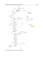
Operative Technique (Figures 8.1 to 8.3)
The patient is placed in a high lithotomy posi-
tion using candy-cane stirrups, and the lower
abdomen, perineum, and vagina are prepped
and draped. A Scott ring retractor (Lone Star
Medical Products) is placed to aid exposure.
A 16-French urethral Foley catheter with the
balloon infl ated to 10 cc allows for identifi cation
of the bladder neck by palpation, and this site is
marked transversely with an ink pen to identify
the distal limits of the anterior vaginal wall
plate. The cervix or vaginal apex/cuff (if prior
hysterectomy) will comprise the proximal limit
of the plate, and marking sutures are placed at
this site in the midline and 1.5 to 2 cm on either
side. A longitudinal incision is initiated 1.5 cm
lateral to the bladder neck on the vaginal wall
and extended proximally to the apical/cervix
marking suture on the same side. The same
incision is made on the contralateral side. These
incisions deviate slightly lateral more proxi-
mally, resulting in a trapezoidal confi guration
of the anterior vaginal wall plate. The dimen-
sions of the anterior vaginal wall between the
incisions are approximately 3 cm in width and
4 to 8 cm in length, accounting for normal ana-
tomic variability. In the presence of a moderate
98 Vaginal Surgery for Incontinence and Prolapse
2 cm
SP
60˚
Preoperative straining
5 cm
60˚
Preoperative straining
SP
BN
Midline of
cystocele
1
.
5
-
2
c
m
1
.
5
-
2
c
m
1
.
5
-
2
c
m
1
.
5
-
2
c
m
Vaginal apex
BN
Midline of
cystocele
1
.
5
-
2
c
m
1
.
5
-
2
c
m
1
.
5
-
2
c
m
1
.
5
-
2
c
m
Vaginal apex
Figure 8.1. Surgical technique of the anterior vaginal wall suspension.
Intraoperative photos and pre- and postoperative standing voiding cys-
tourethrogram (VCUG) images illustrating the technique and anatomic
outcomes after anterior vaginal wall suspension (AVWS) performed for
incontinence with urethral hypermobility in the presence of a small/
moderate-size (left) or large (right) cystocele.
A,B: lateral standing VCUG images objectively demonstrate urethral
hypermobility on resting and straining lateral views (straining view only
shown). The urethral angle at straining is indicated. A moderate-size
cystocele is shown in A and a large cystocele in B. Cystocele grade on
VCUG is measured on the straining views (grade I <2 cm, grade II 2–5 cm,
grade III >5 cm below the symphysis pubis [SP]).
C,D: Superimposed markings on preoperative photos indicate key ana-
tomic landmarks: the bladder neck (BN), vaginal apex, and lateral vaginal
sulci. The midline of the anterior vaginal wall between the lateral sulci is
marked from the BN to vaginal apex to allow precise measurement of
the in situ anterior vaginal wall plate, which is created between two
longitudinal incisions, each 1.5 to 2 cm lateral to the midline. For a small/
moderate size cystocele, only the lateral incisions are made, while for a
large cystocele, redundant AVW skin (between the lateral sulci and lon-
gitudinal incisions of the in situ AVW plate) is marked for excision so as
to prevent redundancy after closure.
Transvaginal Surgery for Stress Urinary Incontinence 99
3-4 cm
3-4 cm
vaginal apex
b
a
1
.
5
-
2
c
m
M
i
d
l
i
n
e
o
f
v
a
g
i
n
a
1
.
5
-
2
c
m
3-4 cm
vaginal apex
Figure 8.1. E–H: The BN and vaginal apex are marked with a marking
pen and three chromic sutures (midline and 1.5–2 cm on either side),
respectively, for both small/moderate and large cystoceles (E). Redun-
dant lateral AVW skin between point a and b is excised bilaterally for
large cystoceles only (F,H). The lateral aspect of the incision is under-
mined only enough to facilitate closure of the incision later, while no
undermining is performed medially on the in situ AVW plate.
I: The final midline AVW plate measures 3 to 4 cm in total width and
extends from the BN to vaginal apex. The AVW plate is the same for the
small/moderate-size cystocele and large cystocele, regardless of whether
of not lateral AVW excision is performed.
b
a
vaginal apex
BN
100 Vaginal Surgery for Incontinence and Prolapse
1.5-2 cm
symphysis
Figure 8.1. J,K: Four No. 1 polypropylene sutures (taper CT-2 needle,
Ethicon D-4412) are placed, one in each equal quadrant of the in situ
AVW plate (J). Each suture is passed 3 to 5 mm below the AVW epithelium
in a broad, overlapping manner, such that the entire AVW plate is
included in the four sutures. Using a double-pronged ligature carrier,
each suture is transferred through a 2- to 3-cm suprapubic incision after
development of the retropubic space using finger dissection (K). Sutures
are tied without tension. A right-angled clamp ensures that each knot is
tied at a level of 1.5–2 cm above the insertion of the tendinous rectus
sheath on the symphysis pubis.
L–O: Final anatomic results shown on intraoperative photos and stand-
ing lateral VCUG views (at 6 months postoperative) demonstrate a well-
supported AVW through its entire length for both small/moderate-size
(left) and large cystoceles (right). Note the anatomic vaginal axis, absence
of secondary cystocele and correction of urethral hypermobility on
VCUG.
Transvaginal Surgery for Stress Urinary Incontinence 101
SP
18˚
Postoperative straining
SP
Postoperative straining
15˚
Figure 8.1. Continued.
3
-
4
c
m
3
-
4
c
m
Apex
BN
bb
aa
small
post-repair
large
Figure 8.2. Surgical technique of the anterior vaginal wall suspen-
sion—schematic. The surgical setup for the anterior vaginal wall suspen-
sion (AVWS) with retractor in place is illustrated on the left for the case
of a large cystocele in which lateral AVW skin is marked for excision
between points a and b. The final AVW plate is 3 to 4 cm in total width
and extends from the bladder neck (BN) to vaginal apex. On the right,
cross-sectional diagrams illustrate the AVWS with and without resection
of lateral AVW skin (between a and b) for small and large cystoceles. The
final AVW plate and final anatomic result with suspension sutures in
place are the same in both cases.
102 Vaginal Surgery for Incontinence and Prolapse
to large cystocele (Baden-Walker grade >II or
POP-Q stage >II) with lateral detachments and
central defect, excision of a segment of vaginal
wall lateral to each of the two longitudinal inci-
sions (corresponding to the lateral detachment)
is performed to eliminate redundancy and
reconstitute the lateral vaginal sulci. It is impor-
tant to preserve 0.5 to 1 cm of anterior vaginal
wall lateral to each incision to ensure a tension-
free closure.
BN
Cervix 3 cm
proximal
to introitus
BN
Rectus fascia
PVS graft
}
Cervix
Cervix
Figure 8.3. Versatility of the anterior vaginal wall suspension, with
uterine preservation and “classic” pubovaginal sling for intrinsic sphinc-
teric deficiency. Operative photos demonstrate the use of the anterior
vaginal wall suspension (AVWS) in a patient with stress urinary inconti-
nence due to intrinsic sphincteric deficiency as well as a moderate-size
cystocele. In addition, this patient desired uterine preservation. Preopera-
tive photos show a grade II/III cystocele (A; BN, bladder neck) and cervical
position after placing distal traction (B). The cervix does not descend
beyond the distal third of the vagina—the criterion for uterine preserva-
tion. The upper anterior vaginal wall (cystocele) is supported by a single
pair of helically placed sutures, which incorporate the cardinal ligaments
at the proximal aspect. Distally, an autologous fasical pubovaginal sling is
placed beneath an inverted-U vaginal flap, which extends off the proximal
AVWS longitudinal incisions bilaterally. Both pairs of sutures are passed
suprapubically. Final anatomic result demonstrating a well-supported
AVW through its entire length, from the bladder neck to the cervix (D).
PVS, Pubovaginal Sling.
Transvaginal Surgery for Stress Urinary Incontinence 103
In preparation for placement of the suspen-
sion sutures into the in situ anterior vaginal wall
plate, the plate, is divided into equal quadrants
using an ink pen. One suture (No. 1 polypropyl-
ene on a taper CT-2 needle, Ethicon D-4412, New
Brunswick, New Jersey (USA)) is placed in each
quadrant. For a right-handed surgeon, the fi rst
suture is placed at the proximal extent of the
vaginal wall incision on the left side into the scar
at the vaginal cuff or cardinal ligament, and run
in a helical fashion toward the mid-vagina (half
the length of the vaginal wall plate). The helical
bites are taken to the midline of the vaginal plate
and securely incorporated approximately 3 to
5 mm deep to the epithelium. Each subsequent
bite overlaps the preceding one, for a total of
three to four passes per suture on average. The
second suture on the same side is placed at the
mid-vagina, overlapping the end of the fi rst
suture, and run in the same manner to the distal
extent of the lateral vaginal wall incision. On the
right side, two additional sutures are placed sim-
ilarly, but starting at the distal extent of the inci-
sion (bladder neck) and running proximally.
A 2 to 3 cm wide suprapubic incision is made
approximately one fi ngerbreadth above the
pubic symphysis in the midline. The incision is
deepened to the level of the tendinous insertion
of the rectus fascia on the symphysis pubis. The
subcutaneous space is developed enough to
allow for accurate retropubic passage of the
ligature carrier (guiding the instrument with a
retropubic fi nger tip), and for soft tissue cover-
age of the suture knots. It is important to avoid
more lateral dissection to prevent injury to the
genital branch of the genitofemoral and ilioin-
guinal nerves.
To minimize bleeding, development of the
retropubic space is performed after both place-
ment of the vaginal wall plate sutures and cre-
ation of the suprapubic incision have been
completed. With the bladder on full drainage,
the dissection is started laterally at the level of
the bladder neck. Blunt or sharp dissection may
be used, although in our experience the former
is adequate unless there has been extensive scar-
ring from prior anti-incontinence surgery. After
the endopelvic fascia is perforated, the retropu-
bic space is developed further using a sweeping
motion of the index fi nger in a lateral-to-medial
direction. Because one follows the plane of the
tear that resulted in the lateral defect, develop-
ment of this space proceeds without signifi cant
resistance and usually with minimal bleeding.
After prior surgery, such as a Burch or MMK
bladder neck suspension, the risk of bladder
injury is greater during both the retropubic dis-
section and passage of the ligature carrier. When
the space is adequately developed, the dissecting
fi nger can be palpated through the suprapubic
incision with only rectus fascia and muscle
intervening, thus allowing safe passage of the
Raz double-pronged ligature carrier under fi n-
gertip-guidance. The ends of the suspension
sutures are threaded through the eyes of the liga-
ture carrier, which is then withdrawn suprapubi-
cally. The procedure is performed twice on each
side, allowing transfer of all four suspension
sutures from the vagina to the suprapubic inci-
sion. Although a Raz double-pronged ligature
carrier is preferred, when the patient is signifi -
cantly obese or there is dense scarring either in
the abdominal wall or retropubically, a sharper,
single-pronged instrument (Raz or Stamey) may
be necessary.
After administration of intravenous indigo
carmine, cystoscopy with 30- and 70-degree
lenses is performed to confi rm the absence of
bladder perforation or ureteral injury. If sutures
are present in the bladder, they are pulled out
via the vaginal incision and repositioned. In case
of a small bladder perforation, conservative
management with simple urethral Foley drain-
age for several days is recommended. A larger
perforation may necessitate a multiple layer
closure in addition to placement of a suprapubic
catheter. Duration of drainage should take into
consideration the location of the perforation.
There is a higher risk of vesicovaginal fi stula
formation in more dependent cystotomies close
to the bladder neck than for more lateral and
bladder dome injuries. The former situation is
one in which interposition of a fat pad graft may
be considered after primary closure and verifi ca-
tion of water-tightness. When there is intravesi-
cal bleeding, endoscopic fulguration is usually
suffi cient along with upsizing the Foley catheter
or changing to a three-way irrigation catheter in
preparation for potential bladder irrigation to
prevent clot formation.
The vaginal wall incisions are closed with
running 2–0 or 3–0 absorbable sutures. The sus-
pension sutures are tied suprapubically 1.5 to
2 cm above the rectus fascia without tension.
This is achieved by securing the suture at the
appropriate level with a rubber-shod right-angle
clamp. The goal is to support the vaginal wall
plate in a horizontal position, preserving the
104 Vaginal Surgery for Incontinence and Prolapse
130-degree, “banana-shaped” normal vaginal
axis. In this way, restoration of anatomy without
overcorrection of either the bladder neck or
apex is achieved and risk of secondary entero-
cele or rectocele is minimized. An antibiotic-
soaked vaginal pack is inserted and the
suprapubic incision is closed, ensuring that the
polypropylene knots are well buried. On postop-
erative day one, the vaginal pack is removed and
a voiding trial is performed before discharging
the patient the same day.
Outcomes and Advantages
The effi cacy of the AVWS has been assessed
with both subjective and objective outcome
measures. In 2000, Lemack and Zimmern (44)
reported the results of questionnaire analysis
with a mean follow-up duration of 25 months in
61 of 102 patients who responded. Subjective
cure or improvement of stress incontinence was
obtained in 77%, and response to a quality of
life question (analog scale response to the ques-
tion “If you were to spend the rest of your life
with your urinary condition just the way it is
now, how would you feel about that?”) was sig-
nifi cantly improved compared to preopera-
tively, decreasing from a median score of 6.7 to
2 (0 = pleased, 10 = terrible). De novo inconti-
nence occurred in 8% and diuretic use was the
only poor prognostic indicator. Objective out-
comes for the AVWS were assessed using the
standing voiding cystourethrogram (VCUG) in
a study by Showalter et al (42), in which VCUG
fi ndings were compared in 76 continent con-
trols and 52 women who had undergone an
AVWS for urethral hypermobility and concom-
itant grade I or II cystocele. At 3 to 6 months
postoperatively, there was no difference in ure-
thral angle between the two groups, and a sig-
nifi cant reduction in the lateral cystocele height
was observed, comparable to that in a group of
36 women who had undergone conventional
anterior colporrhaphy for grade III cystocele.
Lemack and Zimmern (45) reported on sexual
function following AVWS with or without pos-
terior repair of rectocele. Using a mailed ques-
tionnaire developed by the authors with a 60%
response rate, questionnaire information was
obtained for 29 patients who underwent AVWS
alone and 27 who underwent both AVWS and
posterior repair. There was no difference in the
proportion of sexually active women preopera-
tively compared to postoperatively and a 20%
postoperative rate of dyspareunia was noted
compared to 29% preoperatively. Overall, the
AVWS did not adversely affect sexual function
and symptomatic vaginal narrowing was rare.
Beyond the advantages of vaginal procedures
over abdominal procedures described elsewhere
in this chapter, there are many advantages spe-
cifi c to the AVWS that derive from the tech-
nique’s rationale. First, the procedure restores
anterior vaginal wall anatomy and eliminates
UHM without producing secondary anatomic
defects. Although prevention of secondary pro-
lapse initially depends on accurate preoperative
staging (i.e., identifi cation of existing or
potential defects), attention intraoperatively to
avoid alteration of the vaginal axis by providing
support with minimal tension to the entire
length of the anterior vaginal wall is critical to
restoration of anatomy. The VCUG outcomes
following AVWS have objectively demonstrated
no difference in urethral angle and bladder base
descent between cystograms of postoperative
patients and age-matched controls (42). In terms
of restoring function, the midterm continence
results of the AVWS are comparable to the gold
standard of bladder neck suspensions, the Burch
colposuspension. Like the Burch procedure, the
vaginal wall support of the AVWS is broadly
based, secured to a strong anchor (the rectus
muscle tendon as it inserts on the posterior
pubic symphysis), and involves retropubic dis-
section to further enhance vaginal support by
promoting scar formation. Of equal importance
is that voiding function is not sacrifi ced at the
expense of establishing continence. This is
achieved by (1) more lateral placement of the
vaginal suspension sutures (similar to the Burch
procedure) so as to avoid outlet obstruction (as
in the MMK), and (2) providing support to the
upper vagina to avoid a secondary angulation at
the urethrovesical junction. Although results of
the AVWS thus far indicate a low reoperation
rate for incontinence and cystocele, it is impor-
tant to recognize that secondary vaginal proce-
dures (e.g., conventional pubovaginal sling) or
abdominal (e.g., mesh sacrocolpopexy) are not
made more diffi cult after AWVS as the planes of
dissection, particularly the vesicovaginal space,
are not signifi cantly altered.
Preservation of sexual function is facilitated
by preserving normal vaginal confi guration—
not only axis, but also length and width.
Vaginal support without overtensioning, both
Transvaginal Surgery for Stress Urinary Incontinence 105
proximally and distally, allows for a normal
vaginal axis, whereas vaginal narrowing and
shortening are avoided by minimizing vaginal
skin excision or advancement and midline plica-
tion, which contrasts to other procedures, par-
ticularly the anterior colporrhaphy. The option
of uterine support in the appropriate patient
allows fl exibility in the decision for or against
vaginal hysterectomy. Finally, the AVWS pro-
vides a relatively cost-effective procedure based
on operative materials, procedure time (approx-
imately 1 hour), and patient recovery. In this
day and age when the use of new technology is
often at odds with cost-effi ciency demands, a
technique such as the AVWS, which uses no
expensive materials such as synthetic grafts, het-
erologous materials, or bone anchors should be
a welcome addition to the surgical options avail-
able to treat incontinence and cystocele.
Although the procedures utilizing in situ ante-
rior vaginal wall to treat incontinence and ante-
rior prolapse as originally described by Raz in
the 1980s are less commonly used today, these
basic concepts have been applied to procedures
other than the AVWS described in more con-
temporary series (41,46–53). These “vaginal wall
slings” and “four-corner suspensions,” despite
the common name, can vary considerably in
both technical detail and specifi c clinical indica-
tions depending on the author. For example, Su
et al (46) applied a modifi ed Raz vaginal wall
sling with a broader suture anchor and no retro-
pubic dissection to patients with recurrent
genuine stress urinary incontinence without
urethral hypermobility, whereas Mikhail and
coworkers (53) used a different modifi cation of
the Raz vaginal wall sling, eliminating the vaginal
U advancement fl ap, in patients with primary
genuine stress urinary incontinence and ure-
thral hypermobility. Others, such as Kaplan et al
(50,51) and Appell’s group (41) have reported on
results after the Raz vaginal wall sling and “in
situ sling,” respectively, in series that included
both patients with anatomic stress incontinence
and intrinsic sphincteric defi ciency. Variations
in outcome measures, in addition to those in
technique and indications, make direct com-
parison of effi cacy diffi cult. Generally, however,
these series suggest that continence outcomes
are best in women with mild to moderate stress
incontinence owing to either urethral hypermo-
bility or ISD, whereas anterior prolapse results
are best in those with low- to moderate-grade
cystocele. Urodynamic prognostic factors have
been identifi ed by several authors. For example,
Goldman et al (41) found that a Valsalva leak
point pressure of greater than 50 cmH
2
O was
associated with a continence success rate of 93%
after the in situ sling, which was later supported
by Kilicarslan et al (52), who found that a
Valsalva leak point pressure ≥50 cmH
2
O and
maximum urethral closure pressure ≥30 cmH
2
O
were associated with a 91% success rate after the
in situ sling.
Conclusion
The AVWS is a reliable surgical option for treat-
ing stress urinary incontinence due to urethral
hypermobility alone or associated with a
cystocele. The transvaginal approach has clear
advantages with respect to performing concom-
itant vaginal procedures as well as short dura-
tion of recovery and minimal overall morbidity.
In addition, the procedure is short, simple, easy
to teach, and cost-effective. The midterm conti-
nence and cystocele outcomes are comparable
to more well-established bladder neck suspen-
sion techniques, and longer-term outcome
results are forthcoming.
References
1. Chaikin DC, Rosenthal J, Blaivas JG. Pubovaginal
fascial sling for all types of stress urinary incontinence:
long-term analysis. J Urol 1998;160:1312–1316.
2. Cross CA, Cespedes RD, McGuire EJ. Our experience
with pubovaginal slings in patients with stress urinary
incontinence. J Urol 1998;159:1195–1198.
3. Pereyra AJ. A simplifi ed surgical procedure for the cor-
rection of stress urinary incontinence in women. West
J Obstet Gynaecol 1959;67:223–226.
4. Zimmern PE, Hadley HR, Leach GE, Raz S. Transvagi-
nal closure of the bladder neck and placement of
a suprapubic catheter for destroyed urethra after
long-term indwelling catheterization. J Urol 1985;134:
554–557.
5. Pereyra AJ, Lebherz TB. Combined urethrovesical
suspension and vaginourethroplasty for correction of
urinary stress incontinence. Obstet Gynecol 1967;30:
537–546.
6. Crist T, Singleton H, Robertson W. Urethrovesical
needle suspension: postoperative loss of vesical neck
support demonstrated by chain cysography. Obstet
Gynecol 1969;34:489–494.
7. Kursh ED, Wainstein M, Persky L. The Pereyra
procedure and urinary stress incontinence. J Urol
1972;108:591–593.
8. Pereyra A, Lebherz T. The revised Pereyra procedure.
In: Buchsbaum H, ed. Gynecologic and Obstetric
Urology. Philadelphia: WB Saunders, 1978:208–222.
106 Vaginal Surgery for Incontinence and Prolapse
9. Pereyra AJ, Lebherz TB, Growdon WA, Powers JA.
Pubourethral supports in perspective: modifi ed pereyra
procedure for urinary incontinence. Obstet Gynecol
1982;59:643–648.
10. Raz S. Modifi ed bladder neck suspension for female
stress incontinence. Urology 1981;17:82–85.
11. Raz S, Sussman EM, Erickson DB, Bregg KJ, Nitti VW.
The Raz bladder neck suspension: results in 206
patients. J Urol 1992;148:845–850.
12. Leach GE. Bone fi xation technique for transvaginal
needle suspension. Urology 1988;31:388–390.
13. Trockman BA, Leach GE, Hamilton J, Sakamoto M,
Santiago L, Zimmern PE. Modifi ed Pereyra bladder neck
suspension: 10-year mean followup using outcomes
analysis in 125 patients. J Urol 1995;154:1841–1847.
14. Kelly MJ, Knielsen K, Bruskewitz R, Roskamp D, Leach
GE. Symptom analysis of patients undergoing modifi ed
Pereyra bladder neck suspension for stress urinary
incontinence. Pre- and postoperative fi ndings. Urology
1991;37:213–219.
15. Korman HJ, Sirls LT, Kirkemo AK. Success rate of
modifi ed Pereyra bladder neck suspension determined
by outcomes analysis. J Urol 1994;152:1453–1457.
16. Sirls LT, Keoleian CM, Korman HJ, Kirkemo AK. The
effect of study methodology on reported success rates
of the modifi ed Pereyra bladder neck suspension.
J Urol 1995;154:1732–1735.
17. Stamey TA. Endoscopic suspension of the vesical neck
for urinary incontinence. Surg Gynecol Obstet 1973;
136:547–554.
18. Ginsberg D, Rovner E, Raz S. Stamey and gittes bladder
neck suspension, In: Graham S Jr, ed. Glenn’s Urologic
Surgery. Philadelphia: Lippincott-Raven, 1998:313–335.
19. Stothers L, Raz S, Chopra A. Stamey needle suspension
for female stress incontinence. In: Raz S, ed. Female
Urology. Philadelphia: WB Saunders, 1996:333–337.
20. Stamey TA. Endoscopic suspension of the vesical neck
for urinary incontinence in females. Report on 203 con-
secutive patients. Ann Surg 1980;192:465–471.
21. Ashken MH. Follow-up results with the Stamey opera-
tion for stress incontinence of urine. Br J Urol 1990;
65:168–169.
22. Gaum L, Ricciotti NA, Fair WR. Endoscopic bladder
neck suspension for stress urinary incontinence. J Urol
1984;132:1119–1121.
23. Hilton P, Mayne CJ. The Stamey endoscopic bladder
neck suspension: a clinical and urodynamic investiga-
tion, including actuarial follow-up over four years. Br
J Obstet Gynaecol 1991;98:1141–1149.
24. Kelly MJ, Leach GE. Long term results of bladder neck
suspension procedures. Problems Urol 1991;5:94.
25. Spencer JR, O’Conor VJ Jr, Schaeffer AJ. A comparison
of endoscopic suspension of the vesical neck with
suprapubic vesicourethropexy for treatment of stress
urinary incontinence. J Urol 1987;137:411–415.
26. O’Sullivan DC, Chilton CP, Munson KW. Should
Stamey colposuspension be our primary surgery for
stress incontinence? Br J Urol 1995;75:457–460.
27. Knispel HH, Klan R, Siegman K, Miller K. Results of the
Stamey procedure in 251 consecutive patients with
genuine urinary stress incontinence (Abstract 812).
J Urol 1995;153:431A.
28. Clemens JQ, Stern JA, Bushman WA, Schaeffer AJ.
Long-term results of the Stamey bladder neck
suspension: direct comparison with the Marshall-
Marchetti-Krantz procedure. J Urol 1998;160:372–376.
29. Wang AC. Burch colposuspension vs. Stamey bladder
neck suspension. A comparison of complications with
special emphasis on detrusor instability and voiding
dysfunction. J Reprod Med 1996;41:529–533.
30. Gittes RF, Loughlin KR. No-incision pubovaginal
suspension for stress incontinence. J Urol 1987;138:
568–570.
31. Loughlin KR, Whitmore WF III, Gittes RF, Richie JP.
Review of an 8-year experience with modifi cations of
endoscopic suspension of the bladder neck for female
stress urinary incontinence. J Urol 1990;143:44–45.
32. Kil PJ, Hoekstra JW, van der Meijden AP, Smans AJ,
Theeuwes AG, Schreinemachers LM. Transvaginal
ultrasonography and urodynamic evaluation after
suspension operations: comparison among the Gittes,
Stamey and Burch suspensions. J Urol 1991;146:132–
136.
33. Elkabir JJ, Mee AD. Long-term evaluation of the Gittes
procedure for urinary stress incontinence. J Urol
1998;159:1203–1205.
34. Benderev TV. A modifi ed percutaneous outpatient
bladder neck suspension system. J Urol 1994;152:
2316–2320.
35. Leach GE, Appell R. Percutaneous bladder neck
suspension. Urol Clin North Am 1996;23:511–516.
36. Schultheiss D, Hofner K, Oelke M, Grunewald V, Jonas
U. Does bone anchor fi xation improve the outcome of
percutaneous bladder neck suspension in female stress
urinary incontinence? Br J Urol 1998;82:192–195.
37. Madjar S, Beyar M, Nativ O. Transvaginal bone
anchored sling. Urology 2000;55:422–426.
38. Bruskewitz RC, Nielsen KT, Graversen PH, Saville WD,
Gasser TC. Bladder neck suspension material investi-
gated in a rabbit model. J Urol 1989;142:1361–1363.
39. Zimmern PE, Leach GE, Sirls L. Four-corner bladder
neck suspension. In: Leach GE ed. Atlas of the Urologic
Clinics of North America. Philadelphia: WB Saunders,
1995:29–36.
40. Dmochowski RR, Zimmern PE, Ganabathi K, Sirls L,
Leach GE. Role of the four-corner bladder neck suspen-
sion to correct stress incontinence with a mild to mod-
erate cystocele. Urology 1997;49:35–40.
41. Goldman HB, Rackley RR, Appell RA. The in situ ante-
rior vaginal wall sling: predictors of success. J Urol
2001;166:2259–2262.
42. Showalter PR, Zimmern PE, Roehrborn CG, Lemack
GE. Standing cystourethrogram: an outcome measure
after anti-incontinence procedures and cystocele repair
in women. Urology 2001;58:33–37.
43. Raz S, Stothers L, Chopra A. Raz techniques for ante-
rior vaginal wall repair. In: Raz S, ed. Philadelphia:
WB Saunders, Female Urology, Second Edition 1996:
344–366.
44. Lemack GE, Zimmern PE. Questionnaire-based
outcome after anterior vaginal wall suspension for
stress urinary incontinence. J Urology 2000;163:73
(abstract 321).
45. Lemack GE, Zimmern PE. Sexual function after vaginal
surgery for stress incontinence: results of a mailed
questionnaire. Urology 2000;56:223–227.
46. Su TH, Huang JP, Wang YL, Yang JM, Wei HJ, Huang
CL. Is modifi ed in situ vaginal wall sling operation the
treatment of choice for recurrent genuine stress incon-
tinence? J Urol 1999;162:2073–2077.
47. Appell RA. In situ vaginal wall sling. Urology 2000;
56:499–503.
Transvaginal Surgery for Stress Urinary Incontinence 107
48. Constantini E, Pajoncini C, Zucchi A, et al. Four-corner
colposuspension: clinical and functional results. Int
Urogynecol J 2003;14:113–118.
49. Couillard DR, Deckard-Janatpour KA, Stone AR. The
vaginal wall sling: a compressive suspension procedure
for recurrent incontinence in elderly patients. Urology
1994;43:203–208.
50. Kaplan SA, Santarosa RP, Te AE. Comparison of fascial
and vaginal wall slings in the management of intrinsic
sphincter defi ciency. Urology 1996;47:885–889.
51. Kaplan SA, Te AE, Young GP, Andrade A, Cabelin MA,
Ikeguchi EF. Prospective analysis of 373 consecutive
women with stress urinary incontinence treated with a
vaginal wall sling: the Columbia-Cornell University
experience. J Urol 2000;164:1623–1627.
52. Kilicarslan H, Gokce G, Ayan S, Guvenal T, Kaya K,
Gultekin EY. Predictors of outcome after in situ ante-
rior vaginal wall sling surgery. Int Urogynecol J Pelvic
Floor Dysfunct 2003;14:339–341.
53. Mikhail MS, Rosa H, Packer P, Palan P, Lazarou G. A
modifi ed vaginal wall patch sling technique as a fi rst-
line surgical approach for genuine stress incontinence
with urethral hypermobility: long-term follow up. Int
Urogynecol J Pelvic Floor Dysfunct 2004;15:132–136.