Vaginal Surgery for Incontinence and Prolapse - part 5 pdf
Bạn đang xem bản rút gọn của tài liệu. Xem và tải ngay bản đầy đủ của tài liệu tại đây (714.24 KB, 30 trang )
114 Vaginal Surgery for Incontinence and Prolapse
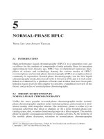
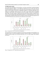
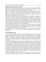
Figure 9.3. Traditional technique for sling placement. A: Retropubic
space is entered laterally through the vaginal incision. B: Curved clamp
passed through the retropubic space with direct finger guidance. C: Sling
pulled up to the suprapubic incision by clamp (From Hinman F. Atlas of
Urologic Surgery, 2nd ed., pp. 566–567. Copyright 1998, with permission
from Elsevier.)
Stress Urinary Incontinence Secondary to Intrinsic Sphincteric Deficiency 115
synthetic mesh to avoid the morbidity of fascial
harvest, varying lengths of slings, from full-
length to patch grafts, and different sites of
fi xation for the sling, such as Cooper’s ligament,
suprapubic or transvaginal pubic bone anchor
fi xation, and passive fi xation by tissue adher-
ence to the mesh within the retropubic space or
obturator foramen. Currently there are no ran-
domized trials comparing the different varia-
tions of slings with respect to treatment of ISD.
The choices of which sling material and which
method of sling placement are at the discretion
of the surgeon (130).
For all types of SUI, urethral hypermobility,
and ISD, autologous fascial pubovaginal slings
are reported to have cure rates of 70% to
90% and cure/improved rates of 85% to 95%
(15,16,18,23,24). Continence rates for patients
with pure ISD appear to be slightly lower than
that of patients with urethral hypermobility.
With a mean follow-up of 22 months, Cross and
colleagues (25) reported continence rates con-
fi rmed by videourodynamics of 96% (43/45) in
patients with preoperative urethral hypermobil-
ity vs. 89% (65/73) in patients with preoperative
ISD. With a mean follow-up of 51 months,
Morgan and associates (16) reported continence
rates of 91% for SUI due to urethral hypermobil-
ity vs. 84% for SUI owing to ISD. Results of
various sling procedures as treatment for SUI
owing to ISD are noted in Table 9.2.
Allograft/Xenograft Slings
To decrease operative time and avoid the
morbidity of autologous fascial harvest, the use
of cadaveric allografts and xenografts has
increased in recent years. The different nonau-
tologous grafts are listed in Table 9.3 (26). These
nonautologous grafts exhibit different elasticity
and tensile strength. Kubricht et al (27) found
that freeze-dried, gamma-irradiated cadaveric
fascia lata had a tensile strength twice that of
freeze-dried porcine small intestine submu-
cosa. When comparing freeze-dried cadaveric
fascia lata to solvent-dehydrated fascia lata,
Lemer et al (28) found the solvent-dehydrated
fascia to be signifi cantly stronger by tensiome-
try. Although it is apparent that these grafts
have different properties, it remains unclear
which provides the best long-term results for
sling surgery.
Results using nonautologous grafts for sling
surgery have been comparable to autologous
slings with short- to intermediate-term follow-
up. Brown and Govier (29) found a SUI cure rate
of 74% and cured/improved rate of 93% with a
mean follow-up of 12 months after freeze-dried
cadaveric fascia lata sling, which was not signifi -
cantly different from the 73% cured and 100%
cured/improved after autologous slings at the
same institution with mean follow-up of 44
months. In another comparison of allograft vs.
Table 9.2. Results for suburethral sling series as treatment for ISD
Sling Mean follow-up, %
Type of sling Author, year (ref.) material n months (range) success
Autologous Mason, 1996 (95) Rectus 63 12 (3–27) 94
Zaragoza, 1996 (23) Rectus 60 25 (11–34) 100
Barbalias, 1997 (96) Rectus 32 32 (30–38) 66
Hassouna, 1999 (98) Rectus 82 41 (6–96) 89
Kreder, 1996 (24) Rectus/FL 27 22 (9–32) 96
Haab, 1997 (81) Rectus/FL 37 48 (24–60) 73
Wright, 1998 (99) Rectus/FL 33 16 (15–28) 94
Richter, 2001 (100) Rectus/FL 57 42 (0.5–134) 84
Govier, 1997 (101) FL 30 14 (3–33) 70
Synthetic Barbalias, 1997 (97) PTFE 24 30 83
Staskin, 1997 (102) PTFE 122 24 88
Yamada, 2001 (103) PTFE 72 67 (50–75) 78
Morgan, 1985 (104) Marlex 208 >60 77
Rezapour, 2001 (40) TVT 49 48 86
Allograft Wright, 1998 (99) Cad FL 59 10 98
Ruiz, 2000 (105) Cad FL 105 18 93
FL, Fascia lata; PTFE, polytetrafluoroethylene; TVT, transvaginal tape.
116 Vaginal Surgery for Incontinence and Prolapse
autologous sling with 2 years’ minimum follow-
up, Flynn and Yap (30) found a cure rate of 71%
and cured/improved rate of 84% with mean
follow-up of 29 months in the allograft group vs.
77% cured rate and 90% cured/improved rate in
the autologous sling group with mean follow-
up of 44 months. The use of allograft in their
series resulted in less postoperative pain and
disability.
A question that has been raised concerning
the use of allografts in sling surgery is graft dura-
bility. Although most allograft sling series report
success rates comparable to autologous slings,
the length of follow-up in the allograft series has
been relatively short. Carbone and colleagues
(31) reported disappointing results in 154
patients treated with freeze-dried cadaveric
fascia lata and transvaginal bone anchor fi xa-
tion. They found a high SUI recurrence rate of
38% within 1 year and attributed these early
failures to cadaveric allograft degeneration based
on fi ndings at reoperation. Fitzgerald and asso-
ciates (32) noted that upon reoperating on
freeze-dried cadaveric fascial sling failures, the
slings had undergone some form of degenera-
tion or autolysis, and in some cases the sling
could not be identifi ed. Elliot and Boone (33)
found no evidence of rapid graft degeneration
following solvent-dehydrated cadaveric fascia
lata sling, with a 96% cured/improved rate using
12 months as the minimum follow-up. After per-
forming over 400 sling procedures using solvent-
dehydrated cadaveric fascia lata, the authors
have noted no evidence of rapid graft degenera-
tion as well. When comparing the various mate-
rials used in sling surgery after 12 weeks of
subcutaneous implantation in the rabbit model,
Dora et al (34) found that human cadaveric
fascia and porcine xenografts showed a marked
decrease (60–89%) in tensile strength and stiff-
ness, whereas polypropylene mesh and autolo-
gous fascia did not differ in tensile strength
from baseline. With intermediate-term out-
comes using nonautologous grafts for sling
surgery reported in most series as comparable
to that of autologous slings, the signifi cance of
these fi ndings is not known.
Another concern with the use of allografts has
been the risk of disease transmission. Measures
used to prevent disease transmission in tissue
allografts include donor screening and a multi-
step tissue sterilization process. Despite these
measures, the presence of intact DNA material
has been reported (35). Another potential
concern is for the transmission of prion disease,
such as Creutzfeldt-Jakob disease. Prions are
protein molecules that can resist conventional
means of sterilization. Although there is a theo-
retical risk, to date there have been no reported
cases of disease transmission with the use of
cadaveric allografts in continence surgery.
Synthetic Slings
In recent years the use of synthetic mesh slings
has gained popularity. Many of the early mesh
slings, such as Marlex, Mersilene, silicone, and
Protogen (Boston Scientifi c, Natwick, MA), were
shown to have increased complication rates,
such as urethral and vaginal erosions requiring
mesh removal (18,36,37). In 1996 Ulmsten and
associates (38) introduced the tension-free
vaginal tape (TVT) procedure as a sling proce-
dure performed with local anesthetic using a
loosely woven polypropylene mesh. This sling
Table 9.3. Allograft and xenograft materials used in sling surgery
Sling material Trade name (manufacturer) Processing technique
Cadaveric allografts Fascia lata FasLata (CR Bard, Inc., Murray Hill, New Jersey) Freeze-dried, gamma irradiated
Suspend (Mentor, Santa Barbara, CA) Solvent-dehydrated, gamma-irradiated
Decellularized dermis Duraderm (CR Bard, Inc., Murray Hill, New Jersey) Freeze-dried
Alloderm (LifeCell Corp., Branchburg, NJ) Freeze-dried
Acellular dermal matrix Repliform (Boston Scientific, Natick, Freeze-dried
Massachusetts)
Urogen (American Medical Systems, Inc., Gamma-irradiated
Minnetoka, MN)
Xenografts Acellular porcine dermis Dermatrix (Advanced UroScience, Saint-Paul,
MN)
Pelvicol (CR Bard, Inc., Murray Hill, New Jersey)
Acellular matrix from Stratasis (Cook Urological, Bloomington,
porcine small intestine IN)
submucosa Fortaflex (Organogenisis, Canton, MA)
Stress Urinary Incontinence Secondary to Intrinsic Sphincteric Deficiency 117
aims to re-create the pubourethral ligament
and support of the suburethral vagina, by its
placement at the mid-urethra without tension,
and does not require suture fi xation. It. With
follow-up out to 5 years in some series, the
success rates are similar to that of autologous
slings (39,40), and the previous problems of
mesh erosion have been minimal. Modifi cations
to the TVT procedure include the Suprapubic
arc (AMS; Minnetouka, MN) procedure, where
needle passage for sling placement is performed
from the suprapubic incisions down to the
vagina, and the newer transobturator slings, in
which there is no retropubic needle passage and
the ends of the mesh sling are brought through
the obturator foramen on either side.
Early results using a transobturator technique
are promising. Delorme and associates (41)
reported 91% cured and 100% cured/improved
rates for all types of SUI using the transobturator
technique (TOT) in 32 patients with a minimum
follow-up of 12 months (mean 17 months). In a
prospective randomized trial comparing 1-year
outcomes of TVT (31 patients) to transobturator
suburethral sling (30 patients), deTayrac et al
(42) found comparable cure rates, 84% for TVT
vs. 90% for TOT, with signifi cantly lower operat-
ing times, 15 minutes vs. 27 minutes, and lower
incidence of intraoperative bladder injuries, 0 vs.
10%, in the TOT group. Further discussion on
mid-urethral slings is provided in Chapter 10.
Bone-Anchored Slings
Another method for securing the suburethral
sling by transvaginal pubic bone anchor fi xa-
tion has been described (43). Advantages of
using transvaginal bone anchors include the
ability to perform a sling procedure completely
transvaginally without retropubic needle
passage, minimal postoperative pain, and a
consistent, stable point of fi xation. Nonautolo-
gous allograft or synthetic sling materials are
employed, obviating the need for fascial harvest.
The theoretical disadvantage of bone anchor
fi xation is the potential for osseous complica-
tions, such as osteitis pubis or osteomyelitis.
Results for the treatment of SUI using bone
anchor slings have been variable. As stated pre-
viously, Carbone and associates (31) experienced
a high early failure rate using freeze-dried cadav-
eric fascia lata. In a later report on their experi-
ence with transvaginal bone anchor gelatin-coated
Dacron sling, they reported a 95% cured/
improved rate for SUI, but patients with signifi -
cant ISD were excluded from this study (44).
Giberti and Rovida (45) reported on 63 patients
receiving gelatin-coated Dacron bone anchored
slings. With 17 months mean follow-up, the
cured rate was 82% and the cured/improved rate
was 91%, but they noted that all of the patients
with preoperative ISD failed.
In the authors’experience using solvent-
dehydrated, nonfrozen cadaveric fascia lata with
bone anchor fi xation in 330 patients with a mean
follow-up of 25 months (maximum follow-up of
63 months), the cured rate for all types of SUI
was 59% and the cured/improved rate was 80%.
When comparing those patients in our series
who had ISD preoperatively to those who did
not, with Intrinsic sphincteric defi ciency (ISD)
defi ned as VLPP < 50, the failure rate was 24%
vs. 18%, respectively. This difference was not
statistically signifi cant.
Complications of Suburethral Slings
The most common complication of suburethral
sling procedures is voiding dysfunction/inade-
quate bladder emptying requiring intermittent
catheterization or suprapubic catheterization
drainage to avoid urinary retention. These
symptoms are usually transient and resolve
within the fi rst week postoperatively. Prolonged
urinary retention >3 months postoperatively
has been reported to occur 2% to 10% in most
sling series, with a procedure to loosen an
obstructing sling or formal urethrolysis being
required in 1% to 5%.
Another common cause of postoperative mor-
bidity following sling surgery is urinary urgency/
urge incontinence. De novo urinary urgency has
been reported to occur in 5% to 30% of patients,
and de novo urge incontinence has been reported
in up to 10% of patients. The etiology of these
symptoms is not clear, but may include an unmask-
ing of undiagnosed preoperative detrusor overac-
tivity, a direct effect of increased bladder outlet
resistance on detrusor function, or denervation
from surgical dissection. These symptoms are
usually transient in the absence of overt bladder
outlet obstruction, and respond well to anticho-
linergic therapy and behavioral modifi cation/bio-
feedback. Interestingly, many patients who suffer
from mixed urinary incontinence preoperatively
have resolution of their urge incontinence follow-
ing sling surgery. Schrepferman and associates
(46) reported that after pubovaginal sling,
118 Vaginal Surgery for Incontinence and Prolapse
preoperative urge symptoms resolved in 91% of
patients with low pressure (<15 cmH
2
O) detrusor
instability preoperatively, in 32% of patients with
sensory instability (no unstable detrusor contrac-
tions) preoperatively, and in 28% of patients with
high pressure (>15 cmH
2
O) detrusor instability
preoperatively. In the authors’ experience, after
transvaginal suburethral sling, 35% of patients
with preoperative urge incontinence have persis-
tent urge incontinence postoperatively.
Injury to the urethra or bladder may occur dur-
ing suburethral sling surgery. Patients who have
had previous retropubic or anti-incontinence
operations are at increased risk. With careful
dissection, these complications can usually be
avoided. It is important that any injury to the
bladder or urethra during sling surgery is identi-
fi ed with intraoperative cystoscopy to prevent the
subsequent morbidities of erosion, fi stula forma-
tion, or infection. If a small, uncomplicated ure-
thral injury occurs in a patient with healthy
urethral tissues, it can be repaired primarily in
layers and the sling operation can be completed
followed by urethral catheter drainage. If the ure-
thral injury is more extensive or the patient has
poor tissues, such as those with previous radia-
tion, it is prudent to repair the urethra, augment
the repair with a Martius graft, and postpone sling
placement until healing has occurred. If a bladder
injury occurs during sling passage, on the side of
the injury, the sling is pulled back down into the
vaginal incision and sling passage is repeated.
The surgeon may choose to provide continuous
bladder drainage by suprapubic tube or urethral
catheter for 2 to 7 days postoperatively depending
on the degree of injury.
Some infrequent, but potentially serious com-
plications have been reported with the mid-ure-
thral polypropylene procedures that pass the
sling blindly through the retropubic space.
Because the vaginal and suprapubic dissections
are limited and passage of the sling through the
retropubic space is performed without direct
guidance, major vascular injuries (47), bowel
perforations (48–50), and seven deaths have
been reported by the manufacturer, six of which
were due to unrecognized bowel injury (51).
Kobashi and Govier (49) noted a mean decrease
in hematocrit level of 7.1% on the fi rst postop-
erative day following the SPARC procedure, and
4 of 140 patients (2.9%) required blood transfu-
sions postoperatively.
When using bone anchors for sling fi xation,
concern has been raised about the potential for
osseous complications. In our 5-year experience
of over 400 transvaginal bone anchor slings
using solvent-dehydrated cadaveric fascia lata
(52), we reported two cases of postoperative
osteitis pubis that resolved within 3 months with
conservative treatment, no cases of osteomyeli-
tis, and no bone anchors have required removal.
Infectious osseous complications experienced
previously with suprapubic bone anchor fi xation
for suspension procedures (53) have not been
experienced with transvaginal techniques.
Injectable Bulking Agents
As an alternative to open surgery, injectable
bulking agents have become a common therapy
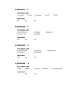
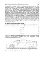
for SUI owing to ISD. The purpose of this form
of therapy is to increase the volume or bulk
within the proximal urethral wall, between the
external sphincter and bladder neck, thereby
compressing the urethral mucosa into the lumen
and providing better coaptation, thus increas-
ing outfl ow resistance (Figure 9.4). Historically,
bulking agents have not been used to treat ure-
thral hypermobility, as it provides no external
support to return the bladder neck or proximal
urethra to their normal anatomic position.
The fi rst reported periurethral injection
therapy was reported in 1938, when Murless (54)
injected sodium morrhuate (a sclerosing agent)
through the anterior vaginal wall in an attempt
to obtain scarring of the periurethral tissues to
achieve continence. Subsequently, Quackles (55)
injected paraffi n wax transperineally and Sachse
(56) injected Dondren (a sclerosing agent). In
these early experiences, results were not opti-
mistic and signifi cant complications, such as
pulmonary embolism and urethral sloughing,
were reported. In the last 30 years, with the
development of more suitable materials for
injection, like polytetrafl uoroethylene (PTFE)
(57), glutaraldehyde cross-linked collagen (58),
and carbon-coated zirconium beads (59), this
minimally invasive therapy has seen increas-
ingly widespread use.
Indications
The ideal candidate for injectable therapy has
been described as one with diminished urethral
function (ISD), a well-supported urethra,
and normal bladder function (60). Despite the
Stress Urinary Incontinence Secondary to Intrinsic Sphincteric Deficiency 119
general perception that injectable bulking
therapy should be used to treat isolated ISD,
several series have included patients with and
without urethral hypermobility and have
reported no signifi cant difference in outcomes
(61–64). Patients with comorbidities that are
prohibitive of, or who refuse, more invasive
surgery are good candidates for injectable
therapy, as well as those patients with recurrent
SUI and a well-supported urethra after a previ-
ous anti-incontinence operation.
In a randomized controlled trial comparing
collagen vs. open surgery (modifi ed Burch, sub-
urethral sling, or bladder neck suspension) as
fi rst-line treatment for SUI, Collet and associates
(65) found that at 12 months follow-up, collagen
was 53% successful vs. 72% success in the surgi-
cal group, with success being defi ned as 24-hour
pad test <2.5 g. They additionally noted no
statistical difference between the groups with
respect to improvement in quality of life or
patient satisfaction, whereas complications were
signifi cantly less frequent and severe in the col-
lagen group. Prior to conducting the trial, a large
survey of urologists and gynecologists revealed
that a 20% difference in results would be accept-
able for considering bulking therapy as fi rst-line
treatment for SUI.
Techniques of Injection
Prior to performing a proximal urethral bulking
procedure, the patient should have sterile urine
and be taught how to perform self-catheteriza-
tion in the event that urinary retention occurs
in the early postoperative period. The proce-
dure may be performed in the offi ce setting with
local anesthetic (topical and injectable lido-
caine), or in an ambulatory surgical center or
operating room if intravenous sedation is
preferred by the patient or the surgeon. Two
techniques for injection have been described:
transurethral and periurethral. The authors
routinely perform bulking procedures under
monitored sedation, providing optimal patient
comfort while avoiding patient movement
during needle placement and injection, using
the periurethral technique, which avoids
mucosal disruption and bulking agent extru-
sion through the injection site. Faeber and
colleagues (66) compared transurethral to peri-
urethral injection techniques and found no sig-
nifi cant difference in continence outcomes,
complications, or number of injections per
patient, but did note that a signifi cantly higher
volume of collagen was injected when the
procedure was performed periurethrally.
For transurethral injection the patient is
placed in the lithotomy position and a 12-degree,
blunt-tipped cystoscope with an injection needle
port is introduced into the patient’s urethra. A
syringe of the desired bulking agent is attached
to the needle and the needle is primed. The scope
is positioned at the mid-urethra, and rotated for
needle placement at the 4 o’clock position. The
needle is advanced with the bevel toward the
urethral lumen, and the urethral mucosa is
Figure 9.4. Anatomy and cystoscopic views of the bladder neck and urethra. (Courtesy of Carbon Medical Technologies, Inc., St. Paul, MN, with per-
mission.) A: Open bladder neck prior to injection of bulking material. B: Coaptation of the bladder neck and proximal urethra after injection.
120 Vaginal Surgery for Incontinence and Prolapse
punctured at a 45-degree angle until the bevel of
the needle is covered (Figure 9.5A). Keeping the
needle in place the scope is re-angled back paral-
lel with the urethra, and the needle is advanced
1 to 2 cm so that the tip is located in the submu-
cosa of the proximal urethra. The bulking mate-
rial is injected with consistent, moderate thumb
pressure on the plunger (Figure 9.5B). With
correct needle placement, the fl ow should be
even and smooth. Viewed cystoscopically, the
urethral mucosa should rise as the material
is introduced. Injection is continued until
the resulting submucosal bleb has crossed the
midline. If circumferential closure is not obtained
from the initial injection site, the procedure is
repeated at the 8 o’clock position. The objective
is to obtain complete coaptation of the urethral
mucosa when viewed cystoscopically with the
irrigation on. Care should be taken not to
advance the cystoscope proximal to the mid-
urethra once injection is initiated, so that
mucosal disruption is avoided. The bladder
may be drained by passing a well-lubricated,
10-French red rubber catheter.
For the periurethral technique, the patient is
placed in lithotomy position and the cystoscope
is introduced into the bladder. With the scope
held in the neutral position, parallel to the fl oor,
the periurethral groove is identifi ed approxi-
mately 0.5 to 1 cm lateral to the meatus. An 18-
gauge, 1.5-inch, angled needle is attached to a
syringe fi lled with normal saline or lidocaine
that will be used for hydrodissection. The needle
is then inserted at the 3 o’clock position in the
urethral groove and advanced 2 to 3 cm, keeping
the needle hub parallel to the scope (Figure
9.6A). The 15-degree angle of the needle guides
the tip into the correct submucosal plane. To
verify placement, the cystoscope is withdrawn to
the mid-urethra and the needle tip is wiggled,
causing tenting of the overlying urethral mucosa.
Hydrodissection is performed by injecting 1 to
2 cc of fl uid. A mucosal bleb should be visualized
during hydrodissection if the needle is in the
correct position. If no bleb is seen, the needle
should be withdrawn and repositioned. With
correct needle placement confi rmed, the needle
is held in place while switching the syringe to
one fi lled with bulking material. The material is
injected under direct cystoscopic visualization
as previously described with transurethral injec-
tion (Figure 9.6B). Once an adequate amount of
material has been delivered, a fi gure-of-eight
absorbable suture is placed around the needle
puncture site in the urethral groove. The suture
is tied down as the needle is removed to prevent
extrusion of bulking material and bleeding from
the puncture site.
Bulking Agents
Currently, the ideal bulking agent has not
been found. The ideal agent should be hypoal-
lergenic, biocompatible, nonimmunogenic,
noncarcinogenic, and durable without biodeg-
radation or migration (67). Other important
considerations for bulking agents include ease
Figure 9.5. Transurethral injection technique. (Courtesy of Carbon Medical Technologies, Inc., St Paul, MN, with permission.) A: Needle puncture at
the mid-urethra, at a 45-degree angle. B: Needle advanced submucosally, parallel to the urethra, to the proximal urethra for injection.
Stress Urinary Incontinence Secondary to Intrinsic Sphincteric Deficiency 121
of injection (agents that require higher pres-
sures to inject, have higher extravasation rates),
requirement for specialized injection equip-
ment, need for preparation or special handling
of the material before injection, and cost. A list
of approved and investigational injectable
agents is found in Table 9.4. Presently, autolo-
gous fat, cross-linked collagen, and carbon-
coated beads are the only Food and Drug
Administration (FDA)-approved bulking agents
for the treatment of SUI owing to ISD in the
United States.
Autologous Fat
In 1989, the periurethral injection of autologous
fat was fi rst reported by Gonzalez et al (68).
Using a liposuction technique, subcutaneous fat
was harvested from the anterior abdominal
wall, washed to remove debris, and injected
using a transurethral technique. Autologous fat
has the advantages of being biocompatible,
readily available, and inexpensive. The primary
disadvantage of using autologous fat as a
bulking agent appears to be poor durability
related to a high rate of resorption. Within 6
months, 50% to 60% volume loss of free fat
grafts has been demonstrated by magnetic reso-
nance imaging (69). This rapid resorption rate
is thought to be a result of inadequate neovas-
cularity to the central portion of the graft and
destruction of the normal adipocyte architec-
ture during the retrieval and washing process
(70,71). Other available bulking agents have
been shown to be more effective for the
Figure 9.6. Periurethral injection technique. (Courtesy of Carbon Medical Technologies, Inc., St Paul, MN, with permission.) A: Needle puncture in
the groove lateral to the urethral meatus. B: With needle placement confirmed, bulking material is injected.
Table 9.4. Currently available and investigational injectable bulking agents
Agent Trade name Company Approval status
Autologous fat
Bovine cross-linked collage Contigen Bard, Covington, GA FDA approved 1993
Carbon-coated zirconium beads Durasphere Boston Scientific, Boston, MA FDA approved 1999, no longer available
Graphite-coated zirconium beads Durasphere EXP Boston Scientific, Boston, MA FDA approved 2003
PTFE (Teflon) Urethrin Mentor, Santa Barbara, CA Approved in Canada/Europe
Silicone Macroplastique Uroplasty, Minneapolis, MN FDA trials ongoing
Dimethylsulfoxide and ethylene Uryx Genyx Medical Inc., FDA submission
vinyl alcohol copolymer San Diego, CA
Hyaluronic acid and dextranomer Zuidex Q-med, Uppsala, Sweden FDA trials ongoing
microspheres
Calcium hydroxyapatite Coaptite Bioform, Franksville, WS FDA trials ongoing
FDA, Food and Drug Administration; PTFE, polytetrafluoroethylene.
122 Vaginal Surgery for Incontinence and Prolapse
treatment of female SUI, making the use of
autologous fat less desirable.
Cross-Linked Collagen
Glutaraldehyde cross-linked (GAX)-collagen is
derived from bovine dermis, purifi ed into an
acellular derivative, enzymatically treated to
eliminate antigenicity, and fi nally cross-linked
with glutaraldehyde for resistance to host col-
lagenases (72). More than any other bulking
agent, there have been numerous studies looking
at the effi cacy and safety of collagen as treat-
ment for female SUI. Because collagen is well
tolerated with proven safety, it is currently the
most widely used injectable bulking agent. Pre-
operative skin testing must be performed as a
4% allergy rate has been reported. Once injected,
there is minimal host infl ammatory response
and no migration (134).
Graphite-Coated Zirconium Beads
Durasphere EXP is a synthetic bulking agent
composed of graphite-coated zirconium beads
that are suspended in a water-based carrier.
This material is nonreactive, nonantigenic (no
skin test is required), and nonbiodegradable,
making it the authors’ agent of choice for
bladder neck injection. Durasphere EXP is
similar to its predecessor, Durasphere (carbon-
coated zirconium beads), with two exceptions:
it is not visualized on plain radiographs, and
the particle size is slightly smaller (90–212 μm).
There was one prior report of possible carbon-
coated zirconium bead migration to local and
regional lymph nodes, as evidenced on x-rays
obtained 3 months after injection (73). These
patients suffered no resultant sequelae, and
tissue examination was not performed to
confi rm that what was seen on the postopera-
tive radiographs was indeed particles that had
migrated.
Polytetrafl uoroethylene (PTFE, Tefl on)
Polytetrafl uoroethylene is a colloidal suspen-
sion of microparticles varying in size, the
majority of which are <50 μm. It is commonly
used as a urethral bulking agent in Europe and
Canada, but has never gained approval in the
United States due to safety concerns. Because of
the small particle size, a propensity for migra-
tion has been noted to local and distant sites
with resultant foreign-body granulomatous
reaction (74,75). Polytetrafl uoroethylene is
locally reactive as well with cases of urethral
granuloma formation, urethral fi brosis, and
periurethral abscess reported (76). Claes and
associates (77) reported a case of febrile alveo-
litis believed to be attributed to pulmonary par-
ticle migration after PTFE for SUI. Other than
this case, signifi cant clinical sequelae of PTFE
particle migration have not been reported.
An additional drawback to PTFE as an inject-
able therapy for SUI is the high viscosity of the
substance, making it more diffi cult to inject. A
high-pressure injection syringe or gun is neces-
sary for agent delivery. The pressures required
to inject PTFE increase the risk of injection site
extrusion and/or urethral mucosal disruption
during placement.
Silicone
Macroplastique is composed of silicone mic-
roparticles, ranging in size from 50 to 300 μm,
suspended in a water-soluble carrier. Its use was
fi rst reported in 1992 (78). Like PTFE, with a
portion of the particles being <70 μm in size,
migration of silicone particles has been demon-
strated (79). Unlike PTFE, there is no granulo-
matous reactive response to silicone particles.
Owing to the uncertain etiologic role of silicone
in the development of collagen vascular disor-
ders, and the implant’s propensity to migrate
after injection, approval for this agent in the
United States is not imminent.
Dimethylsulfoxide (DMSO) and
Ethylene Vinyl Alcohol Copolymer
Uryx is an injectable solution that was origi-
nally developed as an embolic agent for the
treatment of vascular anomalies. When this
solution contacts body tissues or fl uid, the
DMSO diffuses away from the copolymer,
resulting in precipitation of a soft, solid mass.
Studies have demonstrated that Uryx is biocom-
patible and nonmigratory, without signifi cant
adverse reactions in human studies for embolic
purposes (80). This substance is currently
undergoing trials for FDA approval as a ure-
thral bulking agent.
Stress Urinary Incontinence Secondary to Intrinsic Sphincteric Deficiency 123
Hyaluronic Acid and Dextranomer
Microspheres
This substance was approved in the United
States for subureteric injection for the treatment
of vesicoureteral refl ux in 2001. Both constitu-
ents, cross-linked dextran and hyaluronic acid,
are biocompatible and biodegradable. Tolera-
bility and safety have been demonstrated in the
pediatric population. Presently, trials evaluat-
ing the effi cacy and durability of dextranomer
as a bulking agent for the treatment of SUI are
ongoing (129).
Calcium Hydroxyapatite
This is a synthetic injectable consisting of
hydroxyapatite spheres, 75 to 125 μm in size,
suspended in a gel of sodium carboxylmethyl-
cellulose. Calcium hydroxyapatite is naturally
found in bone and teeth, and has been used
safely for orthopedic and dental procedures for
many years. The microspheres do not migrate,
and are biocompatible, nonimmunogenic, and
nonantigenic. This substance can be visualized
radiographically. The effi cacy and durability of
calcium hydroxyapatite as a urethral bulking
agent is currently in clinical trials.
Results
Published continence results of various
injectable agents are found in Table 9.5. Inject-
able agents have attained sufficient conti-
nence improvement to be declared a success
(by varying definitions) by the evaluating
physicians 60% to 80% of the time at varying
lengths of follow-up. Strict continence,
defined as no urinary leakage (not uniformly
reported in published series), is achieved in
the minority of patients after injectable
therapy, with rates in the 20% to 50% range
typically reported. With the exception of
autologous fat, which has been shown to have
poor efficacy durability (81), the results of the
various agents have been comparable. All of
the available agents may require more than
one injection to achieve initial success, and
subsequent injections later to maintain the
continence improvement.
The only large randomized, controlled trial
comparing bulking agents, carbon-coated zirco-
nium beads to collagen, was published by Light-
ner and associates (59). At 12 months’ follow-up,
they showed a modestly superior cure/improved
continence rate in the Durasphere group, but
this difference was not statistically signifi cant.
In a recent follow-up study of this cohort (82),
Table 9.5. Continence results for the different injectable bulking agents
Injectable No. Mean Mean no. of % %
Author, year (ref.) material of pts. follow-up injections Cured (C) Improved (I) % Failed
Trockman, 1995 (106) AF 32 6 1.6 12 44 44
Haab, 1997 (81) AF 45 7 1.7 13 30 57
Collagen 22 7 1.9 24 62 14
Winters, 1995 (107) Collagen 160 24 NR (1–3) 50 28 22
Monga, 1995 (61) Collagen 60 24 1.6 48 20 32
Richardson, 1995 (108) Collagen 42 46 2 40 43 17
Herschorn, 1996 (62) Collagen 187 22 2.5 23 52 25
Homma, 1996 (109) Collagen 78 24 1.9 7 65 28
Smith, 1997 (110) Collagen 94 14 2.1 38 29 33
Swami, 1997 (111) Collagen 111 39 NR 25 40 35
Corcos, 1999 (112) Collagen 48 48 2.2 30 40 30
Politano, 1982 (113) PTFE 51 6 1.8 51 20 29
Lopez, 1993 (114) PTFE 128 31 1.3 54 19 27
Herschorn, 2000 (115) PTFE 46 12 2 30 41 29
Lightner, 2001 (59) Carbon 61 12 1.7 80% (C/I) 20
Collagen 68 12 1.6 69% (C/I) 31
Harriss, 1996 (116) Silicone 40 36 1 40 18 42
Barranger, 2000 (117) Silicone 21 24 1 19 29 52
Radley, 2001 (118) Silicone 56 19 NR (1–3) 20 41 39
Tamanini, 2003 (119) Silicone 21 12 1.4 38 29 33
Mayer, 2001 (120) Coaptite 10 12 1.7 70% (C/I) 30
Stenberg, 2003 (121) Dextranomer 16 >60 NR 56% (C/I) 44
124 Vaginal Surgery for Incontinence and Prolapse
Durasphere remained effective in 33% of patients
at 24 months and in 21% of patients at 36 months
compared to 19% and 9% with the same follow-
up in the collagen group. Neither bulking agent
was shown to provide durable improvement in
continence.
Complications
Complications following injectable therapies
for SUI are uncommon and, when they occur,
typically short-lived. Immediate postoperative
urinary retention rates of 5% to 25% have been
reported. Indwelling urethral catheters should
be avoided, so that molding of the newly injected
material around the catheter does not occur.
Urinary retention is always transient, with
resolution typically occurring within the fi rst
2 days.
Irritative voiding symptoms may develop in
up to 20% of patients. Stothers and associates
(83) found de novo urinary urgency and urge
incontinence to be the most common complica-
tion after transurethral injection of collagen in
337 patients, occurring in 12.6%. These symp-
toms usually resolve within the fi rst week post-
operatively, but a minority will persist.
Sweat and Lightner (84) reported three cases
of sterile abscess formation following transure-
thral injection of collagen and one case of
pulmonary embolism following autologous fat
injection. Other rare complications include
delayed bladder outlet obstruction (85,86), ure-
thral prolapse (87), delayed skin hypersensitiv-
ity and systemic arthralgia (83), and pseudocyst
formation (88).
Artificial Urinary Sphincter
Another treatment option for SUI owing to ISD
is placement of an artifi cial urinary sphincter.
Because of the proven effi cacy, durability, and
comparatively low morbidity of suburethral
slings, the artifi cial urinary sphincter has never
gained popularity as a fi rst-line treatment for
ISD in females in the United States. The artifi -
cial sphincter is a novel therapy, in that it
provides circumferential compression at the
level of the proximal urethra/bladder, and
minimizes the risk of postoperative urinary
retention by its ability to lower outlet resistance
during voiding.
Scott (89) fi rst reported the use of implanted
prosthetic sphincters for the treatment of
urinary incontinence. Since that time, several
series have been reported using the artifi cial
sphincter for the treatment of urinary inconti-
nence of various etiologies, including post-
prostatectomy incontinence in men, congenital
incontinence owing to epispadias or exstrophy,
neuropathic dysfunction, traumatic urethral
injuries, and SUI owing to severe urethral
incompetence in women.
Device modifi cations over the last 30 years
have simplifi ed placement of the sphincter,
decreased the morbidity and revision rates, and
improved the duration of proper device func-
tion. Important sphincter modifi cations include
development of a narrow-backed cuff, an easily
palpable deactivation button that allows delayed
activation without another procedure, kink-
resistant tubing that is color coded for easy
intraoperative identifi cation, and “quick-
connect” tubing connectors that eliminate the
reliance on sutures for device continuity (131).
Indications
In female patients, an artifi cial urinary sphinc-
ter is indicated for the treatment of SUI owing
to ISD. Women with severe urethral incompe-
tence are the most suitable candidates, such as
those with a fi brotic, pipe-stem urethra from
previous failed anti-incontinence surgery or a
history of bladder neck/urethral reconstruc-
tion. Prior to consideration for artifi cial sphinc-
ter placement, candidates should be evaluated
for evidence of detrusor overactivity or poor
detrusor compliance, as these conditions could
result in upper tract deterioration after device
implantation. If present, high-pressure detru-
sor dysfunction should be controlled medically,
or in refractory cases surgically, prior to place-
ment of an artifi cial sphincter.
Patients who have impaired detrusor contrac-
tility or elevated postvoid residuals may have a
sphincter implanted, but should be advised of
the potential need for intermittent catheteriza-
tion postoperatively. Urinary tract infection
must be eradicated prior to sphincter implanta-
tion to prevent device contamination at the time
of placement. Candidates must be medically able
to tolerate a surgical procedure with general
anesthetic. Uncontrolled high-pressure detrusor
dysfunction is an absolute contraindication to
Stress Urinary Incontinence Secondary to Intrinsic Sphincteric Deficiency 125
artifi cial sphincter implantation. Female patients
with a history of pelvic irradiation are thought
to be unsuitable candidates for artifi cial sphinc-
ter, because the risk of cuff erosion is too high
(90).
It is particularly important to establish that
the patient has the physical ability to use the
device and is motivated to do so properly. Can-
didates must have adequate mental capacity and
manual dexterity. They should understand that
pump manipulation will be necessary every time
they need to urinate.
American Medical Systems (AMS) 800
Artificial Urinary Sphincter
The AMS 800 artifi cial urinary sphincter con-
sists of a control pump, a cuff, and a pressure-
regulating reservoir balloon (Figure 9.7)
(American Medical Systems, Minnetoka, MN).
The device is composed primarily of solid sili-
cone elastomer. The components are fi lled with
either normal saline or isotonic contrast media,
and the device is assembled by the surgeon
intraoperatively.
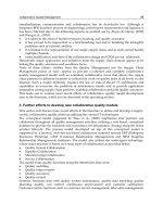
The cuff is placed around the proximal
urethra/bladder neck in the female patient
(Figure 9.8). Cuff sizes range from 4.0 to 11.0 cm,
with 7 to 9 cm being the typical cuff sizes used in
women. It is imperative to implant the appropri-
ately sized cuff, as cuffs that are too large will
not provide adequate urethral compression to
prevent incontinence, and cuffs that are too
small will cause urethral atrophy and are more
likely to erode.
The reservoir is placed in the retropubic
space. The balloon wall tension provides the
pressure that pushes fl uid back into the cuff
after voiding, and maintains outlet resistance
during bladder fi lling. Two reservoir pressure
ranges are available, 51 to 60 cmH
2
O and 61 to
70 cmH
2
O.
The control pump is placed subcutaneously in
the labia majora in female patients. The upper
part of the pump contains a resistor and valves
to transfer fl uid to and from the cuff. There is a
small button on the upper part of the pump that
should be palpable through the labial skin. This
button, when pressed with the cuff open, pre-
vents fl uid from traveling back into the cuff, thus
deactivating the device. The lower part of the
pump forms a bulb that, when squeezed, opens
the cuff to allow voiding.
Technique for Artificial Urinary Sphincter
Placement in Females
When placing an artifi cial urinary sphincter,
strict precautions should be used to avoid con-
tamination or damage to the device. The patient
should receive 24 hours of intravenous antibiot-
ics with the fi rst dose administered just prior to
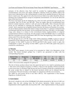
Figure 9.7. AMS-800 artificial sphincter: consisting of a urethral cuff,
pump with deactivation button, and balloon reservoir. (AMS 800
TM
Urinary Control System, courtesy of American Medical Systems, Inc.,
Minnetonka, MN, www.AmericanMedicalSystems.com.)
Figure 9.8. The AMS-800 artificial sphincter after placement in the
female. (AMS 800
TM
Urinary Control System, courtesy of American
Medical Systems, Inc., Minnetonka, MN, www.AmericanMedicalSystems.
com.)
126 Vaginal Surgery for Incontinence and Prolapse
starting the procedure. Oral antibiotics should
be continued for 1 week postoperatively. The
sphincter components should be soaked in anti-
biotic irrigation on the fi eld prior to placement
and device handling should be limited. Rubber-
shodded mosquito clamps should be used to
clamp the tubing. Care must be taken not to
puncture the device components or tubing with
surgical instruments or suturing needles during
wound closures. Blood must not enter the device
tubing, as it can obstruct the free fl ow of
fl uid within the device required for proper
function.
Historically, artifi cial urinary sphincters were
placed entirely through a suprapubic approach
for fear of device contamination from a clean-
contaminated vaginal wound. To facilitate the
dissection between the urethra/bladder neck
and the vaginal wall, Appell (91) described a
transvaginal approach for cuff placement. In his
series of patients, there was no increase in the
incidence of cuff erosions or implant infections,
and these results were later corroborated by
Hadley and associates (92).
When a suprapubic approach is elected, a
transverse, muscle-cutting incision is made 3 cm
above the pubic symphysis. Dissection is carried
down anterior to the bladder in the retropubic
space. In patients who have a history of multiple
previous retropubic operations, dense fi brosis
may be encountered, making dissection diffi cult.
In this situation, one may consider a formal cys-
totomy to facilitate the dissection. Once the
bladder is freed down to the level of the bladder
neck/proximal urethra as identifi ed by the Foley
balloon, longitudinal incisions are made in the
endopelvic fascia on each side of the bladder
neck.
In a plane distal to the ureteral orifi ces, careful
dissection is performed to develop a plane
between the bladder neck and the vaginal wall.
A long Babcock placed around the Foley cathe-
ter, right-angle scissors, and palpation of the
vaginal wall aid in this part of the dissection.
Once a suburethral tunnel is made, it is widened
using a right-angle clamp to enable passage of
the cuff. The cuff-sizer is passed around the
bladder neck and cinched down so that it lays
fl ush around the bladder neck without com-
pressing it. The appropriate size cuff is passed
around the urethra with a right-angle clamp and
it is secured in place by pulling the perforated
tab over the tubing insert on the cuff. The tubing
from the cuff is then passed through the lower
abdominal fascia just over the pubic symphysis
into the subcutaneous space.
The reservoir is then placed in the retropubic
space lateral to the bladder on the same side the
labial pump is to be implanted. The reservoir is
fi lled with normal saline or isotonic contrast and
the tubing is brought through the abdominal fascia
in the same manner as the cuff tubing. A Hegar
dilator (Medical Resources Lewis Center, OH) is
then used to bluntly dissect a path in the subcuta-
neous space into the labia for pump placement.
The pouch created in the labia must be superfi cial
so that the pump and the deactivation button lay
just beneath the skin for easy palpation.
Once all of the components are in place, the
tubing is connected in the subcutaneous space
using the “quick-connect” tubing connectors
according to the manufacturers instructions.
Device function is confi rmed by squeezing the
labial pump and then the device is deactivated
for the next 6 weeks. A urethral Foley is left in
place overnight. If inadvertent bladder injury or
formal cystotomy occurred, a suprapubic tube is
placed on the side opposite the reservoir and left
to drain for 10 to 14 days before removal. If an
injury to the bladder neck or urethra occurs
during dissection, this should be repaired pri-
marily, and sphincter placement should be
delayed a minimum of 12 weeks.
When the transvaginal approach is elected, an
inverted U-shaped incision is made in the ante-
rior vaginal wall. The vaginal wall is dissected off
of the urethra and bladder neck, and the retro-
pubic space is entered laterally on the undersur-
face of the pubic bone as previously described
for pubovaginal sling placement. Circumferen-
tial dissection around the bladder neck is per-
formed using a Babcock clamp around the Foley
catheter and Metzenbaum scissors, keeping the
plane of dissection on the surface of the pubic
bone. Once an adequate space is created around
the bladder neck, the cuff-sizer is passed and the
appropriate size cuff is placed in the same
manner as previously described.
A transverse incision is made just above the
pubic symphysis and carried down to the
abdominal fascia. With the bladder and urethra
retracted contralaterally through the vaginal
incision, the cuff tubing is passed through the
retropubic space and brought through the fascia
into the suprapubic incision. The vaginal inci-
sion is closed and a betadine soaked vaginal
pack is placed. A 2-cm transverse incision is
made in the fascia on the same side that the
Stress Urinary Incontinence Secondary to Intrinsic Sphincteric Deficiency 127
labial pump is to be implanted. The rectus
muscle is split using a curved clamp and a ret-
ropubic pocket is made for the reservoir with
blunt fi nger dissection. The reservoir is placed
and fi lled. Pump placement and tubing connec-
tions are made subcutaneously as previously
described. If there is any concern about he integ-
rity of the vaginal wall tissue or the vaginal
wound closure, a Martius labial fat pad graft
should be interposed from the labium contralat-
eral to the pump.
Results and Complications
Results for the artifi cial urinary sphincter in
females with ISD have been good, particularly
when one considers that the patients have
usually failed multiple other anti-incontinence
operations. Continence results, revision rates,
and removal rates from published series of arti-
fi cial sphincters are noted in Table 9.6. Conti-
nence rates of 70% to 90% can be expected in
those patients who do not develop early (within
the fi rst 6 months) cuff erosion or infection.
Revision rates for the artifi cial urinary sphinc-
ter in females have decreased with advances in
device technology. Revision or replacement of
the device should be expected in 10% to 20% of
patients over 10 years.
Erosion rates, vaginal and urethral, are higher
in females than males after artifi cial urinary
sphincter placement (93). In series that have
included previously irradiated women, the
majority of the devices erode (93,94) into the
urethra. Consequently, most would agree that in
women with a history of pelvic irradiation, an
artifi cial urinary sphincter should not be consid-
ered a viable treatment option. Early device
erosion/infections are likely the result of ure-
thral or vaginal injury during dissection. These
injuries are more common in women with mul-
tiple previous anti-incontinence procedures,
particularly suburethral slings (94). It is diffi cult
to generalize the risk of erosion/infection of arti-
fi cial urinary sphincter in females with the avail-
able published series, as there are differences in
length of follow-up, etiology of incontinence,
technique of sphincter placement, and patient
comorbidities (previous surgery or radiation). If
previously irradiated women are excluded from
sphincter placement, the risk of sphincter
removal owing to infection/erosion is approxi-
mately 30% to 40% over 10 years.
Because there is only one location to place the
cuff in women, when an artifi cial urinary sphinc-
ter erodes or becomes infected management can
be complicated. The device must be removed
and urethral reconstruction with Martius or
omental grafts is required. In cases where sig-
nifi cant urethral tissue loss occurs, urinary
diversion may be required. Because of the rela-
tively high risk of artifi cial urinary sphincter
infection/erosion in females and the potentially
morbid management of these complications
when they occur, we stress that patients consid-
ering this option of treatment for their SUI
should be appropriately counseled.
Conclusion
Our understanding of the etiology of SUI and the
options for SUI treatment have evolved over the
last 30 years. The preoperative evaluation should
document the contributing factors to SUI:
Table 9.6. Female artificial urinary sphincter series
Mean
follow-up % Success % Removal
Author, year (ref.) No. of pts. (months) (≤1 pad per day) % Revision (infection/erosion)
Scott, 1985 (122) 139 36 84 NR 8
Donovan, 1985 (123) 31 24 68 29 33
Diokno, 1987 (124) 32 30 91 21 3
Appell, 1988 (91) 34 36 100 9 0
Parulkar, 1990 (125) 24 40 71 50 17
Webster, 1992 (126) 24 31 100 17 0
Duncan, 1992 (94) 29 65 7 28
Costa, 2001 (127) 206 47 87 NR 6
Thomas, 2002 (90) 68 144 (median) 82* 18 46
* Success rate calculated for patients with sphincter remaining in situ.
128 Vaginal Surgery for Incontinence and Prolapse
urethral hypermobility, ISD, and detrusor dys-
function. When ISD is present, three treatment
options are indicated: suburethral sling, inject-
able bulking agents, and artifi cial urinary
sphincter. In patients who are surgical candi-
dates, suburethral slings offer the best effi cacy
and durability, with low morbidity. Injectable
bulking agents are useful in patients who are not
surgical candidates, or have recurrent SUI with
a well-supported urethra. Currently available
bulking agents have demonstrated good effi cacy
and minimal morbidity in the short-term, but
have not been shown to be durable. Additional
treatment sessions may be necessary for the
maintenance of continence. The artifi cial urinary
sphincter may offer the minority of women with
severe ISD, having failed other treatment options,
a viable option to achieve continence. In the
experienced surgeons’ hands, continence results
have been good with the artifi cial urinary sphinc-
ter, but relatively high complication rates have
prohibited its generalized use.
Surgical treatment for SUI owing to ISD
should be individualized for each patient based
on the several factors, including concurrent
medical comorbidities, the patient’s goals and
quality of life, history of previous failed conti-
nence surgery, and the need for additional
concurrent vaginal or pelvic surgery. Pelvic
reconstructive surgeons should be able to recog-
nize the contribution of ISD to a patient’s SUI,
and be familiar with the surgical techniques,
cure rates, and the diagnosis and management
of complications of the treatment options for
ISD outlined in this chapter.
References
1. McGuire EJ, Lytton B, Kohorn EI, Pepe V. The value
of urodynamic testing in stress urinary incontinence.
J Urol 1980;124:256–258.
2. Ghoniem GM, Elgamasy AN, Elsergany R, Kapoor DS.
Grades of intrinsic sphincteric defi ciency (ISD) asso-
ciated with female stress urinary incontinence. Int
Urogynecol J 2002;13:99–105.
3. Haab F, Zimmern PE, Leach GE. Female stress urinary
incontinence due to intrinsic sphincteric defi ciency:
recognition and management. J Urol 1996;156:3–17.
4. Bergman A, McCarthy TA, Ballard CA, Yanai J. Role
of the Q-tip test in evaluating stress urinary inconti-
nence. J Reprod Med 1987;32:273–275.
5. Kayigil O, Ahmed SI, Metin A. The coexistence of
intrinsic sphincter defi ciency with type II stress incon-
tinence. J Urol 1999;162:1365–1366.
6. McGuire EJ, Fitzpatrick CC, Wan J, et al. Clinical
assessment of urethral sphincter function. J Urol
1993;150:1452–1454.
7. McGuire EJ. Urodynamic fi ndings in patients after
failure of stress incontinence operations. Prog Clin
Biol Res 1981;78:351–360.
8. Sand PK, Bowen LW, Panganiban R, Ostergard DR.
The low pressure urethra as a factor in failed retropu-
bic urethropexy. Obstet Gynecol 1987;69:399–402.
9. Bump RC, Elser DM, McClish DK. Valsalva leak point
pressures in women with genuine stress incontinence:
reproducibility, effect of catheter caliber, and correla-
tions with other measures of urethral resistance. Am
J Obstet Gynecol 1995;173:551–557.
10. DeLancey JO. Structural support of the urethra as it
relates to stress urinary incontinence: the hammock
hypothesis. Am J Obstet Gynecol 1994;170:1713–
1723.
11. Francis LN, Sand PK, Hamrang K, et al. A urodynamic
appraisal of success and failure after retropubic ure-
thropexy. J Reprod Med 1987;32:693–699.
12. Deming CL. Transplantation of the gracilis muscle for
incontinence of urine. JAMA 1926;86:822.
13. Goebell R. Zur operativen beseitigunug der ange-
bornen incontinentia vesicae. Ztschr Gynak 1910;2:
187.
14. Aldridge AH. Transplantation of rectus fascia for the
relief of urinary stress incontinence. Am J Obstet
Gynecol 1942;44:398.
15. Chaikin DC, Rosenthal J, Blaivas JG. Pubovaginal
fascial sling for all types of stress urinary incontinence:
long-term analysis. J Urol 1998;160:1312–1316.
16. Morgan TO, Westney OL, McGuire EJ. Pubovaginal
sling: 4-year outcome analysis and quality of life
assessment. J Urol 2000;163:1845–1848.
17. McGure EJ, Bennett CJ, Konnak JA, et al. Experience
with pubovaginal slings for urinary incontinence at
the University of Michigan. J Urol 1987;138:525–526.
18. Leach GE, Dmochowski RR, Appell RA, et al. Female
stress urinary incontinence clinical guidelines panel
summary report on surgical management of female
stress urinary incontinence. J Urol 1997;158:875–880.
19. Blaivas JG. Pubovaginal sling. In: Kursh ED, McGuire
EJ, eds. Female Urology. Philadelphia: JB Lippincott,
1994:239.
20. Latini JM, Lux MM, Kreder KJ. Effi cacy and morbidity
of autologous fascia lata sling cystourethropexy. J
Urol 2004;171:1180–1184.
21. Kohli N, Karram MM. Surgery for genuine stress
incontinence: vaginal procedures, injections, and the
artifi cial urinary sphincter. In: Walters MD, Karram
MM, eds. Urogynecology and Reconstructive Pelvic
Surgery, 2nd ed. St. Louis: Mosby, 1999:181.
22. McGuire EJ, O’Connell HE. Surgical treatment of
intrinsic urethral dysfunction: slings. Urol Clin North
Am 1985;22(3):657–664.
23. Zaragoza MR. Expanded indications for the pubovagi-
nal sling: treatment of type 2 or 3 stress incontinence.
J Urol 1996;156:1620–1622.
24. Kreder KJ, Austin CA. Treatment of stress urinary
incontinence in women with urethral hypermobility
and intrinsic sphincteric defi ciency. J Urol 1996;156:
1995–1998.
25. Cross CA, Cespedes RD, McGuire EJ. Our experience
with pubovaginal slings in patients with stress urinary
incontinence. J Urol 1998;159:1195–1198.
26. Wilson TS, Lemack GE, Zimmern PE. Management of
intrinsic sphincteric defi ciency in women. J Urol
2003;169:1662–1669.
Stress Urinary Incontinence Secondary to Intrinsic Sphincteric Deficiency 129
27. Kubricht WS III, Williams BJ, Eastham JA, Venable
DD. Tensile strength of cadaveric fascia lata compared
to small intestine submucosa using suture pull through
analysis. J Urol 2001;165:486–490.
28. Lemer ML, Chaikin DC, Blaivas JG. Tissue strength
analysis of autologous and cadaveric allografts for
the pubovaginal sling. Neurourol Urodyn 1999;18:
497–503.
29. Brown SL, Govier FE. Cadaveric versus autologous
fascia lata for the pubovaginal sling: surgical outcome
and patient satisfaction. J Urol 2000;164:1633–1637.
30. Flynn BJ, Yap WT. Pubovaginal sling using allograft
fascia lata versus autograft fascia for all types of stress
urinary incontinence: 2-year minimum follow-up. J
Urol 2002;167:608–612.
31. Carbone JM, Kavaler E, Hu JC, Raz S. Pubovaginal
sling using cadaveric fascia and bone anchors: disap-
pointing early results. J Urol 2001;165:1605–1611.
32. Fitzgerald MP, Mollenhauer J, Brubaker L. Failure of
allograft suburethral slings. BJU Int 1999;84:785–788.
33. Elliot DS, Boone TB. Is fascia lata allograft material
trustworthy for pubovaginal sling repair? Urology
2000;56:772–776.
34. Dora CD, Dimarco DS, Zobitz ME, et al. Time depen-
dent variations in biochemical properties of cadaveric
fascia, porcine dermis, porcine small intestine submu-
cosa, polypropylene mesh and autologous fascia in the
rabbit model: implications for sling surgery. J Urol
2004;171:1970–1973.
35. Hathaway JK, Choe JM. Intact genetic material is
present in commercially processed cadaver allografts
used for pubovaginal slings. J Urol 2002;168:1040–
1043.
36. Duckett JR, Constatine G. Complications of silicone
sling insertion for stress urinary incontinence. J Urol
2000;163:1835–1837.
37. Kobashi KC, Dmochowski RR, Mee SL, et al. Erosion
of woven polyester pubovaginal sling. J Urol 1999;162:
2070–2072.
38. Ulmsten U, Henriksson L, Johnson P, et al. An ambu-
latory surgical procedure under local anesthesia for
treatment of female urinary incontinence. Int Urogy-
necol J 1996;7:81–85.
39. Nilsson CG, Kuuva N, Falconer C, et al. Long-term
results of tension-free vaginal tape (TVT) procedure
for surgical treatment of female stress urinary incon-
tinence. Int Urogynecol J Pelvic Floor Dysfunct
2001;12:S5.
40. Rezapour M, Falconer C, Ulmsten U. Tension-free
vaginal tape (TVT) in stress incontinent women with
intrinsic sphincter defi ciency (ISD)—a long-term
follow-up. Int Urogynecol J Pelvic Floor Dysfunct
2001;suppl 12:S12.
41. Delorme E, Droupy S, de Tayrac R, et al. Transobtura-
tor tape (Uratape): a new minimally-invasive proce-
dure to treat female urinary incontinence. Eur Urol
2004;45:203–207.
42. De Tayrac R, Deffi eux X, Droupy S, et al. A prospective
randomized trial comparing tension-free vaginal tape
and transobturator suburethral tape for surgical treat-
ment of stress urinary incontinence. Am J Obstet
Gynecol 2004;190:602–608.
43. Kobashi KC, Mee SL, Leach GE. A New technique for
cystocele repair and transvaginal sling: the cadaveric
prolapse repair and sling (CaPS). Urology 2000;56(suppl
6A):9–14.
44. Carbone A, Palleschi G, Ciavarella P, et al. Experience
with a bone anchor sling for treating stress urinary
incontinence: outcome at 30 months. BJU Int 2004;
93:780–787.
45. Giberti C, Rovida S. Transvaginal bone-anchored syn-
thetic sling for the treatment of stress urinary incon-
tinence: an outcomes analysis. Urology 2000;56:
956–961.
46. Schrepferman CG, Griebling TL, Nygaard IE, et al.
Resolution of urge symptoms following sling urethro-
pexy. J Urol 2000;164:1628–1631.
47. Meschia M, Pifarotti P, Bernasconi F, et al. Tension-
free vaginal tape: analysis of outcomes and complica-
tions in 404 stress incontinent women. Int Urogynecol
J Pelvic Floor Dysfunct 2001;12:S24–27.
48. Leboeuf L, Tellez CA, Ead D, et al. Complication of
bowel perforation during insertion of tension-free
vaginal tape. J Urol 2003;170:1310–1311.
49. Kobashi KC, Govier FE. Perioperative complications:
the fi rst 140 polypropylene pubovaginal slings. J Urol
2003;170(5):1918–1922.
50. Peyrat L, Boutin JM, Bruyere F, et al. Intestinal perfo-
ration as a complication of tension-free vaginal tape
procedure for urinary incontinence. Eur Urol 2001;
39:603–605.
51. Bent A. Sling and bulking agent placement proce-
dures. Rev Urol 2004;6(suppl 5):26–46.
52. Frederick RW, Leach GE. Osseous complications fol-
lowing transvaginal bone anchor fi xation in female
pelvic reconstructive surgery: a report from the single
largest prospective series and review of the literature.
Urology 2004;64(4):669–674.
53. Rackley RR, Abdelmalak JB, Madjar S, et al. Bone
anchor infections in female pelvic reconstructive pro-
cedures: a literature review of series and case reports.
J Urol 2001;165:1975–1978.
54. Murless BC. The injection treatment of stress inconti-
nence. J Obstet Gynaecol Br Emp 1938;45:521–524.
55. Quackles R. Deux incontinences après adenomecto-
mie gueries par injection de paraffi ne dans le perinee.
Acta Urol Belg 1955;23:259–262.
56. Sachse S. Treatment of urinary incontinence with scle-
rosing solutions: indications, results, complications.
Urol Int 1963;15:225–229.
57. Berg S. Polytef augmentation urethroplasty. Correc-
tion of surgically incurable urinary incontinence by
injection technique. Arch Surg 1973;107:379–381.
58. Shortliffe LM, Freiha FS, Kessler R, et al. Treatment of
urinary incontinence by periurethral implantation of
glutaraldehyde cross-linked collagen. J Urol 1989;141:
538–541.
59. Lightner D, Calvosa C, Andersen R, et al. A new inject-
able bulking agent for the treatment of stress urinary
incontinence: results of a multicenter, randomized,
controlled, double-blind study of Durasphere. Urology
2001;58:12–15.
60. Appell RA. Injection therapy for urinary incontinence.
In: Walsh PC, Retik AB, Vaughan ED, Wein AJ, eds.
Campbell’s Urology, 8th ed. Philadelphia: WB
Saunders, 2002:1172–1186.
61. Monga AK, Robinson D, Stanton SL. Periurethral col-
lagen injections for genuine stress incontinence: a 2-
year follow-up. Br J Urol 1995;76:156–160.
62. Herschorn S, Steele DJ, Radomski SB. Follow-up of
intraurethral collagen for female stress urinary incon-
tinence. J Urol 1996;156:1305–1309.
130 Vaginal Surgery for Incontinence and Prolapse
63. Steele AC, Kohli N, Karram MM. Periurethral collagen
injection for stress incontinence with and without
urethral hypermobility. Obstet Gynecol 2000;95:
322–331.
64. Bent AE, Foote J, Siegel S, et al. Collagen implant for
treating stress urinary incontinence in women with
urethral hypermobility. J Urol 2001;166:1354–1357.
65. Collet JP, Shapiro S, Echick E, et al. Surgery vs. colla-
gen for the treatment of female stress urinary incon-
tinence (SUI): results of a multicentric randomized
trial. J Urol 2001;165(suppl 5):198(abstr 819).
66. Faerber GJ, Belville WD, Ohl DA, et al. Comparison of
transurethral versus periurethral collagen injection in
women with intrinsic sphincter defi ciency. Tech Urol
1998;4(3):124–127.
67. Dmochowski RR, Appell RA. Injectable agents in the
treatment of stress urinary incontinence in women:
where are we now? Urology 2000;56(suppl 6A):32–40.
68. Gonzalez GS, Jimeno C, York M, et al. Endoscopic
autotransplantation of fat tissue in the treatment of
urinary incontinence in the female [in French]. J Urol
(Paris) 1989;95:363–366.
69. Horl HW, Feller AM, Bierner E. Technique for lipo-
suction fat reimplantation and long-term volume
evaluation by magnetic resonance imaging. Ann Plast
Surg 1991;26:248–258.
70. Bartynski J, Marion MS, Wang TD. Histopathologic
evaluation of adipose autografts in a rabbit ear model.
Otolaryngology 1990;102:314–321.
71. Nguyen A, Pasyk KA, Bouvier TN. Comparative study
of survival of autologous adipose tissue taken and
transplanted by different techniques. Plast Reconstr
Surg 1990;85:378–386.
72. Kershen RT, Dmochowski RR, Appell RA. Beyond col-
lagen: injectable therapies for the treatment of female
stress urinary incontinence in the new millennium.
Urol Clin North Am 2002;29:559–574.
73. Pannek J, Brands FH, Senge T. Particle migration after
transurethral injection of carbon-coated beads for
stress urinary incontinence. J Urol 2001;166:1350–
1353.
74. Malizia AA, Reiman HM, Myers RP, et al. Migration
and granulomatous reaction after periurethral injec-
tion of polytef (Tefl on). JAMA 1984;251:3277–3281.
75. Mittleman RE, Marraccini JV. Pulmonary Tefl on
granulomas following periurethral Tefl on injection
for urinary incontinence. Arch Pathol Lab Med 1983;
107:611–612.
76. McKinney CD, Gaffey MJ, Gillenwater JY. Bladder
outlet obstruction after multiple PTEE injections.
J Urol 1995;153(1):149–151.
77. Claes H, Stroobants D, Van Meerbeek J, et al. Pulmo-
nary migration following periurethral polytetrafl uo-
roethyelne injection for urinary incontinence. J Urol
1989;142:821–822.
78. Buckley JF, Lingham K, Meddings RN, et al. Injectable
silicone macroparticles: a new treatment for female
stress incontinence. J Urol 1992;147:280A.
79. Henly DR, Barrett DM, Weiland TL, et al. Particulate
silicone for use in periurethral injections: local tissue
effects and search for migration. J Urol 1995;153:
2039–2043.
80. Lylyk P, Vinuela F, Vinters HV, et al. Use of a new
mixture for embolization of intracranial vascular mal-
formations. Preliminary experimental experience.
Neuroradiology 1990;32:304–310.
81. Haab F, Zimmern PE, Leach GE. Urinary stress incon-
tinence due to intrinsic sphincteric defi ciency: experi-
ence with fat and collagen periurethal injections. J
Urol 1997;157:1283–1286.
82. Chrouser KL, Fick F, Goel A, et al. Carbon coated
zirconium beads in β-glucan gel and bovine glutar-
aldehyde cross-linked collagen injections for
intrinsic sphincteric defi ciency: continence and satis-
faction after extended followup. J Urol 2004;171:
1152–1155.
83. Stothers L, Goldenberg SL. Delayed hypersensitivity
and systemic arthralgia following transurethral colla-
gen injection for stress urinary incontinence. J Urol
1998;159:1507–1509.
84. Sweat SD, Lightner DJ. Complications of sterile abscess
formation and pulmonary embolism following peri-
urethral bulking agents. J Urol 1999;161:93–96.
85. Hartano VH, Lightner DJ, Nitti VW. Endoscopic evac-
uation of Durasphere. Urology 2003;62:135–137.
86. Selahattin B, Kilciler M, Ozgok Y, et al. Long-term
complication due to dextranomer based implant:
granuloma causing urinary obstruction. J Urol 2004;
172:247–248.
87. Harris RL, Cundiff GW, Coates KW, et al. Urethral
prolapse after collagen injection. Am J Obstet Gynecol
1998;178:614–615.
88. Wainstein MA, Klutke CG. Periurethral pseudocyst
following cystoscopic collagen injection. Urology
1998;51:835–836.
89. Scott F, Bradley W, Timm G. Treatment of urinary
incontinence by an implantable prosthetic sphincter.
Urology 1973;1:252–259.
90. Thomas K, Venn SN, Mundy AR. Outcome of the arti-
fi cial urinary sphincter in female patients. J Urol
2002;167:1720–1722.
91. Appell RA. Techniques and results in the implanta-
tion of the artifi cial urinary sphincter in women with
type III stress urinary incontinence by a vaginal
approach. Neurourol Urodynam 1988;7:613–619.
92. Hadley R, Loisides P, Dickinson M. Long-term follow-
up (2–5 years) of transvaginally placed artifi cial
urinary sphincters by an experienced surgeon. J Urol
1985;153(part 2):432A(abstr 816).
93. Venn SN, Greenwell TJ, Mundy AR. The long-term
outcome of artifi cial urinary sphincters. J Urol 2000;
164(3 pt 1):702–706.
94. Duncan HJ, Nurse DE, Mundy AR. Role of the artifi -
cial urinary sphincter in the treatment of stress incon-
tinence in women. Br J Urol 1992;69:141–143.
95. Mason RC, Roach M. Modifi ed pubovaginal sling for
treatment of intrinsic sphincteric defi ciency. J Urol
1996;156:1991–1994.
96. Barbalias G, Liatsikos E, Barbalias D. Use of slings
made of indigenous and allogenic material (Goretex)
in type III urinary incontinence and comparison
between them. Eur Urol 1997;31:394–400.
97. Barbalias GA, Liatsikos EN, Athanasopoulos A. Gore-
tex sling urethral suspension in type III female urinary
incontinence: clinical results and urodynamic changes.
Int Urogynecol J 1997;8:344–350.
98. Hassouna ME, Ghoneim GM. Long-term outcome and
quality of life after modifi
ed pubovaginal sling for
intrinsic sphincteric defi ciency. Urology 1999;53:287–
291.
99. Wright EJ, Iselin CE, Carr LK, et al. Pubovaginal sling
using cadaveric allograft fascia for the treatment
Stress Urinary Incontinence Secondary to Intrinsic Sphincteric Deficiency 131
of intrinsic sphincteric defi ciency. J Urol 1998;160:
759–762.
100. Richter HE, Varner E, Sanders E, et al. Effects of pubo-
vaginal sling procedure on patients with urethral
hypermobility and intrinsic sphincteric defi ciency:
Would they do it again? Am J Obstet Gynecol 2001;
184:14–19.
101. Govier FE, Gibbons RP, Correa RJ, et al. Pubovaginal
slings using fascia lata for the treatment of intrinsic
sphincter defi ciency. J Urol 1997;157:117–121.
102. Staskin DR, Choe JM, Breslin DS. The Gore-tex sling
procedure for female sphincteric incontinence: indi-
cations, technique, and results. World J Urol 1997;
15(5):295–299.
103. Yamada T, Kamata S, Nagahama K, et al. Polytetra-
fl uoroethylene patch sling for type 2 or 3 stress urinary
incontinence. Int J Urol 2001;8:675–680.
104. Morgan JE, Farrow GA, Stewart FE. The Marlex sling
operation for the treatment of recurrent stress urinary
incontinence: a 16-year review. Am J Obstet Gynecol
1985;151:224–226.
105. Ruiz H, Amundsen CL, Webster G. Cadaveric fascia
slings. Atlas Urol Clin North Am 2000;8:41–50.
106. Trockman BA, Leach GE. Surgical treatment of intrin-
sic urethral dysfunction: injectables (fat). Urol Clin
North Am 1995;22:665–671.
107. Winters JC, Appell RA. Periurethral injection of col-
lagen in the treatment of intrinsic sphincteric defi -
ciency in the female patient. Urol Clin North Am
1995;22:673–678.
108. Richardson TD, Kennelly MJ, Faerber GJ. Endoscopic
injection of glutaraldehyde cross-linked collagen for
the treatment of intrinsic sphincteric defi ciency in
women. Urology 1995;46:378–381.
109. Homma Y, Kawabe K, Kageyama S. Injection of glu-
taraldehyde cross-linked collagen for urinary inconti-
nence: two-year effi cacy by self-assessment. Int J Urol
1996;3:124–127.
110. Smith DN, Appell RA, Winters JC, et al. Collagen
injection therapy for female intrinsic sphincteric defi -
ciency. J Urol 1997;157:1275–1278.
111. Swami S, Batista JE, Abrams P. Collagen for female
genuine stress incontinence after a minimum 2-year
follow-up. Br J Urol 1997;80:757–760.
112. Corcos J, Fournier C. Periurethral collagen injection for
the treatment of female stress urinary incontinence:
4-year follow-up results. Urology 1999;54:815–818.
113. Politano VA. Periurethral polytetrafl uoroethylene
injection for urinary incontinence. J Urol 1982;127:
439–442.
114. Lopez AE, Padron OF, Patsias G, et al. Transurethral
polytetrafl uoroethylene injection in female patients
with urinary incontinence. J Urol 1993;150:856–858.
115. Herschorn S, Glazer AA. Early experience with small
volume periurethral polytetrafl uoroethylene for
female stress urinary incontinence. J Urol 2000;163:
1838–1842.
116. Harriss D, Iacovou J, Lemberger R. Peri-urethral sili-
cone microimplants (Macroplastique) for the treat-
ment of genuine stress incontinence. Br J Urol 1996;
78:722–725.
117. Barranger E, Fritel X, Kadoch O, et al. Results of trans-
urethral injection of silicone microimplants for
females with intrinsic sphincteric defi ciency. J Urol
2000;164(5):1619–1622.
118. Radley SC, Chapple CR, Mitsogiannis IC, et al.
Transurethral implantation of macroplastique for
the treatment of female stress urinary incontinence
secondary to urethral sphincter defi ciency. Eur Urol
2001;39:383–389.
119. Tamanini JT, D’Ancona CA, Tadini V, et al. Macro-
plastique implantation system for the treatment of
female stress urinary incontinence. J Urol 2003;169(6):
2229–2233.
120. Mayer R, Lightfoot M, Jung I. Preliminary evaluation
of calcium hydroxylapatite as a transurethral bulking
agent for stress urinary incontinence. Urology
2001;57:434–438.
121. Stenberg AM, Larsson G, Johnson P. Urethral injec-
tion for stress urinary incontinence: long-term results
with dextranomer/hyaluronic acid copolymer. Int
Urogynecol J 2003;14:335–338.
122. Scott FB. The use of the artifi cial sphincter in the treat-
ment of urinary incontinence in the female patient.
Urol Clin North Am 1985;12:305–315.
123. Donovan MG, Barrett DM, Furlow WL. Use of the
artifi cial urinary sphincter in the management of
severe incontinence in females. Gynecol Obstet
1985;161:17–20.
124. Diokno AC, Hollander JB, Alderson TP. Artifi cial
urinary sphincter for recurrent female urinary incon-
tinence: indications and results. J Urol 1987;138:
778–780.
125. Parulkar BG, Barrett DM. Application of the AS-
800 artifi cial sphincter for intractable urinary
incontinence in females. Gynecol Obstet 1990;171:
131–138.
126. Webster GD, Perez LM, Khoury JM, et al. Management
of type III stress urinary incontinence using artifi cial
urinary sphincter. Urology 1992;39:499–503.
127. Costa P, Mottet N, Rabut B, et al. The use of an artifi -
cial urinary sphincter in women with type III inconti-
nence and a negative Marshall test. J Urol 2001;
165:1172–1176.
128. Amundsen CL, Visco AG, Ruiz H, et al. Outcome in
104 pubovaginal slings using freeze-dried allograft
fascia lata from a single tissue bank. Urology 2000;56(6
suppl 1):2–8.
129. Bedir S, Kilciler M, Ozgok Y, et al. Long-term compli-
cation due to dextranomer based implant: granuloma
causing urinary obstruction. J Urol 2004;172:247–
248.
130. Dora CD, Dimarco DS, Zobitz ME, et al. Time depen-
dent variations in biomechanical properties of cadav-
eric fascia, porcine dermis, porcine small intestine
submucosa, polypropylene mesh, and autologous
facia in the rabbit model: implications for sling
surgery. J Urol 2004;171:1970–1973.
131. Elliot DS, Barrett DM. The artifi cial urinary sphincter
in the female: indications for use, surgical approach
and results. Int Urogynecol J 1998;9:409–415.
132. Haab F, Trockman BA, Zimmern PE, et al. Results of
pubovaginal sling for the treatment of intrinsic
sphincteric defi ciency determined by questionnaire
analysis. J Urol 1997;158:1738–1741.
133. McGuire EJ, Clemens JQ. Pubovaginal slings. In:
Walsh PC, Retik AB, Vaughan ED, Wein AJ, eds.
Campbell’s Urology, 8th ed. Philadelphia: WB Saun-
ders, 2002:1151–1171.
134. Stothers L, Goldenberg SL, Leone EF. Complications
of periurethral collagen injection for stress urinary
incontinence. J Urol 1995;159:806–807.
The tension-free vaginal tape (TVT) Gynecare
was fi rst described in 1996 by Ulmsten (1) as
a minimally invasive procedure to treat female
stress urinary incontinence (SUI). This tech-
nique quickly gained a major place in incon-
tinence surgery in Europe and is now being
used more and more in North America. Prior
to TVT, the gold standard technique to treat
SUI was the Burch procedure. Several ran-
domized controlled trials have compared the
effi cacy and safety of TVT and the Burch pro-
cedure. These studies demonstrated that TVT
had a lower morbidity and an equal or supe-
rior effi cacy (on midterm follow-up), justify-
ing the widespread use of this technique (2).
The long-term results (5-year outcome) of the
procedure are also known (82% to 85% of
patients are cured), and they justify the use of
this technique (3,4). However, there are con-
cerns regarding its operative safety. A Finnish
series (5) of 1455 women treated for SUI dem-
onstrated few vascular injuries (venous lacer-
ations were the most frequent injury reported),
10
The Mid-Urethral Tapes
Bruno Deval and François Haab
133
Surgical Techniques . . . . . . . . . . . . . . . . . . . . . 134
The TVT Technique . . . . . . . . . . . . . . . . . . . 134
The Outside-In Procedure . . . . . . . . . . . . . 135
The Inside-Out Surgical Procedure . . . . . . 136
Results . . . . . . . . . . . . . . . . . . . . . . . . . . . . . . . . . 138
The TVT Procedure . . . . . . . . . . . . . . . . . . . 138
The Outside-In Procedure . . . . . . . . . . . . . . 139
The Inside-Out Surgical Procedure . . . . . . 139
Comments . . . . . . . . . . . . . . . . . . . . . . . . . . . . . 139
Conclusion . . . . . . . . . . . . . . . . . . . . . . . . . . . . . 140
whereas Zilbert and Farrell (6) reported a case
of right external iliac artery injury. In addi-
tion, two deaths due to serious vascular inju-
ries have been reported to the manufacturers
(7). Three bowel perforations have also been
reported (8). Postoperative voiding diffi culties
such as transient urine retention are present
in 8% to 17%, and urgency in 5% to 15%. Most
of these complications seem to be related
to the penetration of the retropubic space.
Keeping the principle of a minimally invasive
procedure to reinforce the structures support-
ing the urethra, E. Delorme introduced a pro-
cedure that would avoid these complications.
In 2001, the transobturator procedure was
described, in which the tape is inserted
through the obturator foramen from outside
to inside (in extenso from the thigh folds
to underneath the urethra) (9). Even though
the transobturator out-in TVT technique is
claimed to be a safe procedure, it may cause
urethra and bladder injuries (Figure 10.1). In
2003, De Leval (10) described a novel surgical
technique that allows the passage of a tape
through the obturator foramens, from inside
to outside, with the use of newly designed
surgical instruments. This technique avoids
damage to the urethra and bladder and, for
this reason, makes cystoscopy unnecessary.
However, the long-term safety of this type of
tape is not known, particularly in relation to
changes in the synthetic material and changes
in bladder and urethral functioning caused by
the tape, such as voiding disorders and bladder
overactivity.
134 Vaginal Surgery for Incontinence and Prolapse
Surgical Techniques
The TVT Technique
The overall concept was to place a synthetic
sling at the level of the mid-urethra in order
to restaure the physiologic support provided
by the pubourethral ligaments. After initial
research, the tape proposed was a monofi la-
ment polypropylene woven mesh. This material
appeared from the begining to be safer than
other materials like Dacron or polyester. Origi-
nally the technique was described as an ambu-
latory surgery performed under local anesthesia.
The tape is inserted from the vagina using a
very limited dissection, and positioned into the
retropubic space immediately behind the pubic
bone. The tension of the tape had to be adjusted
using an intraoperative cough test in order to
avoid an excessive tightening of the tape. Overall
the procedure appeared very easy to perform
with a high reproducibility after an initial
proper training.
Since the original description of the technique,
some changes have been introduced and some
controversies still remain. Several randomized
trials have assessed the role of the type of anes-
thesia chosen for the procedure, considering the
fact that the intraoperative cough test could be
done only when the procedure is performed
under local anesthesia or eventually under
general anesthesia. Most of these comparative
trials concluded that the success rate of the TVT
procedure is not correlated with the mode of
anesthesia chosen or with the performance of an
intraoperative cough test.
Concerning the intraoperative complications,
most of them (e.g., bladder injury, bowel or
vessels injuries) could be prevented when the
technique has been done according to the origi-
nal procedure. First, the patient should be placed
in the lithotomy position on the operating table,
but any excessive fl exion of the legs over the
abdomen should be avoided in order to main-
tain easy access to the retropubic space. The tip
of the needles should absolutely follow the pubic
bone and stay medial, with ideally a skin perfo-
ration located 1 to 2 cm apart from the midline.
Owing to the risk of bladder injury, intraopera-
tive cystoscopy is mandatory. The bladder
examination should be carried out using either
a rigid cystoscope with a 70-degree lens of a
fl exible cystoscope. In case of bladder perfora-
tion, the needle is simply removed and reposi-
tioned immediately.
The mean duration of the operation is between
20 and 30 minutes, and most patients can be
discharged the same day. Postoperatively, heavy
lifting should be avoided for 3 to 4 weeks to
allow tape incorporation into the surrounding
tissues.
Obturator artery
Femoral artery
Internal pudendal artery
Epigastric artery
Figure 10.1. The obturator hole is
free of major vessels in its anteroin-
ferior part, allowing safe needle
passage.
The Mid-Urethral Tapes 135
The Outside-In Procedure
The procedure was originally described with
the use of a special tape, Obtape (Mentor-Porges,
Plessis-Robinson, France), made of nonwoven,
non-knitted, thermally bonded polypropylene
(TBP). There is a black line down the middle of
the tape, on the vaginal side, to allow the tape
to be positioned the right way around and along
the midline. The other specifi c item of equip-
ment is a tunneler, a specially curved needle
with a blunt tip and an eye for the tape to be
passed through. More recently, other compa-
nies have introduced procedures allowing
a transobturator placement of suburethral
meshes. The most popular one is the Monarc-
AMS (American Medical Systems, Minnetonka,
MN) technique using a monofi lament polypro-
pylene nonwoven mesh and an helical needle.
The procedure is done under general, spinal,
or local anesthesia. The patient is placed on the
operating table in the lithotomy position with
hyperfl exion of the legs. After hydrodissection
of the vagina is performed, a vertical midline
vaginal incision is made in the middle third of
the urethra, passing through the entire thickness
of the vaginal wall. Starting at the incision, the
vagina is released laterally on either side of the
urethra with Mayo scissors over a width of
approximately 15 mm. The dissection stops
against the ischiopubic ramus. The dissection
must be in the deep tissue layer between the
vesicovaginal fascia and the urethra, and not too
superfi cially between the vesicovaginal fascia
and the vaginal skin. The lateral margin of the
ischiopubic ramus is identifi ed between an index
fi nger placed in the laterovaginal fornix and
thumb placed in front of the obturator foramen.
A puncture incision is made 15 mm lateral to the
ischiopubic ramus on a horizontal line level with
the preputium clitoridis (Figure 10.2). The tun-
neler is held in the same hand as the side on
which the operator is working. Two types of tun-
neler can be used: a simple curved Emeth needle
or a helical needle. The choice between these two
options is made according to the surgeon’s
preference.
The tunneler is held vertically with the handle
downward; it is then introduced through the
skin incision, and it crosses the obturator mem-
brane. As the membrane is crossed, a specifi c
resistance is felt, which is easily recognized. The
tunneler is then turned to a horizontal posi-
tion, with the handle pointing medially. The tip
of the tunneler is led medially toward the
urethra, aiming above the urethral meatus and
Figure 10.2. After an incision is made at the level of the mid-urethra, the needle is introduced in the obturator hole. A helicoidal or simple curved
needle can be used.
136 Vaginal Surgery for Incontinence and Prolapse
underneath the symphysis pubis. The safest
method is to lead the tunneler around the ischio-
pubic ramus while remaining in contact with it.
The aim of this procedure is to trace a perineal
route with the instrument below the superior
fascia of the levator ani. A fi nger is placed in the
incision to check that the tunneler is not pierc-
ing the vagina and is passing well above the lat-
erovaginal fornix some distance away from it.
The index fi nger is introduced into the vaginal
incision to fold the urethra up and protect it
from the needle. The fi nger will then make
contact with the tip of the tunneler laterally
underneath the symphysis pubis. The tunneler
is then guided by the fi nger into the vaginal inci-
sion (Figure 10.3). Once this procedure has been
completed, it is prudent to check that the vagina
and urethra have not been pierced by the tun-
neler. The end of the tape is introduced into the
eye of the needle and then pulled through to
place it in position. The texture of the tape allows
the tape to be pulled hard without risk of break-
ing. The tape is inserted tension-free behind the
urethra (Figure 10.4). The low elasticity of ther-
mally bonded polypropylene makes it possible
to adjust the position of the tape precisely.
There are two important points to remember
during this adjustment to reduce the risk of
compressing the urethra, which causes voiding
disorders:
• Leave a visible space between the tape and the
urethra.
• Avoid adjusting the tape with the patient in
the Trendelenburg position, as the cervical
and urethral region is at its highest in this
position. It is therefore better to place the
patient in the horizontal position or even
tilted to ensure that the urethra is at its
lowest.
No study has assessed the potential benefi ts
of an intraoperative cough test during this
procedure.
The Inside-Out Surgical Procedure
Three specifi c surgical instruments were created
for the procedure: helical passers, plastic tubes,
and an introducer. The helical passers are pairs
of instruments, specifi c for the left and right
sides. They are stainless steel instruments
with a spirally shaped section and a handle.
The spiral section comprises an open circular
segment having a 3-cm radius terminated by
Figure 10.3. The tape is grasped and pulled through the vaginal inci-
sion in the obturator hole.
Figure 10.4. The tape is positioned at the level of the mid-urethra without any excessive tension.
The Mid-Urethral Tapes 137
two linear segments. On a horizontal plane per-
pendicular to the handle’s axis, the gap between
the extremities of the spiral section is 2 cm. The
element supported by the helical passer is a
polyethylene tube with a sharp pointed distal
end. It bears a lateral opening, which allows the
insertion of the spiral segment of the helical
passer into its lumen. The proximal end of the
tube is opened and it can be attached intraop-
eratively to a nonabsorbable synthetic tape.
The introducer is a stainless steel device that
has two segments: a proximal tubular hollow
segment and a distal, semicircular, 7-cm-long
gutter. The introducer acts as a shoehorn to ease,
without danger, the slipping in of the passer,
introduced alongside the gutter, from the peri-
neal space through the obturator foramen.
The surgical procedure is generally carried
out under spinal anesthesia but may also be per-
formed under general or local anesthesia. Two
grams of third-generation cephalosporin are
administered intravenously at the time of anes-
thesia induction. The patient is fi rst placed in the
gynecologic position, legs on stirrups and thighs
in hyperfl exion. The patient’s buttocks reach the
edge of the table. The operative fi eld is cleaned
with a standard antiseptic agent and draped with
multiple drapes rather than a single trousers-
shaped drape, with care being taken to keep the
groin folds in the operative fi eld. Labia minor
are suspended by fi xation to the skin with nylon
suture a few centimeters above the vulvar ostium,
inside the thigh folds, in order to expose the
vulvar vestibulum. A 16-French Foley catheter is
inserted into the bladder.
The points where the needles will exit at the
skin level are identifi ed by tracing a horizontal
line at the level of the urethral meatus. The exit
points are located 2 cm above this line and 2 cm
outside the thigh folds. A 5-mm skin incision is
made at each exit point.
The anterior vaginal wall is suspended with
two Allis clamps on either side of the midline,
1 cm proximally to the urethral meatus. A median
sagittal incision of the vaginal wall is started at
this level and is continued proximally (toward
the vaginal pouches) over a 1-cm distance. Both
vaginal mucosal and submucosal tissues are
incised. Minimal paraurethral subvaginal dis-
section is then carried out laterally with the
blade, over a few millimeters distance, on either
side. One Allis clamp grasps the right minor and
major labia, while another Allis clamp holds the
left margin of the suburethral vaginal incision,
to clearly expose the most posterior aspect of the
right vulvar vestibulum. Fine dissection scissors
are introduced through the blade-initiated dis-
section path, and then further, on a horizontal
plane with a 45-degree angle relative to the ure-
thral sagittal plane, toward the upper part of
ischiopubic ramus. It is important to correctly
expose the vulvar vestibulum and to respect the
specifi c direction of the dissection in order to
avoid any perforation of the vaginal wall.
Once the upper part of the ischiopubic ramus
is reached—a bone contact is perceived—the
right obturator membrane is perforated with the
tips of the scissors, which are then slightly
opened. During the dissection, bleeding can
occur but is never important and only occasion-
ally requires a blood-aspirating device. The
introducer is then pushed in the preformed dis-
section pathway until it reaches and perforates
the obturator membrane (Figure 10.5). The open
side of the introducer’s gutter must be facing the
operator. The distal end of the tube is mounted
onto the spiral segment of the needle and the
assembled device is gently slipped along the
gutter of the introducer so as to pass through the
obturator foramen. The introducer and Allis
clamps are removed. At this step, the handle of
the passer must be aligned in a parallel manner
with the sagittal axis of the vulvar slit. Then,
owing to a rotational movement of the passer,
the pointed tip of the tube appears at the previ-
ously incised skin exit points at the level of the
thigh folds. The tube is pulled from the support-
ing passer, which is removed by a backward-
rotational movement, until the fi rst centimeters
of the tape become externalized. The same tech-
nique is applied to the left side. It is important
to take care not to twist the tape.
When both tubes have been extracted through
the skin incisions, the ends of the tape are cut.
The tape is then aligned under the junction
between the mid- and distal urethra and the
tension of the tape is adjusted by exerting trac-
tion on its two ends and by interposing a pair of
scissors between the tape and the urethra so as
to leave a space, avoiding any tension of the tape.
The plastic sheaths are then removed simultane-
ously. An alternative procedure for correctly
aligning the tape under the urethra is to grasp
the tape at its middle with Babcock forceps so
as to create a small, 5-mm-long tape loop. As
described above, traction is exerted on the distal
ends of the tape, avoiding compression of the
urethra.
138 Vaginal Surgery for Incontinence and Prolapse
Results
The TVT Procedure
The original TVT procedure has been widely
assessed, with more than 300 publications in
international journals. Most published results
report stress incontinence owing to a hypermo-
bile urethra. The cure rate in these patients is
above 85%. The failure rate usually ranges from
5% to 10%, mainly owing to persistent or de
novo urgency symptoms. However, one remark-
able thing is that the overall results are remark-
ably consistent throughout the literature. This
suggests that the procedure is highly repro-
ducible and well standardized, and no doubt
accounts for its rapid and widespread prolifera-
tion. Moreover, unlike laparoscopic Burch col-
posuspension or suburethral slings, which are
not easy to position, the TVT procedure does
not appear to be operator dependent.
Few studies have evaluated long-term follow-
up. Nilsson et al (1) recently published 7-year
results of the procedure. A total of 80 women
have been evaluated at a mean follow-up of 91
months (range 78–100 months). Assessment
variables included a 24-hour pad weighing test,
a stress test, a visual analog scale for assessing
the degree of bother, and a questionnaire assess-
ing the subjective perception of the women
about their continence status. At 7-year follow-
up, both objective and subjective cure rates were
81.3%. Asymptomatic pelvic organ prolapse
was found in 7.8%, de novo urge symptoms in
6.3%, and recurrent urinary infections in 7.5%.
No other long-term adverse events have been
described in that series.
Only one study has compared the results of
the Burch colposuspension and those of the TVT
procedure (2). The trial was conducted in 14
centers in the United Kingdom and Ireland. A
total of 344 patients were included in the study
and randomized; 170 patients underwent a TVT
procedure and 146 a Burch colposuspension. At
6 months’ follow-up, the continence rates based
on objective and subjective criteria were not sta-
tistically different in the two groups. Regarding
the complications, the authors found an advan-
tage for the Burch over the TVT for the bladder
perforation rate (2% versus 9%). However, the
estimated blood loss was the same in both
groups. The TVT procedure appeared signifi -
cantly better than the Burch on the following
items: postoperative opiate analgesia (21%
versus 91%), mean duration of hospital stay (2.2
days versus 6.5 days), and number of patients
rehospitalized (9 versus 18 patients). Moreover,
a medico-economic analysis has been derived
from that study showing that TVT is a more
cost-effi cient procedure compared to the Burch
colposuspension.
Currently, there are no identifi ed prognostic
factor for success. Whether sphincter defi ciency as
given by the measurement of urethral pressure or
of the Valsalva leak point pressure is a cause of
Figure 10.5. The TVT-O technique. The tape is introduced in the obturator hole from the vaginal incision. After an incision is made beneath the
urethra with limited dissection laterally, the special curved needle is pushed on a guide through the obturator hole.
The Mid-Urethral Tapes 139
failure remains a moot point. Even if this remains
a controversial issue, the results of the TVT pro-
cedure appeared inferior in a context of intrinsic
sphincteric defi ciency defi ned either by a low
closure pressure of the urethra or by a low Val-
salva leak point pressure. Furthermore, it appeared
that patients who have had multiple surgeries for
SUI with a subsequent rigid urethra have poorer
results. Among other pronostic factors, age and
obesity have been identifi ed has having a negative
impact on postoperative results.
Based on these results, the TVT procedure has
become a fi rst-line therapy when surgery is con-
sidered to cure stress urinary incontinence.
The Outside-In Procedure
Delorme et al (11) published in 2004 the results
on 32 consecutive patients; 90.6% were cured
and three (9.4%) were improved. The mean
operating time was 15 minutes. No intraopera-
tive complications were recorded. One patient
had complete postoperative bladder retention,
which resolved after 4 weeks of self-catheteriza-
tion. There were no problems with urethral
erosion, residual pain, or functional impair-
ment related to the tape. Five patients had
voiding disorders, suggesting bladder outfl ow
obstruction. Two patients developed de novo
urge incontinence.
Costa et al (12) published in 2004 the results of
a multicenter trial on 183 women with SUI asso-
ciated with urethral hypermobility. The mean
follow-up was 7 months (range 1–21 months). At
1-year follow-up, 80.5% of the patients were
completely cured and 7.5% were improved. The
overall perioperative complication rate was 2.2%
with no vascular, nerve, or bowel injury. Six
patients (3.3%) had postoperative urinary reten-
tion. Cindolo et al (13) published in 2004 the
results on 80 females affected by SUI associated
with urethral hypermobility and without severe
urogenital prolapse. The mean operative time
was 16 minutes (range 11–36 minutes). No major
intraoperative complications were observed.
One bladder neck laceration occurred and was
treated intraoperatively. No cystoscopy was per-
formed. The mean hospital postoperative stay
time was 1.1 (1–6) days. All patients were exam-
ined periodically at 7, 30, and 90 days from inter-
vention (mean follow-up 4 months, range 1–8).
There was no urethral erosion. One vaginal
erosion with inguinal abscess was diagnosed and
treated without removing the sling. Two patients
with de novo urge incontinence were observed.
The objective and subjective cure rates were 92%
and 97%; 96% expressed good quality of life (sat-
isfi ed/very satisfi ed).
Few results have been published using the
Monarc system. The largest report on this tech-
nique has been presented at the International
Continence Society-International Urogyneco-
logical Association (ICS-IUGA) meeting in 2004
(14). The authors reviewed the results of two
prospective trials conducted in nine countries
and including 204 patients. The follow-up was
rather short and most of the data were related to
the morbidity issues. In this series no major
intraoperative complications have been found.
Groin pain occurred in 1.6% of the patients, and
vaginal erosion occurred in 1.0%. Six patients
(2.9%) had surgical revision to release sling
tension or remove the sling.
The Inside-Out Surgical Procedure
De Leval (10) published in 2003 the results on
107 consecutive patients (mean age: 62 years)
using the same operative protocol in all case
subjects, independently of the patient’s size and
weight. The mean operative time was 14 minutes
(range 7–20) in cases of isolated SUI treatment.
No bladder or urethral injuries and no vascular
(hematoma or bleeding) or neurologic compli-
cations were encountered. To date, no other
reports have been published on this new prom-
ising approach.
Comments
De Lancey’s (15) theories on pelvic support for
the bladder and urethra help to explain the
mechanism of action of urethral suspension in
the treatment of stress urinary incontinence.
The new minimally invasive suspension tech-
niques using a polypropylene tape satisfy the
requirements for functional surgery. In the
medium term, the results in the treatment of
female stress urinary incontinence are satisfac-
tory. Unlike the retropubic tapes, the purely
perineal local location of the transobturator
minimizes the risk of trauma to internal organs
(bladder perforation, damage to the intestine
or to blood vessels and nerves). The position of
the transobturator tape is similar to that of
the natural hammock supporting the urethra