Vaginal Surgery for Incontinence and Prolapse - part 6 ppsx
Bạn đang xem bản rút gọn của tài liệu. Xem và tải ngay bản đầy đủ của tài liệu tại đây (844.98 KB, 30 trang )
Anterior Compartment 147
Vaginal Paravaginal Repair
Technique
The patient is placed in the lithotomy position,
and the bladder is drained via a catheter. The
procedure is performed through a midline, an
inverted U or V, or bilateral parallel incisions in
the anterior vaginal wall. The pubocervical
(perivesical) connective tissue should be dis-
sected off of the vaginal epithelium sharply to
the medial border of the descending pubic
ramus. The retropubic space is entered sharply
using Metzenbaum scissors through the endo-
pelvic fascia. The pubocervical fascia is sepa-
rated from the sidewall of the pelvis, exposing
the obturator fascia and the arcus tendineus
fascia pelvis. The arcus tendineus can be fol-
lowed from the back of the pubic ramus to the
ischial spine by retracting the bladder and
urethra medially using a Briesky-Navratil
retractor. Four to six interrupted permanent
sutures are placed between the arcus tendineus
with underlying obturator membrane laterally
and the pubocervical fascia medially. The
sutures extend from the back of the pubis dis-
tally at the level of the urethrovesical junction
to the ischial spine proximally. The sutures
should be left untied. The process is repeated on
the other side. The stitches are then tied sequen-
tially in a distal to proximal direction, alternat-
ing from one side to the other. If a central defect
exists, traditional anterior colporrhaphy sutures
can then be placed to plicate the redundant con-
nective tissue. The vaginal epithelial fl aps are
trimmed and reapproximated once all sutures
have been placed and tied.
Results
A review of seven retrospective cohorts showed
a failure rate of 3% to 39% (Table 11.1). Although
the rate of recurrence of anterior prolapse was
high (39%) in the series from Shull et al (9),
most of the recurrences were mild (32%) and the
prolapse was less than preoperatively.
Four-Corner and Six-Corner
Suspension
The four-corner suspension was devised by Raz
et al (10) for patients with stress incontinence,
urethral hypermobility, and mild to moderate
cystocele with lateral defects. It did not include
anterior colporrhaphy. It was subsequently
modifi ed to a six-corner suspension (11). The
difference is an additional set of proximal
sutures (at the level of the cardinal ligament) to
support the bladder.
Technique
Two oblique incisions or an inverted-U incision
is made from the mid-urethra to the proximal
vagina. The pubocervical fascia is exposed. The
endopelvic fascia on each side is perforated
using curved Mayo scissors (hug underneath
the pubic ramus while pointing toward the
ipsilateral shoulder) to enter into the retropubic
space. The pubocervical fascia connecting the
bladder to the arcus tendineus is separated from
the pelvic sidewall anteriorly. The lateral attach-
ments of the bladder base are exposed proxi-
mally to the cardinal ligaments. Three sets
(six-corner suspension) of 1-0 polypropylene
sutures are placed on each side. Each suture
incorporates multiple passes through the tissue
and is laterally placed to avoid periurethral
scarring and outfl ow obstruction. The proximal
suture is placed through the cardinal ligament
and vaginal wall to support the bladder base.
The middle suture is at the level of the bladder
neck, and the distal suture is at the mid-urethra.
The sutures are passed up individually to a
small suprapubic incision with the double-
pronged ligature carrier. Indigo carmine is
administered intravenously, and cystoscopy
confi rms ureteral patency and the absence of
suture in the bladder or urethra. The sutures are
lifted to ensure adequate anatomic reduction of
Table 11.1. Results of vaginal paravaginal repair for treatment of
anterior vaginal prolapse
Follow-up,
years, range
Author Recurrence (mean)
Shull et al, 1994 (9) 4/56 (7%) severe 0.1–5.5 (1.6)
18/56 (32%) mild
Benson et al, 1996 (20) 12/46 (26%) 1–5.5 (2.5)
Farrell and Ling, 1997 (38) 6/27 (22%) 0.75
Scotti et al, 1998 (39) 3/35 (8.6%) 0.5–4.3 (3.25)
Elkins et al, 2000 (25) 6/25 (24%) 0.5–3
Mallipeddi et al, 2001 (40) 1/35 (3%) 0.7–3 (1.8)
Young, 2001 (24) 22/100 (22%) 0.1–3 (1)
148 Vaginal Surgery for Incontinence and Prolapse
the cystocele and then tied sequentially to
themselves and to the corresponding one from
the opposite side. It is important to avoid tension
on the polypropylene sutures to prevent postop-
erative urinary retention.
Results
Early results of the four-corner suspension were
encouraging (2% recurrence rate), but unfortu-
nately, there were a signifi cant number of late
failures (44). Four- or six-corner suspension
without colporrhaphy for mild to moderate cys-
tocele has not been widely reported. In some
reports, patients had large cystoceles plus ante-
rior colporrhaphy with recurrence rates of 40%
to 59% (12,13). The technique has also been
modifi ed with the addition of mixed fi ber mesh
(14). As a result, the durability of the procedure
is uncertain.
Anterior Colporrhaphy
and Suspensions
Evidence exists that concomitant procedures
at the time of anterior compartment prolapse
repair can adversely affect long-term out-
comes. Kelly et al (15) reported a high cysto-
cele recurrence in 24% of patients at a mean of
62 months. Raz et al (16) reported a recurrence
rate of 11%. In a randomized trial of anterior
colporrhaphy with or without four-corner sus-
pension, Kohli et al (17) reported a recurrence
rate of 33% versus 7% in patients who had
not undergone needle suspension. This effect
was also seen in a randomized, prospective
comparison of needle colposuspension versus
endopelvic fascia plication in women undergo-
ing vaginal reconstruction for stage III or IV
pelvic organ prolapse (18). Especially when
combined with a sacrospinous vaginal vault
suspension, those patients randomized to
receive concomitant needle suspension devel-
oped a high incidence of early, advanced,
recurrent, anterior vaginal prolapse. Sacrospi-
nous vaginal vault suspension has also been
associated with recurrent anterior segment
prolapse (19). Theoretically it is thought to
be caused by altering the vaginal axis (retro-
version) with exposure of the anterior wall to
greater abdominal pressure or a neuropathy
caused by the vaginal dissection (20).
Anterior Colporrhaphy and Sling
Conversely, concomitant suburethral slings at
the time of reconstructive vaginal surgery have
been shown to signifi cantly reduce the recur-
rence of anterior vaginal wall prolapse. Cross et
al (21) reported recurrence rates of 8% (grades
3 and 4) and 15% (grade 1) in 36 of 42 patients.
To improve the long-term failure rate of cysto-
cele repair, Kobashi and Leach (22, 43) described
a transvaginal technique using cadaveric fascia
lata as a sling and support for the cystocele. A
T-shaped segment is incised. The ends of the T
are placed retropubically and fastened to the
pubis using bone anchors; this is the sling
portion of the procedure. The remainder of the
patch is secured to the medial edges of the
levator muscles bilaterally with No.0 polydioxa-
none suture and back to the vaginal cuff or
cervix proximally with absorbable sutures. The
short-term data were excellent (1 to 6 months’
follow-up) with no cystocele recurrence. The
follow-up data on this technique included 132
patients with a mean follow-up of 12.4 months
(range 6–28 months). The recurrence rate of
cystoceles was 12.9%, all grade 1 or 2. There was
a 9.8% rate of apical vaginal prolapse after this
procedure (22). The presence of any type of
suburethral sling was associated with a 54.8%
reduction in prolapse recurrence (23). This
fi nding should be taken into consideration when
planning surgical repair for the woman with
prolapse and stress incontinence or suspected
masked stress incontinence.
Complications
Signifi cant bleeding with cystocele repair is
unusual. Bleeding may occur if dissection is
carried out in the wrong plane during trans-
vaginal procedures; therefore, the vaginal wall
should be taken off of the perivesical fascia
directly on its white shiny surface. Perforation
of the endopelvic fascia to gain access to the
retropubic space is another potential source
of bleeding. Packing with a small laparotomy
sponge can be all that is necessary, but oversew-
ing the area with fi gure-of-eight stitches is often
required. The blood transfusion rate for trans-
vaginal paravaginal repair ranged from 9% to
12% (24,25), in contrast to a transfusion rate of
0% to 4% in series of abdominal paravaginal
defect repair. The limited exposure and techni-
Anterior Compartment 149
cal challenge of the vaginal approach likely
explain this difference.
Bladder or ureteral injuries are rare, but must
not be missed. Intraoperative cystoscopy after
administration of intravenous indigo carmine
will facilitate visualization of the effl ux of blue-
stained urine. Failure to see the effl ux may signify
kinking or ligation of a ureter. The offending
suture must be removed and replaced.
Bladder injuries can be reduced by ensuring
that the bladder is empty prior to dissection or
perforating into the retropubic space. Should
inadvertent injury occur, two-layer closure
needs to be performed. If the tissue quality is
poor, especially in those with a history of pelvic
irradiation, an omental, peritoneal, or labial fl ap
interposition is recommended to prevent fi stula
formation. If a bladder injury is not detected
until after surgery, a trial of conservative therapy
with a catheter may be attempted.
Early postoperative complications for cysto-
cele repair include wound infection, immediate
urinary retention, and irritative voiding symp-
toms. Retention is more likely in cases in which
an anti-incontinence procedure was also per-
formed, but it is usually transient. There was
only one case of prolonged retention requiring
urethrolysis in the cohort of patients who under-
went repair of cystocele using a sling and patch
made of cadaveric fascia (22).
Long-term complications include voiding
dysfunction such as stress urinary incontinence
(SUI), detrusor instability, and incomplete
voiding; SUI can be minimized with proper pre-
operative evaluation and performance of simul-
taneous anti-incontinence procedure. De novo
urge incontinence is a known complication of
all bladder surgery and occurs in 5% to 7%
of patients (10,16). However, preexisting urge
incontinence has been reported to resolve in
63% of cases (26). Other complications include
chronic pain, vaginal shortening or stenosis, and
dyspareunia. Care should be taken not to aggres-
sively excise excessive vaginal wall, causing
vaginal shortening. Finally, a missed or de novo
prolapse of other organs (apical prolapse or
enterocele) can result postoperatively.
Adjunctive Materials
Because of reported long-term recurrences of
anterior vaginal prolapse, classic techniques
modifi ed by the use of surrogate materials have
been tried in an attempt to improve outcome.
These include synthetic mesh (Mersilene,
Marlex (42), Prolene), cadaveric allograft fascia
(Repliform), and xenograft fascia (Pelvicol,
Stratasis).
Julian (27) reported a 66% cure rate for a stan-
dard anterior colporrhaphy for recurrent ante-
rior prolapse compared with a 100% cure rate
when Marlex mesh was used. However, there
was a 25% incidence of mesh-related complica-
tions. This approach was not advocated as a
primary procedure; rather, it was recommended
only for those patients with prior failures. Other
observational studies have subsequently been
published describing the usually successful
experience of using a synthetic mesh, most often
Marlex, in reducing recurrence of anterior
vaginal wall prolapse (28,29). These studies are
most often limited by their small numbers and
lack of long-term follow-up.
In a study by Dora et al (30), rabbits had
implantation of human cadaveric fascia, porcine
dermis, porcine small intestine submucosa,
polypropylene mesh, and autologous fascia in
the anterior abdominal wall. They were sacri-
fi ced at various time points, and tensiometry
and image analysis were performed. Each type
of human cadaveric fascia and porcine allografts
had a marked decrease in tensile strength; in
contrast, polypropylene mesh and autologous
fascia did not experience any change from
baseline.
Xenografts have also been used as reinforce-
ment in prolapse repair. Theoretically these
materials may be better tolerated by the vagina
than synthetics, but they too have been associ-
ated with minor erosions. In addition, there are
concerns regarding transfer of animal disease to
the host human. These materials are also expen-
sive, and there is no published literature proving
their benefi ts or effi cacy.
One prospective randomized controlled trial
was performed using polyglactin 910 mesh to
prevent recurrent anterior vaginal wall prolapse
by Sand et al (31). This mesh is absorbable and
was used as a bulking material folded into the
anterior colporrhaphy stitches. The approach
is thought to enhance scarring just anterior to
the suture line, providing greater protection to
an area potentially more vulnerable to direct
intraabdominal downward forces. Patients with
anterior vaginal wall prolapse to or beyond the
hymenal ring were eligible. At 1 year postopera-
tively, 30 of 70 women (43%) who did not receive
150 Vaginal Surgery for Incontinence and Prolapse
mesh had recurrence versus 18 of 73 women
(25%) who did receive the mesh (p = .02). Pro-
lapse to the hymenal ring occurred in 8 of 70
controls (11.4%) and in two of 73 women (2.7%)
with mesh repair (p = .04). No patient had recur-
rent prolapse past the hymen.
Currently, many materials are available for use,
but the ideal biocompatible material should be
chemically and physically inert, noncarcinogenic,
durable, sterile, readily available, noninfl amma-
tory, and inexpensive. None exists that mimics
autologous tissue, but there are several benefi ts
associated with synthetics that we favor as the
surrogate material of choice. They are available in
any size and can be easily tailored to the surgeon’s
preference. They are durable, permanent, and
maintain their strength over time. In this era of
newly discovered infectious agents, from HIV to
prions, one can be at ease that synthetics are free
of every pathogenic disease. The cost is also sig-
nifi cantly less compared with biomaterials and
cadaveric fascia. The main concern in the past has
been foreign-body reaction, erosion, and infec-
tion that is related to the weave of the mesh and
the size of the pores. Multifi laments (Gore-Tex,
Mersilene) tend to produce a more chronic
infl ammation that can be detrimental compared
with monofi laments, which produce an acute
infl ammatory reaction followed by formation of
fi brous tissue (32). Pore size infl uences the fl exi-
bility of the mesh used, as well as fi broblast and
leukocyte infi ltration and passage (33). We cur-
rently solely use polypropylene (Prolene) because
it is nonabsorbable, macroporous, monofi lamen-
tous, fl exible, and sterile. The large pore size
(>75 μm) allows for the ingrowth of macrophages,
fi broblasts, collagen, and blood vessels. This aids
in rebuilding autologous tissue within the mesh
and allows for the chemotaxis of macrophages in
battling infection.
Authors’ Technique
There have been numerous modifi cations to the
anterior colporrhaphy, including the use of syn-
thetic or allograft materials, variations in suture
placement, and anchoring techniques in order
to improve cystocele repair. The long-term
results of anterior colporrhaphy alone have
been disappointing. The paravaginal repair
addresses the lateral defect in anterior compart-
ment prolapse, but even when paired with
anterior colporrhaphy, recurrence rates were
signifi cant. The main fl aw remains—the
approximation of already attenuated tissue. The
four- and six-corner suspension technique and
its variations were not signifi cantly better in the
rate of recurrence; rather, some studies had a
higher rate of recurrence of mild cystoceles (12).
The quest for a technique that can provide the
strength and characteristics that will contribute
to a lasting repair continued.
Our technique differs from others in that it
is a modifi cation of a transvaginal paravaginal
repair using soft Prolene mesh that addresses
four defects: urethral hypermobility, lateral
bladder support (paravaginal), perivesical fascia
support (central), and separation of sacrouter-
ine ligaments. The four key technical points are
as follows: (1) A distal urethral sling is almost
always performed prior to repair of a high-grade
cystocele. The only exceptions would be prior
sling placement and nonmobile urethra. (2) A
single round mesh (5 × 5 cm) repairs the central
as well as the lateral defect. (3) The mesh attaches
laterally to strong anchoring tissue, the perios-
teum of the descending ramus of the symphysis
and the infralevator obturator fascia, inferior to
the line of arcus tendineus. The retropubic space
is not entered; the sutures are not attached to the
arcus tendineus fascia pelvis or above it as in the
classic paravaginal repairs. (4) The pathologi-
cally separated cardinal ligaments are reapprox-
imated and forms the most proximal support of
the bladder and round mesh.
Procedure
With the patient in the dorsal lithotomy posi-
tion, a 16-French Foley catheter is placed in the
bladder. A suprapubic tube (SPT) is placed after
the bladder has been adequately fi lled. Expo-
sure is maximized with a weighted vaginal
speculum and a Scott ring retractor.
If there is concomitant uterine prolapse and a
hysterectomy is required, a transvaginal hyster-
ectomy is performed at this time prior to the
cystocele repair. We close the cuff but will not
yet tie the vault suspension sutures. If a hyster-
ectomy is not necessary, we start with the distal
urethral sling and prepare the bladder by dis-
secting the bladder away from the vaginal wall
fl aps. Any enterocele defect will be opened, the
cul-de-sac repaired with purse-string sutures,
and the vault secured to the inferior edge of the
sacrouterine ligaments bilaterally. The purse-
Anterior Compartment 151
string sutures are left uncut, to be the base of the
cystocele repair.
We perform a distal urethral prolene sling
(DUPS) in all patients with stage IV cystoceles
(34). The incidence of occult stress urinary
incontinence can be as high as 22% to 80%
among patients with high-stage vaginal vault
prolapse (35). Owing to the known masked
urinary incontinence, and the high incidence of
postoperative de novo stress incontinence, many
authors routinely perform a concomitant anti-
incontinence surgery in all anterior vaginal
reconstruction, independent of the continence
status. Beck and associates (36) reported a 10%
incidence of urinary incontinence after 519 ante-
rior colporrhaphy procedures for prolapse in
continent patients.
An Allis clamp is used to retract the urethra
superiorly. Two parallel incisions are made in
each paravaginal sulcus, carefully avoiding the
inner labia. Metzenbaum scissors are used to
dissect the vaginal wall from the periurethral
fascia. A small window is made in the retropubic
space with a pair of curved Mayo scissors
directed parallel to the urethra. The medial edges
of the urethropelvic ligaments and retropubic
fat can then be seen. A tunnel between the
vaginal wall and periurethral fascia is made at
the level of the distal urethra with a fi ne right
angle, approximately 1.5 cm cephalad from the
urethral meatus. A soft Prolene mesh sling, mea-
suring 1 × 10 cm, is passed through this super-
fi cial tunnel. On each end of the sling, a
0-polyglactin suture has been doubly secured
prior to the beginning of the procedure. The
sling is positioned using the Raz double-pronged
ligature carrier (Cook® Urological, Spencer, IN)
through a 1-cm midline transverse suprapubic
incision (inferior to the SPT). An Allis clamp is
placed on each arm of the sling, on either side of
the urethra, and held in a horizontal plane while
tying down the sutures. This is very important
in preventing tying the sling with too much
tension. Additionally, the ties are only secured
at the level of the superfi cial subcutaneous fat
(3 mm below the skin), not at the level of the
fascia. The vaginal incisions are closed with
running locking 3-0 polyglactin sutures.
An Allis clamp is used to grasp the anterior
vaginal wall at the point of greatest cystocele
descent (about midway between the urethra and
vaginal cuff). A vertical midline incision is made
in the anterior vaginal wall extending from the
bladder neck to the posterior edge of the cysto-
cele. The dissection is directed laterally in the
avascular plane between the vaginal wall and
perivesical connective tissue. The bladder is
exposed laterally to the descending rami of the
symphysis pubis, distally to the bladder neck,
and proximally to the vaginal cuff. This exposes
the perivesical connective tissue that is some-
times referred to as the pubocervical fascia. An
important reminder: this is not true fascia, rather
a meshwork of connective tissue (Figure 11.2).
The main points of anchor include the infra-
levator obturator fascia as it condenses on the
pubic bone anchors laterally on each side. This
is the basis of our vaginal paravaginal defect
repair, acting as an immobile structure to secure
the mesh. Posteriorly, the dissection reaches the
peritoneal fold, exposing the attenuated and
pathologically separated cardinal ligaments as
they fuse with the perivesical fascia. Sutures are
placed through the cardinal ligaments and
approximated midline, to form the most proxi-
mal support of the bladder. This approximation
is an important component of our surgery as the
separation of the cardinal-sacrouterine complex
is a key factor in the formation of cystoceles. The
needle used for the approximation is left in place
to be used later to secure the mesh in place.
The reconstruction starts with the central
defect repair. Horizontal mattress sutures are
placed in the lateral aspects of the perivesical
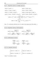
Figure 11.2. The vaginal wall has been opened and dissected off of the
bladder, exposing the pubocervical (perivesical) fascia.
152 Vaginal Surgery for Incontinence and Prolapse
fascia (3-0 polyglactin) from the bladder neck to
the vaginal cuff (Figure 11.3). Once all sutures
have been placed, cystoscopy is performed to
ensure that there is no bladder or ureteral injury;
5 mL of indigo carmine is given 15 minutes prior
to cystoscopy so that ureteral effl ux can be easily
visualized. The centrally imbricating sutures are
then tied in an anterior-to-posterior direction.
Given the presence of the mesh, it is doubtful
these sutures are even necessary. We still reduce
the central hernia in this manner to allow ease
of mesh attachment and placement. To correct
the lateral defect, we aim for the periosteum of
the descending ramus of the symphysis pubis. A
0-polyglactin suture is placed through the previ-
ously dissected infralevator obturator connec-
tive tissue just over the periosteum (Figure 11.4).
We have found this to be a reliable, strong, non-
mobile anchor. A circular soft Prolene mesh is
cut in the shape of a disk (5 × 5 cm). This is
secured to the previously plicated cardinal liga-
ments posteriorly, and the obturator fasciae
laterally. Two additional sutures are placed ante-
riorly, one on each side of the proximal urethra/
bladder neck through the perivesical fascia, to
complete the fi xation of the mesh. The mesh is
trimmed as needed to ensure taut positioning
(Figure 11.5). The excess vaginal wall is then
trimmed.
If a vault repair was also performed, the col-
posuspension sutures (to the sacrouterine liga-
ments) are tied prior to trimming the excess
vaginal wall. The midline vaginal incision is
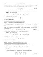
Figure 11.3. Anterior colporrhaphy sutures in place.
Figure 11.4. An 0-polyglactin suture is placed through the obturator
fascia over the periosteum just above the descending ramus of the sym-
physis pubis. This is below the arcus tendineus.
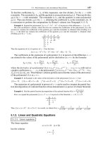
Figure 11.5. The disk-shaped mesh has been trimmed to fit tautly in
position. It is secured anteriorly on each side of the proximal urethra,
laterally to the obturator fascia, and posteriorly to the cardinal
ligaments.
Anterior Compartment 153
closed with a running 3-0 polyglactin suture. If
a rectocele is present, we restore the rectovaginal
fascia, levator hiatus, and perineal defects.
An antibiotic-soaked vaginal pack is placed
until discharge. Most patients go home after 24
hours of observation. The suprapubic tube is
capped, and attempts at voiding are instituted
prior to discharge. Patients are instructed in the
use of the suprapubic catheter in checking post-
void residuals at home. The majority of patients
void within 72 hours, so the placement of a supra-
pubic tube or urethral catheter (and possible pre-
operative teaching of intermittent catheterization)
is the surgeon’s preference. Because many of our
patients are not local residents, we currently
place SPTs in a majority of our patients. We keep
the catheter for at least 1 week to minimize pos-
sible urinary extravasation with its removal.
Our early series of 94 consecutive patients
with stage IV cystocele repairs showed cure or
improvement of the anatomic prolapse in 82%
of patients. The range of follow-up was 8 to 22
months. Our complication rate was 8%. There
was transient retention in two patients and de
novo urinary incontinence in 4% of the patients.
Although no patient developed recurrent high-
grade cystocele, two patients developed mild
grade 2 cystoceles. No complications related
directly to the mesh were seen—specifi cally, no
erosions or graft infections (Urology 66:57–65,
2005 by Rodriguez, LV et al.). We have previ-
ously reported our promising results with the
Prolene sling in treating stress urinary inconti-
nence (34). We now have similar success in the
treatment of anterior compartment prolapse
without any cases of permanent retention.
Conclusion
The diagnosis and treatment of stage IV cysto-
celes is challenging, even to the most experi-
enced pelvic surgeons. Forces that alter the
normal support of the anterior compartment
often result in disorders of the other compart-
ments, resulting in posterior vaginal wall pro-
lapse, which includes uterine or vaginal vault
prolapse, and perineal laxity. Therefore, to
effectively evaluate and treat women with ante-
rior compartment relaxation with or without
urinary incontinence, it is imperative that the
clinician not only understand the normal struc-
ture and function of the lower urinary tract, but
also has a working knowledge of the anatomy
and pathophysiology of pelvic support. The
surgeon will then be able to effectively address
their female patients who present with com-
plaints related to defi ciencies in pelvic support,
and appropriately apply the current methods of
evaluation and treatment discussed in other
chapters of this book.
References
1. Black NA, Downs SH. The effectiveness of surgery for
stress incontinence in women: a systematic review. Br
J Urol 1996;78:497–510.
2. Leach GE, Dmochowski RR, Appell RA, et al. Female
stress incontinence clinical guidelines panel summary
report on surgical management of female stress urinary
incontinence. J Urol 1997;158:875–880.
3. Glazener CM, Cooper K. Anterior vaginal repair for
urinary incontinence in women. Cochrane Database of
Systematic Reviews 2000:CD001755.
4. Weber AM, Walters ME. Anterior vaginal prolapse:
review of anatomy and techniques of surgical repair.
Obstet Gynecol 1997;89:811–818.
5. Weber AM, Walters MD, Piedmonte MR, et al. Anterior
colporrhaphy: a randomized trial of 3 surgical tech-
niques. Am J Obstet Gynecol 2001;185(6):1299–1306.
6. Richardson AC, Edmonds PB, Williams NL. Treatment
of stress urinary incontinence due to paravaginal fascial
defect. Obstet Gynecol 1981;57:357–362.
7. White GR. An anatomic operation for the cure of cys-
tocele. Am J Obstet Gynecol 1912;65:286–290.
8. Richardson AC, Lyon JB, Williams NL. A new look
at pelvic relaxation. Am J Obstet Gynecol 1976;126:
568–573.
9. Shull BL, Benn SJ, Kuehl TJ. Surgical management of
prolapse of the anterior vaginal segment: an analysis
of support defects, operative morbidity and anatomic
outcomes. Am J Obstet Gynecol 1994;171:1429–1439.
10. Raz S, Klutke CG, Golomb J. Four-corner bladder and
urethral suspension for moderate cystocele. J Urol
1989;142:712–715.
11. Albo M, Dupont MC, Raz S. Transvaginal correction of
pelvic prolapse. J Endourol 1996;10:231–239.
12. Dmochowski RR, Zimmern PE, Ganabathi K, et al. Role
of the four-corner bladder neck suspension to correct
stress incontinence with mild to moderate cystocele.
Urology 1997;49:35–40.
13. Miyazaki FS, Miyazaki DW. Raz four-corner suspen-
sion for severe cystocele: poor results. Int Urogynecol
J 1994;5:94–97.
14. Migliari R, Usai E. Treatment results using a mixed
fi ber mesh in patients with grade IV cystocele. J Urol
1999;161:1255–1258.
15. Kelly M, Zimmern PE, Leach GE. Complications of
bladder neck suspension procedures. Urol Clin North
Am 1991;18:339.
16. Raz S, Little NA, Juma S. Repair of severe anterior wall
prolapse (grade IV cystourethrocele). J Urol 1991;146:
988–992.
17. Kohli N, Sze EHM, Roat TW, et al. Incidence of recur-
rent cystocele after anterior colporrhaphy with and
without concomitant transvaginal needle suspension.
Am J Obstet Gynecol 1996;175:1476–1482.
154 Vaginal Surgery for Incontinence and Prolapse
18. Bump RC, Hurt WG, Theofrastous JP, et al. Random-
ized prospective comparison of needle colposuspen-
sion versus endopelvic fascia placation for potential
stress incontinence prophylaxis in women undergoing
vaginal reconstruction for stage II or IV pelvic organ
prolapse. Am J Obstet Gynecol 1996;175:326–333.
19. Holley RL, Varner RE, Gleason BP, et al. Recurrent
pelvic support defects after sacrospinous ligament fi xa-
tion for vaginal vault prolapse. J Am Coll Surg 1995;
180:444–448.
20. Benson JT, Lucente V, McClellan E. Vaginal versus
abdominal reconstructive surgery for the treatment of
pelvic support defects: a prospective randomized study
with long-term outcome evaluation. Am J Obstet
Gynecol 1996;175:1418–1421.
21. Cross CA, Cespedes RD, McGuire EJ. Treatment results
using pubovaginal slings in patients with large cysto-
celes and stress incontinence. J Urol 1997;158:
431–434.
22. Kobashi KC, Leach GE, Chon J, Govier FE. Continued
multicenter followup of cadaveric prolapse repair with
sling. J Urol 2002;168:2063–2068.
23. Goldberg RP, Koduri S, Lobel RW, et al. Protective
effect of suburethral slings on postoperative cystocele
recurrence after reconstructive pelvic operation. Am J
Obstet Gynecol 2001;185:1307–1313.
24. Young SB, Daman JJ, Bony LG. Vaginal paravaginal
repair: one-year outcomes. Am J Obstet Gynecol
2001;185:1360–1367.
25. Elkins TE, Chesson RR, Videla F, et al. Transvaginal
paravaginal repair: a useful adjunctive procedure in
pelvic relaxation surgery. J Pelvic Surg 2000;1:11–15.
26. Nguyen JK, Bhatia NN. Resolution of motor urge
incontinence after surgical repair of pelvic organ pro-
lapse. J Urol 2001;166:2263–2266.
27. Julian TM. The effi cacy of Marlex mesh in the repair of
severe, recurrent vaginal prolapse of the anterior mid-
vaginal wall. Obstet Gynecol 1996;175:1472–1475.
28. Nicita G. A new operation for genitourinary prolapse.
J Urol 1998;160:741–745.
29. Migliara R, De Angelis M, Madeddu G, et al. Tension-
free vaginal mesh repair for anterior vaginal wall pro-
lapse. Eur Urol 2000;38:151–155.
30. Dora CD, Dimarco DS, Zobitz ME, et al. Time-
dependent variations in biomechanical properties of
cadaveric fascia, porcine dermis, porcine small intes-
tine submucosa, polypropylene mesh, and autologous
fascia in the rabbit model: implications for sling
surgery. J Urol 2004;171:1970–1973.
31. Sand PK, Koduri S, Lobel RW, et al. Prospective ran-
domized trial of polyglactin 910 mesh to prevent recur-
rence of cystoceles and rectoceles. Obstet Gynecol
2001;184:1357–1364.
32. Ghoniem GM, Kapoor DS. Nonautologous sling mate-
rials. Curr Urol Rep 2001;2:357–363.
33. Birch C, Fynes MM. The role of synthetic and biological
prostheses in reconstructive pelvic fl oor surgery. Curr
Opin Obstet Gynecol 2002;14:527–535.
34. Rodriguez LV, Raz S. Polypropylene sling for the treat-
ment of stress urinary incontinence. Urology 2001;
58(5):783–785.
35. Gallentine ML, Cespedes RD. Occult stress urinary incon-
tinence and the effect of vaginal vault prolapse on abdom-
inal leak point pressures. Urology 2001;57(1):40–44.
36. Beck RP, McCormick S, Nordstrom L. A 25 year experi-
ence with 519 anterior colporrhaphy procedures.
Obstet Gynecol 1991;78:1011–1018.
37. Nichols DH. Cystoceles. In: Nichols DH, ed. Gyneco-
logic and Obstetric Surgery. St. Louis: CV Mosby, 1993:
334–362.
38. Farrell SA, Ling C. Currycombs for the vaginal para-
vaginal defect repair. Obstet Gynecol 1997;90:845–847.
39. Scotti RJ, Garely AD, Greston WM, et al. Paravaginal
repair of lateral vaginal wall defects by fi xation to the
ischial periosteum and obturator membrane. Am J
Obstet Gynecol 1998;179:1436–1445.
40. Mallipeddi PK, Steele AC, Kohli N, et al. Anatomic and
functional outcome of vaginal paravaginal repair in the
correction of anterior vaginal prolapse. Int Urogynecol
J 2001;12:83–88.
41. Bruce GR, El Galley R, Galloway N. Paravaginal defect
repair in the treatment of female stress urinary incon-
tinence and cystocele. Urology 1999;54:647–651.
42. Canepa G, Ricciotti G, Introini C, et al. Horseshoe-
shaped Marlex mesh for the treatment of pelvic fl oor
prolapse. Eur Urol 2000;39(suppl 2):23–26.
43. Kobashi KC, Mee SL, Leach GE. A new technique for cys-
tocele repair and transvaginal sling: the cadaveric pro-
lapse repair and sling (CaPS). Urology 2000;56:9–14.
44. Raz S, Stothers L, Chopra A. Vaginal reconstructive
surgery for incontinence and prolapse. In: Walsh PC,
Retik AB, Vaughan ED Jr, Wein AJ, eds. Campbell’s
Urology, 7th ed. Philadelphia: WB Saunders, 1998:
1059–1094.
We are what we repeatedly do. Excellence, then, is not
an act, but a habit.
Aristotle
The surgical management of pelvic organ pro-
lapse is more challenging than that for stress
12
Uterine and Vaginal Vault Prolapse
Peggy A. Norton
155
Procedures for Uterine Prolapse . . . . . . . . . . 156
Vaginal hysterectomy . . . . . . . . . . . . . . . . . 157
Technique . . . . . . . . . . . . . . . . . . . . . . . . . 157
Prophylactic Suspension of the Vaginal
Cuff . . . . . . . . . . . . . . . . . . . . . . . . . . . . . 158
McCall Culdoplasty . . . . . . . . . . . . . . . . . 158
Mayo Culdoplasty . . . . . . . . . . . . . . . . . . 159
Resuspension of the Vaginal Apex After
Vaginal Hysterectomy for Uterine
Prolapse . . . . . . . . . . . . . . . . . . . . . . . . . 159
Vaginal Procedures to Preserve the
Uterus . . . . . . . . . . . . . . . . . . . . . . . . . . 160
Laparoscopic Shortening of the
Uterosacral Ligaments . . . . . . . . . . . . 161
Posthysterectomy Vaginal Vault
Prolapse . . . . . . . . . . . . . . . . . . . . . . . . . 161
Suspensory Procedures . . . . . . . . . . . . . . . . 161
High Uterosacral Ligament
Suspension . . . . . . . . . . . . . . . . . . . . . . 161
Iliococcygeus Fascia Fixation . . . . . . . . . 163
Mayo Culdoplasty . . . . . . . . . . . . . . . . . . 163
Sacrospinous Ligament Suspension
(SSLS) or Fixation . . . . . . . . . . . . . . . . 163
Levator Myorrhaphy with Apical
Fixation . . . . . . . . . . . . . . . . . . . . . . . . . 164
Obliterative Procedures . . . . . . . . . . . . . . . . 164
Lefort Colpocleisis/Total Colpectomy
with High Levator Myorrhaphy . . . . . 164
Abdominal Approach to Vaginal Vault
Prolapse . . . . . . . . . . . . . . . . . . . . . . . . . 165
urinary incontinence, and detection and cor-
rection of apical repairs can be the most dif-
fi cult of all pelvic fl oor defects. One-third of
procedures performed for pelvic organ prolapse
are secondary procedures (1). The number of
procedures performed in the United States to
treat posthysterectomy vaginal vault prolapse
increased dramatically from 1437 procedures in
1979 to 22,025 procedures in 1997 (2), while the
overall number of procedures performed for
pelvic organ prolapse declined from 226,000 in
1979 to 205,000 in 1997. Despite this apparent
epidemic of apical prolapse, residency training
for urologists and gynecologists alike favors
repair of cystoceles and rectoceles. Moreover,
defects of the anterior and posterior vaginal
walls are more common and easier to detect
than apical defects such as uterine prolapse and
vaginal vault prolapse (3). For these reasons,
correction of apical defects remains a surgical
challenge for many surgeons.
Suspension of the vaginal apex is the keystone
of surgical repair for pelvic organ prolapse. Good
suspension of the uterus or posthysterectomy
vaginal cuff protects the ventral and dorsal walls
from transabdominal forces that push these
tissues toward the introitus. Recognition that an
apical defect exists remains a major diagnostic
problem in the evaluation of pelvic organ pro-
lapse. While anterior and posterior wall defects
can be demonstrated on vaginal exam with a Sims
speculum or half blade of a bivalve speculum, the
apex may be undeveloped in the supine position
used for examination or surgery. A careful exami-
nation with the patient sitting at a 45-degree angle
156 Vaginal Surgery for Incontinence and Prolapse
or standing often produces the abdominal pres-
sure needed to expose the apical defect. Apical
defects are best demonstrated in the standing
position (4). Clark et al (5) reported that the
highest rates of reoperation for pelvic fl oor disor-
ders in a managed care system occurred in women
undergoing surgery for apical defects (33% reop-
eration) or combined anterior/apical (15%) or
posterior/apical (12%). Thus, failure to appreci-
ate an apical defect prior to surgery may lead to
poor surgical results, and is an important reason
for surgical failure in pelvic organ prolapse.
There is no specifi c degree of descent that
mandates surgical correction. In general, stage
II pelvic organ prolapse is the level of descent
at which prolapse often becomes symptomatic
(6). However, resuspension of the apex may be
considered at levels above this (less descent),
especially with posthysterectomy vaginal vault
prolapse. For example, an anterior wall defect to
2 cm above the hymeneal ring is quite common
(7), while vaginal vault prolapse to the same level
is uncommon and may be much more symptom-
atic, often because there is a large enterocele
inside the vault prolapse. Apical defects rarely
present as an isolated prolapse, and consideration
of whether to repair the apical defect needs to
include sexual function and whether the other
defects will suffer from lack of good apical
support. As another example, a woman with a
large rectocele to the introitus and a vaginal cuff
at 5 cm often needs apical resuspension; there is
often an enterocele pushing the cuff down, and
resuspension of the posterior fi bromuscular wall
of the vagina into such a low apex leads to a vagina
that is too short for comfortable coitus.
Once the degree of descensus has been
assessed (see Chapter 4), a decision is made
about whether repair of the apical defect is indi-
cated, either in isolation or as part of a general
repair for prolapse. Just as with other conditions
that affect quality of life without threatening life,
surgical correction of uterine or vault prolapse
must be a balance between risk and benefi t.
While the risk of surgery is well known to sur-
geons, patients may assume that risks apply to
others and not to themselves. The benefi t of pro-
lapse surgery is not guaranteed: As a general rule
in pelvic organ prolapse, patients should be suf-
fi ciently bothered by their condition to under-
take surgery, knowing that in one third of cases,
the result will not be satisfactory (1,5).
Surgical goals may differ depending on
whether the patient wishes to be sexually active,
her lifestyle, outcomes of prior corrective proce-
dures, comorbidities, and risks for recurrent
prolapse. Patients wishing to continue heavy
lifting or exercise, or with risk factors for recur-
rence such as higher defects (stage III and IV) or
younger age (8) may consider the most durable
procedure, often an abdominal approach such
as sacrocolpopexy (9). Nevertheless, vaginal
repair is to be preferred owing to shorter hospi-
talization and recovery (10). For patients wishing
to be sexually active, the postoperative goal
should be a vaginal length of approximately 9 to
10 cm with good caliber maintained throughout
the vagina. Overcorrection of the anterior wall
leads to signifi cant vaginal shortening, while
overcorrection of the posterior wall leads to a
“shelf” owing to plication of the levators across
the midline, and subsequent dyspareunia. The
apex can be overcorrected, but this usually
occurs with abdominal surgeries in which the
vagina is placed on excessive tension, often with
a result of excessive vaginal length.
Specifi c procedures to address apical defects
can be categorized by the absence or presence of
the uterus, either posthysterectomy vaginal vault
prolapse or uterine prolapse. The latter includes
vaginal hysterectomy used to treat the prolapse,
and prophylactic procedures to prevent future
pelvic organ prolapse or to preserve the uterus
in the presence of support defects. The tech-
nique is described for the procedures that are
widely applicable.
Procedures for Uterine Prolapse
The cervix is located in the anterior (ventral)
vaginal wall, often several centimeters nearer
the hymeneal ring than the posterior cul-de-
sac. Optimally, the cul-de-sac is 10 cm above the
hymeneal ring, with the cervix at 8 to 9 cm
above (11). Descensus with Valsalva to the mid-
vagina, termed stage I in the Baden-Walker
system, corresponds to a cervix located 4 to
5 cm above the hymen, equivalent to C = −4,
stage I in the pelvic organ prolapse quantifi ca-
tion (POPQ) system. Once the uterus descends
to 2 to 3 cm above the hymen, it is still termed
stage I prolapse in the POPQ system, but may be
entirely normal in many women, with adequate
vaginal length for coitus due to the higher cul-
de-sac (12). Descent to a centimeter above or
below the hymeneal ring (POPQ point C = +1 to
−1) is a stage II apical prolapse (Baden-Walker
Enterocele and Rectocele/Perineorrhaphy 157
stage II) and is the level at which most uterine
prolapses become symptomatic (13). If a patient
complains of symptoms due to prolapse above
this level, consideration should be given to other
sources for the complaints (13). Uterine pro-
lapse at this level can still be managed with a
pessary, but consideration should be given to
surgical management if the patient is symptom-
atic. Beyond this level, the prolapsed uterine is
increasingly symptomatic and managed surgi-
cally in many instances.
In a few isolated cases, cervical elongation
occurs and represents good apical support, with
the cervix appearing at the introitus due to elon-
gation of the cervix itself. The measure C is the
cervical descent to symptomatic levels (i.e.,
within 1 cm of the hymeneal ring), while the D
(measuring descent of the cul-de-sac) is at the
level of 8 to 10 cm above the hymeneal ring.
Although such patients have good apical support
for reattachment at hysterectomy, the hysterec-
tomy can be more diffi cult. Occasionally such
patients are managed with a Manchester ampu-
tation of the cervix (14).
Once the decision is made to treat uterine pro-
lapse with surgery, the usual course is to plan
removal of the uterus as part of the treatment.
Any unexplained vaginal bleeding, or any abnor-
mal Pap smear needs to be evaluated before
removal of the uterus. In general, unless the post-
menopausal bleeding can be explained by such
factors as regular withdrawal from progesterone
as part of hormone replacement therapy, an
endometrial biopsy should be performed prior
to hysterectomy. For pre- or perimenopausal
women, any abnormal uterine bleeding needs to
be evaluated similarly: bleeding more often that
every 21 days, bleeding 7 days or longer, or exces-
sive menstrual bleeding, especially in women at
high risk for uterine cancer such as obese or nul-
liparous women. A bimanual exam should be
performed as part of the preoperative assessment
to exclude adnexal enlargement, and the biman-
ual exam repeated at the beginning for surgery
in case of a pelvic mass.
Vaginal Hysterectomy
Vaginal Hysterectomy has been described in
many other texts, but in the case of pelvic organ
prolapse, particular attention should be paid to
preservation of the uterosacral ligaments for
resuspension of the vaginal apex. In the absence
of cervical elongation, uterine prolapse to the
level of the hymeneal ring means that the utero-
sacral ligament is attaching to the cervix just
above the hymen, and simple resuspension of
the vaginal cuff to the ligaments at this level
may not result in a well-supported vaginal apex
posthysterectomy. Factors that favor the vaginal
approach for hysterectomy include a wide pubic
arch, some descensus of the uterus and cervix,
parity, concomitant repairs requiring a vaginal
approach, and any factors that might discour-
age the abdominal approach such as obesity.
Factors that discourage the vaginal approach
include uterine enlargement (beyond the size of
a 12 week pregnancy [15]), narrow pubic arch
(16), a history of cesarean or pelvic adhesions,
and desired prophylactic oophorectomy. While
these factors do not preclude a vaginal approach,
careful thought must be given to these cases and
the patient informed of the possibility of chang-
ing to an abdominal approach during the oper-
ation. The presence of a large adnexal mass
or known dense pelvic adhesions or uterine
enlargement beyond the size of a 16-week preg-
nancy (palpable fundus midway between the
pubic symphysis and the umbilicus) are best
managed abdominally.
Technique
The cervix is grasped, scored circumferentially
with scalpel (Figure 12.1), and the vaginal skin
and underlying fi bromuscular walls will eventu-
ally be sewn together to form the new apex of the
vaginal cuff. Next, the anterior and posterior
Figure 12.1. The cervix is placed on traction and scored with scalpel or
cautery.
158 Vaginal Surgery for Incontinence and Prolapse
refl ections of the peritoneum behind the vaginal
wall are identifi ed. An anterior colpotomy is
performed usually before the posterior colpot-
omy. The surgical plane between the cervix and
the bladder may be obscured by prior cesarean
section, and given this risk of dependent bladder
perforation we usually leave some urine in the
bladder to assist with early diagnosis of cystot-
omy. With posterior colpotomy, the uterosacral
ligaments should be palpated so that the site of
entry into the posterior cul-de-sac does not
unintentionally interrupt either uterosacral
ligament (Figure 12.2). The apex will be resus-
pended to the uterosacral ligaments, so this
pedicle should be ligated at the safest point in a
cephalad direction (Figure 12.3). Each pedicle is
clamped with a curved Heaney clamp, cut, and
ligated with delayed absorbable suture. Pro-
gressive pedicles are taken of the uterosacral
ligaments, the cardinal ligaments, the uterine
arteries, the broad ligament, and the utero-
ovarian ligament. Once the utero-ovarian liga-
ments are transected, the specimen is passed off
and pedicles inspected. If the ovaries are to be
removed, the tube and ovary are grasped with a
Babcock clamp, and the pedicle clamped with
care to remain medial to the ureter.
At any point along the way, the ureter is close
to the plane of dissection. Hurd and colleagues
(17) studied the relationship between the cervix
and the ureter on computed tomography (CT),
and found that at the most dorsal refl ection of
the ureter, the average distance from ureter to
cervical margin was 2.3 ± 0.8 cm (range, 0.1–
5.3 cm) They concluded that fi nding that this
distance is <0.5 cm in 12% of the women studied
may explain the relatively common occurrence
of ureteral injury during hysterectomy. In
uterine prolapse, the ureters have a less predict-
able course and may be pulled caudal and medial
by the prolapsing uterus. Delancey (18) studied
the effect of prolapse on the course of the ureter,
and found that for every 3 cm of cervical descent
the ureters descend 1 cm, thereby widening the
ureterocervical gap and permitting ligament
shortening during vaginal hysterectomy.
Prophylactic Suspension of the
Vaginal Cuff
The support of the vaginal vault after hysterec-
tomy requires some consideration, and may be
achieved by a “prophylactic” procedure in cases
of normal uterine support: attachment of utero-
sacral ligaments to the vaginal cuff, McCall cul-
doplasty, and Mayo culdoplasty (Figure 12.4).
The simplest technique of prophylactic vault
suspension mimics the procedure performed
abdominally: the sutures placed in the uterosac-
ral ligament pedicles are held and reattached to
the vaginal cuff by drawing each end through the
vaginal cuff using a free Mayo needle.
McCall Culdoplasty
With the open cuff, a delayed absorbable suture
is placed at approximately the 5 o’clock position
into the cuff traveling cephalad (Figure 12.5).
The suture is then brought lateral to medial
Figure 12.3. The uterosacral ligament is skeletonized, the ureter pal-
pated against the pubic ramus, and the ligament clamped, cut, ligated,
and held. This results in a uterosacral pedicle that is 4 to 5 cm more
cephalad than if the pedicle is taken at the insertion into the cervix.
Figure 12.2. After the posterior colpotomy, the uterosacral ligaments
can be palpated with the cervix on traction.
Enterocele and Rectocele/Perineorrhaphy 159
through the uterosacral ligament on the patient’s
left, and then in an interrupted fashion across
the peritoneum and into the contralateral utero-
sacral ligament. The suture is then brought back
out through the cuff at approximately the 7
o’clock position and both ends tied down. Several
internal McCall sutures may be placed through
the peritoneum from the uterosacral ligament to
the uterosacral ligament to close the cul-de-sac
prior to tying the external McCall sutures.
Mayo Culdoplasty
Webb and colleagues (19) reported on a large
cohort of women followed for a median 16 years
after Mayo culdoplasty for posthysterectomy
vaginal vault prolapse with 73% follow-up; 15%
of women had symptoms or signs of prolapse,
and the authors commented on the increas-
ing numbers of patients presenting with this
problem over the study period.
Cruikshank and Kovac (20) reported better
support of the apex with the McCall culdoplasty
in a randomized trial comparing this procedure
with simple peritoneal closure or vaginal Mos-
chowitz procedures. In women undergoing hys-
terectomy for benign indications, 25% developed
an apical defect by 3 years, and for those under-
going simple peritoneal closure, almost 40%
developed an apical defect. The best outcomes
were achieved with the McCall culdoplasty,
where only two of 32 subjects developed apical
defects. These discouraging numbers by skilled
vaginal surgeons point to the technique as a
possible explanation for the increasing rates of
posthysterectomy for vault prolapse seen in
the U.S.
Resuspension of the Vaginal Apex After
Vaginal Hysterectomy for Uterine Prolapse
In cases of uterine prolapse at the time of vaginal
hysterectomy, multiple procedures have been
recommended. In addition to the culdoplasty
techniques recommended in textbooks, there
are several reports of uterine preservation with
apical support procedures. These are mostly
retrospective case series using the sacrospinous
ligament fi xation involving fewer than 50 sub-
jects with short follow-up and poorly defi ned
outcome criteria (21,22).
There are two options for suspending the
vaginal cuff to the uterosacral ligaments at the
time of vaginal hysterectomy for uterine pro-
lapse: shortening the ligaments at the time of
transecting the ligament as the fi rst pedicle of
the hysterectomy, or placing sutures higher in
the ligament at the time of attachment to the
apex, essentially as a high uterosacral ligament
suspension. If the shortening of the ligaments
is to be done at the time of transection, con-
siderable shortening must be accomplished to
establish a site appropriately cephalad for
resuspension, optimally at 8 to 10 cm. With a
cervix at the hymeneal ring, the uterosacral
insertion is approximately −2 cm, thus consider-
able shortening needs to occur. The location of
the ureter should be established by palpation
through the anterior colpotomy, pinning the
Figure 12.4. The right uterosacral ligament can be traced from the held
sutures of the pedicle cephalad. The posterior cuff has been sutured to
control bleeding.
Figure 12.5. The external McCall suture is completed by bringing the
suture out at the 7 o’clock position on the posterior cuff. The two ends
are then tied together after completing the internal McCall sutures.
160 Vaginal Surgery for Incontinence and Prolapse
ureter against the pubic ramus. The ligament
should be skeletonized some distance up the
ligament before clamping, as much as 4 to 6 cm.
If resuspension is to be performed after removal
of the uterus, this is usually done after the ante-
rior and possibly posterior wall defects are cor-
rected. With traction on the uterosacral pedicle
in the direction of the ceiling (instead of the
surgeon’s nose, which pulls the ureter toward
the ligament), a suture should be placed into the
ligament lateral to medial (to direct the needle
away from the ureter) at a depth 8 to 10 cm ceph-
alad to the introitus. These sutures should then
be attached into the anterior and posterior
vaginal cuff. If permanent suture is used, the
posterior cuff alone is suffi cient because the
anterior cuff is near the course of the ureter.
Permanent suture is often considered because it
offers additional strength and durability, but has
the additional risk of granulation at the cuff. If
insuffi cient length can be accomplished, then
one of the following apical suspensions should
be considered: high uterosacral ligament sus-
pension or sacrospinous ligament suspension.
These procedures are described below for vaginal
vault suspension, but can be performed at time
of vaginal hysterectomy. There are no reports
comparing each of these procedures. Cruikshank
(20) reported better support of the apex with the
McCall culdoplasty in a randomized trial com-
paring this procedure with simple peritoneal
closure or vaginal Moschowitz procedures.
Vaginal Procedures to Preserve the Uterus
Although most published descriptions of
uterine prolapse involve removal of the uterus,
there are a few descriptions of uterine preserva-
tion. This may be considered if the woman
desires preservation or future childbearing, or
it may be a philosophical decision to preserve
the uterus. The paradigm to remove the uterus
because of pelvic organ prolapse needs to be
challenged: surgeons in France do not routinely
remove the uterus for support defects, and it
may be that surgical education in techniques
for uterine preservation might increase the
options for many women. Two large random-
ized trials compared traditional abdominal
hysterectomy to supracervical hysterectomy, in
which the uterus is removed but the cervix is
conserved along with its ligamentous support
structures (23).
Uterine preservation with apical support
procedures are mostly retrospective case series
(level 3 evidence) using the sacrospinous liga-
ment fi xation involving fewer than 50 subjects
with short follow-up and poorly defi ned outcome
criteria (21,24).
Laparoscopic Shortening of the
Uterosacral Ligaments
This procedure is done with permanent sutures
through the insertion of the ligament into the
cervix, and at a point cephalad where a contigu-
ous ligament can be identifi ed (instead of an
empty sleeve of peritoneum). How high should
the resuspension be? One can push the uterus in
a cephalad direction to accomplish a point of C
= −8 cm or higher, and then suspended to a point
on the ligament to maintain this elevation.
Two additional procedures might be consid-
ered for apical resuspension with uterine pres-
ervation, especially in the case of desired fertility.
A high uterosacral ligament suspension to the
cervix can be performed through a posterior col-
potomy. The posterior cul-de-sac is opened as
with the beginning of vaginal hysterectomy. A
permanent suture may be placed in the utero-
sacral ligaments and attached to the cervix in
the midline. This procedure seems to produce
modest suspension but some prevention of
further prolapse. Likewise, a sacrospinous liga-
ment suspension to the cervix has been described,
with subsequent pregnancies (25). In our hands
this procedure is best performed in a woman
with a fairly large cervix (multiparous) with the
sutures placed more medially (and therefore
more dorsally) on the coccygeus muscle with its
underlying sacrospinous ligament; otherwise,
the cervix is diffi cult to draw laterally in some
individuals.
Posthysterectomy Vaginal
Vault Prolapse
Vaginal vault prolapse occurs after surgical
hysterectomy, and level for level is more symp-
tomatic than uterine prolapse to the same ana-
tomic degree. Correct assessment of the apex
includes direct visualization of the cuff (seen
with two small puckers at the lateral margins in
patients with some suspension) with an open
Graves speculum. The patient is asked to bear
Enterocele and Rectocele/Perineorrhaphy 161
down while in a semisitting supine lithotomy,
and the speculum is withdrawn until further
descent of the apex ceases. The distance from
the apex to the hymeneal ring can then be mea-
sured in centimeters. Similar to uterine pro-
laps e, it may be helpf u l to repe at t he e xa mi nat io n
with the patient in the standing position and
estimating the distance from the apex to the
introitus. Small bowel pushing the apex down
is common, and this enterocele can often be
palpated as loops of bowel in the prolapsing
vault. A vault sitting from a 1 cm above the
hymeneal ring (POPQ C = −1, stage II) or beyond
is usually symptomatic and treatment should be
considered.
More clinical judgment is needed to decide
whether apical defects above this level require
treatment. In isolated apical defects from 2 to
7 cm above the hymeneal ring, deep dyspareunia
is the main concern. Because these defects are
more commonly seen in combination, asymp-
tomatic apical defects may need to be addressed
surgically to suspend and optimize repair of
symptomatic anterior and posterior wall defects.
Thus, asymptomatic apical defects to approxi-
mately midvagina (C = −4 or −5) may need to be
included in the repair of cystoceles and recto-
celes, if these defects are symptomatic. Each case
needs to be individualized; apical defects require
more skill and more complex surgery for repair,
and the risk/benefi t ratio for surgery needs to
refl ect this. But the more common error seen
with treatment of the apex is failure to detect the
apical problem.
In the case of posthysterectomy apical defects,
vaginal procedures can be either supportive
or obliterative. Available data on published
series and trials with more than 50 subjects are
listed for sacrospinous ligament suspension
(Table 12.1), high uterosacral ligament suspen-
sion (Table 12.2) and colpocleisis/colpectomy
(Table 12.3).
Table 12.1. High uterosacral ligament suspension procedures
Reference n Follow-up, months (range) Success rate Complications
Pohl and Frattarelli, 1997 (40) 40 6–40 89%
Jenkins, 1997 (41) 50 6–48 88%
Barber, 2000 (42) 46 15.5 (3.5–40) 90% 11% ureteral
Karram et al, 2001 (43) 168 6–36 89% 2.4% ureteral
Shull, 2000 (44) 289 Not stated 87% 1% ureteral
Amundsen, 2003 (45) 33 28 (6–43) 82%
Table 12.2. Sacrospinous ligament suspension procedures
a
Citation n Follow-up Success rate Outcome measures
Morley and DeLancey, 1988 (46) 78 1 mo–11 yr 78% Subjective, objective
Imparato 1992 (47) 155 ? 90% Objective
Shull 1992 (48) 81 2–5 yr 65% Objective
Pasley, 1995 (49) 156 6–83 mo 94% Subjective, objective
Benson et al, 1996 (50) 42 12–66 mo 29% Objective (third party), RCT
Hardiman, 1996 (51) 125 26.4 mo (98%)
b
Objective
Penalver, 1998 (53) 160 18–78 mo 85% Objective
Colombo, 1998 (54) 62 4–9 yr 73% Subjective, objective
Meschia, 1999 (55) 91 1–6.8 yr (94%)
b
Objective
Sze, 1997 (52) 54 7–72 mo 67%
c
Objective
Lantzsch, 2001 (56) 123 6 mo–9 yr (97%)
d
Objective
RCT, randomized controlled trial.
a
Using publications reporting more than 50 subjects, interpretable data. One RCT is included in which 42 subjects were randomized to SSLS.
b
Apex only; recurrent cystocele 16%, recurrent rectocele 10%, recurrent enterocele 6%.
c
13/18 anterior wall recurrence.
d
Apex only; 10 recurrent cystoceles, one recurrent rectocele, one recurrent enterocele.
162 Vaginal Surgery for Incontinence and Prolapse
Suspensory Procedures
High Uterosacral Ligament Suspension
First reported in 1997, this procedure suspends
the vaginal apex to the remnants of the utero-
sacral ligaments at the level of the ischial spines
and cephalad, with attention to incorporation
of the rectovaginal fascia and pubocervical
fascia into the permanent sutures at the apex.
The procedure maintains the vaginal axis in
the midline, allows adjustment of the vaginal
length, and can include the use of allograft or
xenografts in the suspension.
Technique
Since considerable distortion of the vaginal
walls can occur in pelvic organ prolapse, the
new vaginal apex should be identifi ed and
marked with two silk sutures at that point
where the anterior and posterior walls will have
equal length and tension and the fi nal length of
the vagina will approximately 10 cm. If there
is excessive length, the “toe” of the apex may
be removed; if there is insuffi cient length in a
sexually active woman, an abdominal proce-
dure using mesh may be preferable, since the
vaginal approach is unlikely to result in more
vaginal length.
The vaginal wall is opened in the midline from
perineal body to bladder neck, taking care over
the vaginal apex to avoid opening the enterocele
prematurely. A modest amount of lateral dissec-
tion in the anterior and posterior walls can iden-
tify whether the fi bromuscular wall of the vagina
can be repaired, or whether a tissue graft is
needed. Now the enterocele should be entered
and the bowel packed out of the way with tagged
lap sponges. The right side of the pelvis between
the sigmoid and the side wall should be well
visualized up to the level of S4, and the course of
the ureter appreciated. A lighted retracter
(Miyazaki retractor, Marina Medical, Hollywood
FLA) can improve visualization dramatically.
Beginning at approximately the 8 o’clock posi-
tion on the open peritoneum, a long Allis clamp
is used to place traction on the peritoneum and
is tracked upward toward the sacrum. While the
more caudal portion of the uterosacral ligament
may be an empty peritoneal sleeve, the remain-
ing cephalad portion of the ligament may be
identifi ed with the Allis clamp, optimally at a
location 9 to 10 cm above the hymeneal ring.
Two double-armed sutures of No. 0 Prolene are
placed at 10 and 9 cm on the ligament, and trac-
tion on these sutures should not deviate the
ureter medially. We avoid braided permanent
suture because any suture migration into the
vagina causes granulation tissue, while unbraided
suture is less likely to cause it. A third delayed
absorbable suture is usually placed at the level of
the ischial spine; this suture will be incorporated
into the vaginal skin to re-create the cuff. The
procedure is repeated on the patient’s left side,
beginning at the 4 o’clock position on the open
peritoneum. The six sets of suture, three on each
side, are tagged and held for incorporation in the
anterior and posterior fi bromuscular walls of the
vagina.
Now a standard anterior colporrhaphy is
completed along with any anti-incontinence
procedure. The plicated anterior vaginal wall
should be viewed as a rectangle (more properly,
rhombus) with the wider cephalad end incorpo-
rated into the apical suspension to the uterosac-
ral ligaments. The two permanent uterosacral
Table 12.3. Obliterative vaginal procedures for apical defects
Patient age, Follow-up
Citation n years (mean) (months) Cure (%) Comments
Partial colpocleisis
Fitzgerald, 2003 (57) 64 78 97 *
Moore, 2003 (62) 30 19 90 three reoperations for prolapse
Total colpectomy
DeLancey and Morley, 1997 (59) 33 78 35 100
Cespedes 2001 (58) 38 77 24 100
Harmanli et al, 2003 (60) 41 28.7 100 12 TVH, 10 paravaginal
von Pechmann, 2003 (61) 62 12 97 37 TVH
* 14% takedown rate in patients undergoing concomitant pubovaginal sling.
Enterocele and Rectocele/Perineorrhaphy 163
sutures are brought through the cephalad edge
of the fi bromuscular wall at the lateral margin
and 1 cm medial; the midportion of the wall is
avoided to prevent narrowing of the rectosig-
moid. The third delayed absorbable suture is
brought through the vaginal skin at the marked
new apex, taking into consideration that some
midline trimming of vaginal skin will occur. The
fi bromuscular wall is now attached to the per-
manent sutures on the other side, along with the
delayed absorbable skin suture. At this point,
the anterior vaginal wall should be trimmed and
closed with a running delayed absorbable suture
to the new apex.
The action is repeated on the posterior wall,
fi rst performing a midline colporrhaphy or site
specifi c defect repair, then attaching the poste-
rior arms of the uterosacral sutures to the ceph-
alad edge of the fi bromuscular wall of the
posterior vagina. If the repaired vaginal wall
lacks length or suffi cient strength to be incor-
porated into the apex, a tissue graft (allograft or
xenografts) may be attached to the intact wall
and used for the apical suspension. The other
end of the delayed absorbable suture is brought
out through the marked vaginal apex using a
free needle.
Now the uterosacral sutures are tied down in
sequence, taking care to push the fi bromuscu-
lar walls cephalad and excluding any small
bowel. This brings the anterior vaginal wall in
direct contact with the posterior vaginal wall at
the uterosacral suspension site. The apical
absorbable sutures are tied down to suspend
the vaginal skin. We avoid trimming the suture
until cystoscopy confi rms that the ureters have
not been deviated or kinked by the suspension.
Now the posterior vaginal skin may be trimmed
and the skin closure continued on from the
anterior wall.
Intraoperative ureteric injury with the high
uterosacral ligament suspension has been
reported to be 1% to 11% (26) and intraopera-
tive cystoscopy after these sutures are tied is an
important part of the procedure. Long-term out-
comes have yet to be reported, but Karram and
colleagues (26) reported on 168 of 220 women
with at least 6 months’ follow-up. Eighty-nine
percent of the women expressed satisfaction
with the results of the procedure, and 10 women
(5.5%) underwent a repeat operation (by the
authors) for recurrence of prolapse in one or
more segments of the pelvic fl oor. Bowel dys-
function has been described owing to narrowing
of the rectosigmoid as it passes through the
levator plate. Despite these seeming disadvan-
tages, the procedure has largely replaced the
sacrospinous ligament suspension in many
urogynecologic and female urologic practices
in the U.S. because it optimizes the vaginal
length, restores vaginal axis to its original axis
to the uterosacral ligaments, and provides good
support with permanent sutures (27).
Iliococcygeus Fascia Fixation
This procedure can be used when the intraperi-
toneal approach is not feasible during vaginal
repair of the apex. It sometimes is performed
with a suture-passing device, and is performed
bilaterally. Shull et al (28) reported on 42 women
with 6 weeks to 5 years of follow-up after ilio-
coccygeus fi xation; apical support was optimal
in 39 patients (93%), but eight patients had
apical or other defects (19%). Meeks et al (29)
reported a 96% objective cure in 110 subjects
followed 3 to 13 years. In a retrospective case-
control study, Maher and colleagues (10)
reported similar subjective (94%, 91%) and
objective (67%, 53%) success with the sacrospi-
nous ligament suspension (n = 78) compared to
the iliococcygeus fascial fi xation (n = 50).
Mayo Culdoplasty
This modifi cation of the McCall’s culdoplasty
was used in a large retrospective series from the
Mayo clinic (19), with 82% of patients “satisfi ed”
on subjective follow-up with few intraoperative
complications. It may achieve its suspension in
a similar mechanism to the uterosacral liga-
ment suspension, although no direct compari-
sons exist.
Sacrospinous Ligament Suspension
(SSLS) or Fixation
The popularity of this vaginal apical procedure
has been somewhat superseded by the high
uterosacral ligament suspension, although the
SSLS may still be considered in cases where the
uterosacral ligament approach is not feasible
(such as severe pelvic adhesions preventing
access to the cul-de-sac). The advantage of the
procedure is simultaneous repair of the anterior
and posterior wall defects, ability to excise
164 Vaginal Surgery for Incontinence and Prolapse
excess vaginal skin, and less postoperative
bowel dysfunction. See above for two random-
ized controlled trials (9,10), with similar results
favoring the abdominal approach. The tech-
nique is elsewhere in multiple gyn surgery texts.
The unilateral suspension does not seem to
compromise coital function; however, sacrospi-
nous ligament suspension cannot lengthen an
already shortened vagina. Infrequent complica-
tions include buttock pain or sacral/pudendal
nerve injury. The recurrence of cystocele high
in the vagina has been reported at 20% to 22%
in several studies (30), and as high as 92% in one
series (31). There is some evidence that the
Michigan modifi cation, which draws all four
vaginal walls in direct contact with the coccyg-
eus muscle using absorbable suture, may avoid
this complication (32). Bilateral suspension has
also been described (33).
Levator Myorrhaphy with
Apical Fixation
This procedure has been reported by a single
urology group (34,35) that described an apical
fi xation with closure of the levator ani in the
posterior wall, but 3 of 14 sexually active patients
reported dyspareunia; 42 of 47 patients were
described as “cured,” but subjective follow-up
was available on 35 subjects at a mean of 27.9
months. Five (14%) had undergone subsequent
repairs for symptomatic prolapse, and a further
seven were found to have a signifi cant cystocele
on examination. One patient had a reoperation
for ureteral obstruction, while 5 of 47 had an
intraoperative ureteric compromise requiring
release of suture. The procedure was described
as safe and effective, but compared to other pro-
cedures the rates of dyspareunia and ureteric
injury are high, and the levator myorrhaphy
cannot be recommended for posthysterectomy
vaginal vault prolapse at the present time for
women who wish to preserve vaginal coital
function until other centers can reproduce this
group’s results.
Obliterative Procedures
LeFort Colpocleisis/Total Colpectomy
with High Levator Myorrhaphy
These procedures are offered to women with
stage III to IV pelvic organ prolapse (POP) who
no longer wish to preserve coital function. With
partial colpocleisis, rectangles of vaginal epithe-
lium are excised from the dorsal and ventral sur-
faces of the prolapse, and the vagina is inverted
with the scarring of the raw surfaces (reinforced
with sutured skin edges) acting to obliterate the
vagina. The enterocele is not addressed, and the
uterus is left in situ unless there is separate
pathology. In colpectomy, all vaginal skin is
removed, and a variety of modifi cations have
been reported, including concomitant hysterec-
tomy and/or high levator myorrhaphy.
Technique
The aim of the colpocleisis is to adhere the ante-
rior wall of the vagina to the posterior wall. Two
rectangles of skin are measured from each wall,
leaving enough skin on the side walls to permit
passage of a fi nger up either side (Figure 12.6).
Dissection of the skin is assisted with saline
injection. The apex or “toe of the sock” is left
alone, especially when the cervix is present at
this point. Beginning at the apex midsection of
the two rectangles, a delayed absorbable suture
is begun in a running line in either direction,
inverting the apex (Figure 12.7). As the sutures
are brought along the sides of the rectangles,
the raw surfaces of the two walls can be further
sutured with fi ne delayed absorbable sutures in
interrupted mattress sutures. Once the vagina
is inverted back into the pelvis, the most caudal
portion of the rectangles are sutured with the
Figure 12.6. Marking the rectangles for excision on a colpocleisis. Note
that some skin is left on the lateral walls; these become the vaginal tubes
laterally. The bladder neck is spared, and there is no need to excise the
apex.
Enterocele and Rectocele/Perineorrhaphy 165
remaining running suture lines. A tight perine-
orrhaphy further reinforces the obliteration of
the vagina.
In the United States, the number of LeFort
procedures has declined from a high of 17,200 in
1992 to a low of 900 procedures in 1997 (2), while
the number of vaginectomy procedures ranged
from a high of 3229 procedures in 1989 to a low
of 32 procedures in 1995. Nevertheless, oblitera-
tive procedures have an important role to play
in the management of pelvic organ prolapse: in
many women in their 80s and 90s, the loss of
coital function is balanced by the positive impact
on their daily activities (36). These procedures
are performed on an outpatient basis with an
immediate return to normal activities, and
success rates have been described as high as
100%, but the ventral (anterior) wall of the
vagina is drawn to the dorsal (posterior) wall;
thus, if the bladder neck is incorporated into the
obliteration, the risk of urinary incontinence
after the procedure can be as high as 42% (37),
unless the distal anterior wall is spared or unless
an anti-incontinence procedure is performed
concurrently.
The enterocele as a separate entity has been
reported by few group. Tulikangas et al (38)
reported that of 49 women undergoing vaginal
repair of enterocele using permanent suture at the
time of a variety of concomitant procedures, one
third had a recurrence of stage II prolapse within
the mean follow-up period of 16 months, with a
loss of vaginal length (median 2.5 cm) and introi-
tal caliber (median 2.5 cm) that did not appear to
affect sexual function in most subjects.
Abdominal Approach to Vaginal
Vault Prolapse
The principal abdominal procedure for posthys-
terectomy vaginal vault prolapse is the abdomi-
nal sacrocolpopexy, using permanent mesh or
donor fascia from the apex or both vaginal walls
to the anterior longitudinal ligament at the
level of S2 or S3. The procedure can also be
performed laparoscopically. When considering
whether to use the vaginal approaches described
above, the abdominal approach is often recom-
mended in younger women because of the
perceived durability of the mesh suspension.
The procedure is considered the gold standard
for apical resuspension despite the need for
abdominal surgery and risk of mesh erosion.
There is an extensive review of abdominal
sacrocolpopexy published recently (39).
References
1. Olsen AL, Smith VJ, Bergstrom JO, Colling JC, Clark
AL. Epidemiology of surgically managed pelvic organ
prolapse and urinary incontinence. Obstet Gynecol
1997;89(4):501–506.
2. Boyles S, Weber A, Meyn L. Procedures for pelvic organ
prolapse in the United States, 1979–1997. Am J Obstet
Gynecol 2003;188(1):108–115.
3. Minassian VA, Ross S, Lovatsis D, Al-Badr A, Drutz HP.
Urogynecology experience in Canada’s obstetrics and
gynecology residency programs. Int Urogynecol J Pelvic
Floor Dysfunct 2004;15(5):331–335.
4. Silva WA, Kleeman S, Segal J, Pauls R, Woods SE,
Karram MM. Effects of a full bladder and patient posi-
tioning on pelvic organ prolapse assessment. Obstet
Gynecol 2004;104(1):37–41.
5. Clark A, Gregory T, Smith V, Edwards R. Epidemio-
logic evaluation of reoperation for surgically treated
pelvic organ prolapse and urinary incontinence. Am
J Obstet Gynecol 2003;189(5):1261–1267.
6. Swift SE, Tate SB, Nicholas J. Correlation of symptoms
with degree of pelvic organ support in a general popu-
lation of women: what is pelvic organ prolapse? Am
J Obstet Gynecol 2003;189(2):372–377.
7. Hendrix SL, Clark A, Nygaard I, Aragaki A, Barnabei
V, McTiernan A. Pelvic organ prolapse in the Women’s
Health Initiative: gravity and gravidity. Am J Obstet
Gynecol 2002;186(6):1160–1166.
8. Whiteside JL, Weber AM, Meyn LA, Walters MD. Risk
factors for prolapse recurrence after vaginal repair. Am
J Obstet Gynecol 2004;191(5):1533–1538.
9. Benson JT, Lucente V, McClellan E. Vaginal versus
abdominal reconstructive surgery for the treatment of
Figure 12.7. The cephalad portions of the ventral and dorsal rectangles
can be seen. The inversion begins with two suture lines that connect the
two lines and run to the left and right simultaneously. Note that in this
figure the vaginal skin rectangles have yet to be excised.
166 Vaginal Surgery for Incontinence and Prolapse
pelvic support defects: a prospective randomized study
with long-term outcome evaluation. Am J Obstet Gynecol
1996;175(6):1418–1421.
10. Maher C, Baessler K, Glazener CM, Adams EJ, Hagen
S. Surgical management of pelvic organ prolapse in
women. Cochrane Database Syst Rev 2004;(4):
CD004014.
11. Bland DR, Earle BB, Vitolins MZ, Burke G. Use of the
pelvic organ prolapse staging system of the Interna-
tional Continence Society, American Urogynecologic
Society, and Society of Gynecologic Surgeons in peri-
menopausal women. Am J Obstet Gynecol 1999;181(6):
1324–1327; discussion 1327–1328.
12. Dietz HP, Eldridge A, Grace M, Clarke B. Pelvic organ
descent in young nulligravid women. Am J Obstet
Gynecol 2004;191(1):95–99.
13. Bradley CS, Nygaard IE. Vaginal wall descensus and
pelvic fl oor symptoms in older women. Obstet Gynecol
2005;106(4):759–766.
14. Thomas AG, Brodman ML, Dottino PR, Bodian C,
Friedman F Jr, Bogursky E. Manchester procedure vs.
vaginal hysterectomy for uterine prolapse. A compari-
son. J Reprod Med 1995;40(4):299–304.
15. Doucette R, Sharp H, Alder S. Challenging generally
accepted contraindications to vaginal hysterectomy.
Am J Obstet Gynecol 2001;184(7):1386–1389.
16. Harmanli O, Khilnani R, Dandolu V, Chatwani A.
Narrow pubic arch and increased risk of failure for
vaginal hysterectomy. Obstet Gynecol 2004;104(4):
697–700.
17. Hurd WW, Chee SS, Gallagher KL, Ohl DA, Hurteau JA.
Location of the ureters in relation to the uterine cervix
by computed tomography. Am J Obstet Gynecol 2001;
184(3):336–339.
18. DeLancey JO, Strohbehn K, Aronson MP. Comparison
of ureteral and cervical descents during vaginal hyster-
ectomy for uterine prolapse. Am J Obstet Gynecol
1998;179(6 pt 1):1405–1408; discussion 1409–1410.
19. Webb MJ, Aronson MP, Ferguson LK, Lee RA. Posthys-
terectomy vaginal vault prolapse: primary repair in 693
patients. Obstet Gynecol 1998;92(2):281–285.
20. Cruikshank S, Kovac R. Randomized comparison of
three surgical methods used at the time of vaginal
hysterectomy to prevent posterior enterocele. Am
J Obstet Gynecol 1999;180(4):859–865.
21. Hefni M, El-Toukhy T, Bhaumik J, Katsimani S.
Sacrospinous cervicocolpopexy with uterine conserva-
tion for uterovaginal prolapse in elderly women: an
evolving concept. Am J Obstet Gynecol 2003;188(3):
645–650.
22. Maher C, Murray C, Carey M, Dwyer P, Ugoni A. Ilio-
coccygeus or sacrospinous fi xation for vaginal vault
prolapse. Obstet Gynecol 2001;98(1):40–44.
23. Thakar R, Ayers S, Clarkson P, Stanton S, Manyonda I.
Outcomes after total versus subtotal abdominal hyster-
ectomy. N Engl J Med 2002;347(17):1318–1325.
24. Hoffman M, Harris M, Bouis P. Sacrospinous colpo-
pexy in the management of uterovaginal prolapse.
J Reprod Med 1996;41(5):299–303.
25. Kovac R, Cruikshank S. Successful pregnancies and
vaginal deliveries after sacrospinous uterosacral fi xa-
tion in fi ve of nineteen patients. Am J Obstet Gynecol
1993;168(6 pt 1):1778–1783.
26. Karram M, Goldwasser S, Kleeman S, Steele A, Vasallo
B, Walsh P. High uterosacral vaginal vault suspension
with fascial reconstruction for vaginal repair of entero-
cele and vaginal vault prolapse. Am J Obstet Gynecol
2001;185(6):1339–1342.
27. Flynn BJ, Webster GD. Surgical management of the apical
vaginal defect. Curr Opin Urol 2002;12(4):353–358.
28. Shull BL, Capen CV, Riggs MW, Kuehl TJ. Bilateral
attachment of the vaginal cuff to iliococcygeus fascia:
an effective method of cuff suspension. Am J Obstet
Gynecol 1993;168(6 pt 1):1669–1674.
29. Meeks R, Washburn J, McGehee R, Wiser W. Repair of
vaginal vault prolapse by suspension of the vagina to
iliococcygeus (prespinous) fascia. Am J Obstet Gynecol
1994;171(6):1444–1452.
30. Morley G, DeLancey J. Sacrospinous ligament fi xation
for eversion of the vagina. Am J Obstet Gynecol 1988;
158(4):872–881.
31. Holley RL, Varner RE, et al. Recurrent pelvic support
defects after sacrospinous ligament fi xation for vaginal
vault prolapse. J Am Coll Surg 1995;180(4):444–448.
32. DeLancey JO, Morley GW. Anterior sacrospinous
vaginal vault suspension for prolapse. Obstet Gynecol
2000;96(1):156.
33. Cespedes RD. Anterior approach bilateral sacrospinous
ligament fi xation for vaginal vault prolapse. Urology
2000;56(6 suppl 1):70–75.
34. Lemack G, Blander D, Margulis V, Zimmern P. Vaginal
vault fi xation and prevention of enterocele recurrence
by high midline levator myorraphy: physical examina-
tion and questionnaire-based follow-up. Eur Urol 2001;
40(6):648–651.
35. Lemack G, Zimmern P, Margulis V. The levator myor-
raphy repair for vaginal vault prolapse. Urology 2000;
56(6 suppl 1):50–54.
36. DeLancey JO, Morley GW. Total colpocleisis for
vaginal eversion. Am J Obstet Gynecol 1997;176(6):
1228–1232.
37. Harmanli O, Dandolu V, Chatwani A, Grody M. Total
colpocleisis for severe pelvic organ prolapse. J Reprod
Med 2003;48(9):703–706.
38. Tulikangas PK, Piedmonte MR, Weber AM. Functional
and anatomic follow-up of enterocele repairs. Obstet
Gynecol 2001;98(2):265–268.
39. Nygaard IE, McCreery R, Brubaker L, et al. Pelvic
Floor Disorders Network. Abdominal sacrocolpopexy: a
comprehensive review. Obstet Gynecol 2004;104(4):805–
823.
40. Pohl JF, Frattarelli JL. Bilateral transvaginal sacrospi-
nous colpopexy: preliminary experience. Am J Obstet
Gynecol 1997;177(6):1356–1361.
41. Jenkins VR. Uterosacral ligament fi xation for vaginal
vault suspension in uterine and vaginal vault prolapse.
Am J Obstet Gynecol 1997;177(6):1337–1343.
42. Barber MD, Visco AG, Weidner AC, Amundsen CL,
Bump RC. Bilateral uterosacral ligament vaginal vault
suspension with site-specifi c endopelvic fascia defect
repair for treatment of pelvic organ prolapse. Am J
Obstet Gynecol 2000;183(6):1402–1410.
43. Shull BL, Bachofen C, Coates KW, Kuehl TJ. A trans-
vaginal approach to repair of apical and other associ-
ated sites of pelvic organ prolapse with uterosacral
ligaments. Am J Obstet Gynecol 2000;183(6):1365–
1373.
44. Karram M, Goldwasser S, Kleeman S, Steele A, Vassallo
B, Walsh P. High uterosacral vaginal vault suspension
with fascial reconstruction for vaginal repair of entero-
cele and vaginal vault prolapse. Am J Obstet Gynecol
2001;185(6):1339–1342; discussion 1342–1343.
Enterocele and Rectocele/Perineorrhaphy 167
45. Amundsen CL, Flynn BJ, Webster GD. Anatomical cor-
rection of vaginal vault prolapse by uterosacral liga-
ment fi xation in women who also require a pubovaginal
sling. J Urol 2003;169(5):1770–1774.
46. Morley GW, DeLancey JO. Sacrospinous ligament fi xa-
tion for eversion of the vagina. Am J Obstet Gynecol
1988;158(4):872–881.
47. Imparato E, Aspesi G, Rovetta E, Presti M. Surgical
management and prevention of vaginal vault prolapse.
Surg Gynecol Obstet 1992;175(3):233–237.
48. Shull BL, Capen CV, Riggs MW, Kuehl TJ. Preoperative
and postoperative analysis of site-specifi c pelvic
support defects in 81 women treated with sacrospinous
ligament suspension and pelvic reconstruction. Am J
Obstet Gynecol 1992;166(6 Pt 1):1764–1768.
49. Pasley WW. Sacrospinous suspension: a local practitio-
ner’s experience. Am J Obstet Gynecol 1995;173(2):
440–445.
50. Benson JT, Lucente V, McClellan E. Vaginal versus
abdominal reconstructive surgery for the treatment of
pelvic support defects: a prospective randomized study
with long-term outcome evaluation. Am J Obstet
Gynecol 1996;175(6):1418–1421.
51. Hardiman PJ, Drutz HP. Sacrospinous vault suspen-
sion and abdominal colposacropexy: success rates and
complications. Am J Obstet Gynecol 1996;175(3 Pt
1):612–616.
52. Sze EH, Miklos JR, Partoll L, Roat TW, Karram MM.
Sacrospinous ligament fi xation with transvaginal
needle suspension for advanced pelvic organ prolapse
and stress incontinence. Obstet Gynecol 1997;89(1):
94–96.
53. Penalver M, Mekki Y, Lafferty H, Escobar M, Angioli R.
Should sacrospinous ligament fi xation for the manage-
ment of pelvic support defects be part of a residency
program procedure? The University of Miami experi-
ence. Am J Obstet Gynecol 1998;178(2):326–329.
54. Colombo M, Milani R. Sacrospinous ligament fi xation
and modifi ed McCall culdoplasty during vaginal hys-
terectomy for advanced uterovaginal prolapse. Am J
Obstet Gynecol 1998;179(1):13–20.
55. Meschia M, Bruschi F, Amicarelli F, Pifarotti P,
Marchini M, Crosignani PG. The sacrospinous vaginal
vault suspension: Critical analysis of outcomes. Int
Urogynecol J Pelvic Floor Dysfunct 1999;10(3):155–
159.
56. Lantzsch T, Goepel C, Wolters M, Koelbl H, Methfessel
HD. Sacrospinous ligament fi xation for vaginal vault
prolapse. Arch Gynecol Obstet 2001;265(1):21–25.
57. FitzGerald MP, Brubaker L. Colpocleisis and urinary
incontinence. Am J Obstet Gynecol 2003;Nov;189(5):
1241–1244.
58. Cespedes RD, Winters JC, Ferguson KH. Colpocleisis
for the treatment of vaginal vault prolapse. Tech Urol
2001;Jun;7(2):152–160.
59. DeLancey JO, Morley GW. Total coplocleisis for vaginal
eversion. Am J Obstet Gynecol 1997;176(6):1228–1232;
discussion 1232–1235.
60. Harmanli OH, Dandolu V, et al. Total colpocleisis for
severe pelvic organ prolapse. J Reprod Med 2003;48(9):
703–706.
61. von Pechmann WS, Mutone M, Fyffe J, Hale DS. Total
colpocleisis with high levator plication for the treat-
ment of advanced pelvic organ prolapse. Am J Obstet
Gynecol 2003;Jul;189(1):121–126.
62. Moore RD, Miklos JR. Colpocleisis and tension-free
vaginal tape sling for severe uterine and vaginal
prolapse and stress urinary incontinence under local
anesthesia. J Am Assoc Gynecol Laparosc 2003;10(2):
276–280.
Enterocele
Introduction/Definition
An enterocele is a hernia of the peritoneum-
lined pouch of Douglas, and it may contain
intraabdominal contents including small bowel
and omentum. Most commonly the hernia is at
the vaginal apex or the proximal posterior
vaginal wall on the rectum. Rarely, it is seen at
the apical anterior vaginal wall under the
bladder. Before discussing the pathophysiology
and treatment of enterocele, we will review the
relevant normal pelvic anatomy.
13
Enterocele and Rectocele/Perineorrhaphy
Larry T. Sirls and Matthew P. Rutman
169
Enterocele . . . . . . . . . . . . . . . . . . . . . . . . . . . . . . 169
Introduction/Defi nition . . . . . . . . . . . . . . . 169
Anatomy . . . . . . . . . . . . . . . . . . . . . . . . . . . . 169
Pathophysiology . . . . . . . . . . . . . . . . . . . . . . 169
Evaluation . . . . . . . . . . . . . . . . . . . . . . . . . . . 170
Nonsurgical Treatment . . . . . . . . . . . . . . . . 170
Surgical Indications . . . . . . . . . . . . . . . . . . . 170
Surgical Repairs . . . . . . . . . . . . . . . . . . . . . . 171
Surgical Results and Complications . . . . . 173
Rectocele/Perineal Body Defect . . . . . . . . . . . 174
Introduction/Defi nition . . . . . . . . . . . . . . . 174
Anatomy . . . . . . . . . . . . . . . . . . . . . . . . . . . . 174
Pathophysiology . . . . . . . . . . . . . . . . . . . . . . 174
Evaluation . . . . . . . . . . . . . . . . . . . . . . . . . . . 175
Nonsurgical Treatment . . . . . . . . . . . . . . . . 176
Surgical Indications . . . . . . . . . . . . . . . . . . . 176
Surgical Repairs . . . . . . . . . . . . . . . . . . . . . . 176
New Approaches . . . . . . . . . . . . . . . . . . . . . 177
Surgical Results and Complications . . . . . 178
Conclusion . . . . . . . . . . . . . . . . . . . . . . . . . . . . . 180
Anatomy
See Chapter 1 for a detailed anatomic presenta-
tion. To review briefl y, the levator plate is hori-
zontal and provides signifi cant support for the
pelvic organs, vagina, and rectum. Knowledge
of the normal vaginal axis is critical to under-
standing the pathophysiology of anatomic
failure and ultimately surgical correction. The
distal vagina forms an angle of approximately
45 degrees from the vertical (Figure 13.1). The
proximal vagina forms an angle of 110 degrees
from the distal vagina and lies nearly horizontal
over the rectum on the levator plate. The vaginal
vault is strongly supported by the uterosacral
and cardinal ligament complex and lies poste-
riorly on the rectum. This allows normal
increases in abdominal pressure to compress
the vagina against the rectum.
Pathophysiology
Four subcategories of enterocele have been
described and are conceptually important in
understanding the underlying pathophysiology
(1). Congenital enteroceles occur with failure of
complete fusion of the rectovaginal septum,
resulting in an open cul-de-sac. These are rare,
and may occur with no associated anterior
or posterior compartment prolapse. Pulsion
enteroceles are caused by conditions leading to
chronic increases in intraabdominal pressure.
Traction enteroceles are common and are
“pulled down” with a vaginal vault or uterine
170 Vaginal Surgery for Incontinence and Prolapse
prolapse. Lastly, iatrogenic or acquired entero-
celes occur with surgical alteration of the
vaginal axis or may be seen after hysterectomy
with inadequate closure of the cul-de-sac. An
example is the retropubic bladder neck suspen-
sion that may alter the vaginal axis anteriorly
and vertically, exposing the cul-de-sac to
increases in abdominal pressure and ultimately
enterocele formation (3% to 17% of the time)
(2,3). Therefore, prophylactic closure of the cul-
de-sac at the time of retropubic bladder neck
suspension may minimize the risk of iatrogenic
enterocele.
Evaluation
Patients with small enteroceles are typically
asymptomatic. Symptomatic patients may re-
port the sensation of vaginal or perineal full-
ness or mass, or lower back pain that progresses
during the day and improves in the supine
position.
Physical examination is critical and may be
performed with the patient in the lithotomy posi-
tion, sitting position, or, less commonly, stand-
ing. Two posterior blades of a Graves speculum
facilitate sequential evaluation of the anterior
vaginal wall, vaginal apex, and posterior wall.
Elevation of the anterior vaginal wall with a half
blade of the speculum provides exposure of the
vaginal apex and the posterior vaginal wall. The
patient is asked to cough and perform the Val-
salva maneuver during the exam. The second half
of the speculum can retract posteriorly, further
exposing the cervix or vaginal apex. An entero-
cele may be located posterior to the cervix, or
after hysterectomy at the vaginal apex or high
posterior wall. The vaginal apex must be care-
fully assessed because some degree of vault
descensus is commonly observed with entero-
cele. Clinically it can be diffi cult to differentiate
an enterocele from an apical rectocele and simul-
taneous rectal and vaginal examination may aid
in the diagnosis. The experienced surgeon recog-
nizes that there may be some clinical uncertainty
and must be prepared to correct whatever ana-
tomic defect is confi rmed at the time of surgery.
Additionally, radiologic studies reported in the
diagnosis of enterocele include defecography, fl u-
oroscopy, dynamic cystocolpoproctography, and
magnetic resonance imaging (MRI). The popular-
ity of MRI in the evaluation of pelvic organ pro-
lapse has increased and provides high-quality
imaging of all three compartments and may help
surgical planning (4,5). Although MRI has been
shown to be sensitive and specifi c in identifying
enterocele and is more accurate than physical
examination alone in identifying enterocele, its
routine clinical use may be limited in the presence
of a confi dent physical exam (6). Dynamic MRI
has been used after surgical repair of pelvic organ
prolapse to detect defects in patients with persis-
tent complaints after surgery (7).
Nonsurgical Treatment
Poor surgical candidates and patients with
minimal symptoms and early prolapse may be
candidates for vaginal estrogen and pessary use.
Patients should be educated about dietary modi-
fi cations to avoid constipation, and to avoid
activities such as heavy lifting and straining.
Surgical Indications
Symptomatic patients with signifi cant entero-
cele have a strong indication for surgery. Though
isolated enteroceles may occur, most commonly
an enterocele is associated with additional
pathology including cystocele, rectocele, vaginal
vault/uterine prolapse, and perineal body abnor-
Figure 13.1. Female pelvis—lateral view. The distal vagina is seen
forming an angle of 45 degrees from the vertical, with the proximal
vagina forming an angle of 110 degrees from the distal vagina. Note the
near horizontal lie of the rectum over the levator plate. (From Raz S.
Female Urology, 2nd ed. Philadelphia: WB Saunders, 1996:226. Copyright
1996, with permission from Elsevier.)
Enterocele and Rectocele/Perineorrhaphy 171
malities. Patients with urethral or ureteral
obstruction from prolapse, intractable vaginal
mucosal ulceration, or evisceration mandate
intervention. The presence of stress urinary
incontinence or fecal symptoms, and the patient’s
sexual activity must be considered when choos-
ing the appropriate surgical treatments.
Surgical Repairs
Nichols and Randall (8) described four rules of
enterocele repair: (1) identify the enterocele and
its likely etiology; (2) mobilize and excise or
obliterate the enterocele sac; (3) perform high
ligation of the neck of the sac, providing occlu-
sion; and (4) close the hernia defect by provid-
ing support below the sac, and restore the
normal axis of the vagina. Surgical repair of the
enterocele can be performed vaginally, abdomi-
nally, or laparoscopically.
Coexisting abdominal pathology is the primary
indication for abdominal repair, most commonly
concurrent hysterectomy. The Moschowitz (9)
procedure places multiple purse-string sutures
from the bottom of the cul-de-sac continuing
cephalad to obliterate the cul-de-sac. Careful
attention must be placed on avoiding lateral
suture placement that may medially deviate and
obstruct the ureters. Halban (10) described oblit-
eration of the cul-de-sac placing sutures in the
sagittal plane between the anterior rectal wall
and the posterior vaginal wall/bladder. This
method should avoid the lateral suture place-
ment that may medially deviate and obstruct the
ureters. Finally, if the uterosacral ligaments can
be identifi ed, cul-de-sac closure may be strength-
ened with midline plication of the uterosacral
ligaments. Laparoscopic procedures for entero-
cele repair are performed with the same objec-
tives as the abdominal procedures (11).
Transvaginal enterocele repair is preferred
when possible due to the decreased morbidity,
reduced recovery time, and decreased hospital
costs. In our experience patients rarely have an
isolated enterocele, and the vaginal approach
allows us to repair vaginal vault prolapse, cysto-
cele, rectocele, and stress incontinence with a
single vaginal surgery. In those cases with asso-
ciated vaginal pathology, we usually approach
the vaginal vault and enterocele fi rst.
Patients are placed in lithotomy position after
spinal or general anesthesia, and the lower
abdomen and vagina are prepared and draped
in the standard fashion. An O’Connor transure-
thral resection of the prostate (TURP) drape is
placed to allow repeated rectal examination if
needed (Figure 13.2). A rectal pack may also be
used to help identify the rectum intraoperatively.
A posterior weighted speculum and a fi gure-of-
Figure 13.2. A: Mid-level rectocele
with perineal body laxity, good ante-
rior, and apical support. The O’Connor
drape (TURP drape), useful for repeated
rectal examination, is secured with
sutures at the 3, 9, and 12 o’clock posi-
tions. B: Large enterocele with vault
prolapse after hysterectomy and retro-
pubic bladder suspension. Imaging
would help identify the components of
this postsurgical prolapse.
172 Vaginal Surgery for Incontinence and Prolapse
eight lone-star retractor (Lone Star Medical
Products, Stafford, Texas) are positioned. We
fi nd that wearing a headlight aids visualization.
A Foley catheter is inserted and the bladder is
drained. At this time any associated anatomic
defects are reevaluated and confi rmed. Though
we hydrodissect the anterior and posterior walls
with saline for cystocele and rectocele repair, we
do not hydrodissect the vaginal apex because the
saline bleb may simulate an enterocele sac and
may complicate the dissection. A vertical or
transverse incision is made along the enterocele,
and the vaginal wall is sharply dissected with
scissors from the underlying pubocervical fascia
and enterocele sac (Figure 13.3). Rectal exami-
nation via the TURP drape may help avoid inad-
vertent rectal injury. The peritoneal sac is opened
and a moist laparotomy pad is placed to pack
the abdominal contents cephalad, and Deaver or
right-angle retractors are placed anteriorly and
posteriorly for exposure. We again carefully
reassess vault support, and, if necessary, perma-
nent vault suspension sutures are placed at this
time using the levator myorrhaphy vault repair
(12) (Figure 13.4). We then perform a high liga-
tion of this peritoneal hernia, placing a No. 0
delayed absorbable purse-string suture into the
prerectal fascia posteriorly, continuing in a pli-
cating manner to the uterosacral and cardinal
ligaments laterally, the base of the bladder ante-
Figure 13.3. A: Inspection of the everted vaginal apex identifies the
thick, muscular bladder anteriorly and the wispy, fat-filled enterocele sac
posteriorly. Care is required here, as prerectal fat may look similar. B:
Rectal examination demonstrates the tented rectal wall while the wispy
apical tissue is inspected and confirmed to be enterocele sac. C: The
enterocele sac is opened to prepare for levator myorrhaphy that helps
obliterate the enterocele and provide vault support.