A History of Vascular Surgery - part 3 pps
Bạn đang xem bản rút gọn của tài liệu. Xem và tải ngay bản đầy đủ của tài liệu tại đây (516.15 KB, 24 trang )
CHAPTER 4
John Hunter
35
For faithful life-long study of science you will find no better example than John Hunter, never
satisfied until he had the pericardium of Nature open and her heart throbbing naked in his hand.
(Oliver Wendell Holmes)
John Hunter was born in 1728 and was 10 years William’s junior. During his
childhood, John received little formal education. He disliked books and was
slow to read. From an early age, however, he evidenced an interest in observ-
ing nature: “I watch the ants, bees, birds, tadpoles and caddis worms; I
pestered people with questions about what nobody knew or cared anything
about.”
At the age of 20, Hunter was still without direction and wrote to his older
brother. William invited John to join him in London. From 1748 to 1751, the
younger Hunter spent most of his time in his brother’s Covent Garden
Anatomy School. Under William’s guidance, and that of the famous lithotomist
William Cheselden, John became a proficient anatomist and teacher.
Following the abolition of laws forbidding the practice of private dissection, the
Covent Garden Anatomy School became an important center for the study of
anatomy (Figure 4.1).
In 1751, John Hunter studied with Percival Pott of St Bartholomew’s Hospi-
tal. In 1753, John Hunter was elected Master of Anatomy at Surgeons’ Hall, and
his interest spread to comparative anatomy; he acquired different animals from
many sources available for dissection. From 1754 to 1756, Hunter was house-
surgeon at St George’s Hospital, where he made some of his greatest contribu-
tions to surgery. These included descriptions of lymphatic vessels and placental
circulation.
In 1760, John Hunter joined the British Army under King Frederick and
fought in Portugal during the Seven Years War. This experience laid the ground-
work for his later description of the treatment of gunshot wounds. In 1794, one
of his most famous works would be published: ATreatise on Blood, Inflammation,
and Gun-Shot Wounds.
John Hunter returned to London in 1763 and commenced work on a zoo that
began as a two-story, square, brick building. One can only imagine how often
the house and grounds were expanded in order to accommodate the burgeon-
ing population of birds, fish, animals, and plants that Hunter eventually col-
lected in an attempt to capture the passion of his childhood (Figure 4.2).
In 1767, Hunter was elected Fellow of the Royal Society and a member of the
Corporation of Surgeons. By this time, he had become a celebrated teacher and
had helped to elevate English surgery from a technical trade to a respected pro-
fession. Hunter’s pupils included William Blizzard, John Abernethy, Edward
Jenner, and Astley Cooper. Hunter’s insistence on investigation and experi-
mentation was influential throughout the surgical communities of England
and the United States.
36 Chapter 4
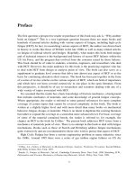
Figure 4.1 John Hunter (from Castiglioni A. A History of Medicine. New York: Alfred A. Knopf,
1947).
John Hunter is probably best known for his treatment of popliteal
aneurysms, even though he did not originate it (Figure 4.3). In the 18th century,
Anel, Desault, and others had ligated aneurysms of the brachial and popliteal
arteries. Nevertheless, many contemporary surgeons of the time condemned
the use of arterial ligation to treat these lesions, preferring instead initial
amputation in light of the gangrene or exsanguinating hemorrhage that some-
times resulted from ligation.
In 1779, Percival Pott stated that, no matter how judiciously performed,
proximal and distal arterial ligation for aneurysm would not save the patient’s
John Hunter 37
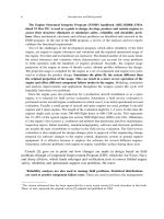
Figure 4.2 The Hunterian Museum (from Causey G. John Hunter’s museum. Surgery 1963;
54:692).
38 Chapter 4
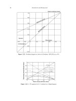
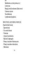
Figure 4.3 Postmortem specimen from John Hunter’s
first case of ligation for a popliteal aneurysm (courtesy of
the Royal College of Surgeons of England).
life. In most cases, “. . . the artery is not only dilated and burst, but it is also dis-
tempered someway above the dilatation.” Hunter reasoned that placement
of a proximal ligature at a distance away from the aneurysm would reduce the
chances of arterial erosion. He also felt that a more remote dissection would
interrupt fewer collaterals, increasing the chances of limb salvage. With this
approach in mind, the stage was set for what would become his most famous
operation.
Hunter’s patient was a 45-year-old London coachman in whom a popliteal
aneurysm had been diagnosed 3 years earlier. It had increased in size dramati-
cally, and in December 1785 the patient was admitted to St George’s Hospital.
The aneurysm could easily be seen as it displaced the hamstrings on either side
of it. In addition, the extremity was swollen and edematous. Hunter’s brother-
in-law and record keeper, Evevard Home, provided the following description of
the operation, which appeared in the London Medical Journal 1 year later:
Mr. Hunter having determined to perform the operation, a tourniquet was previously
applied, but not tightened, that the parts might be left as much in their natural situa-
tion, as possible; and he began the operation by making an incision on the fore and
inner part of the thigh rather than below its middle, which incision was continued
obliquely across the lower edge of the sartorius muscle, and was made large to give
room for the better performing of whatever might be necessary in the course of the
operation; the fascia, which covers the artery, was then laid bare for about three inches
in length, and the artery being plainly felt, a slight incision, about an inch long, was
made through this fascia along the side of the vessel, and the fascia dissected off, by
which means the artery was exposed. Having disengaged the artery from its lateral
connexions by the knife, and from the parts behind it by means of the endave thin
spatula, a double ligature was passed behind it by means of an ide probe, and the
artery tied by both portions of the ligature, but so slightly as only to compress its sides
together; a similar application of ligature was made a little lower; and the reason for
passing four ligatures was to compress such a length of artery as might make up for the
want of tightness, as he chose to avoid great pressure on the vessel at any one part.
The patient remained in St George’s Hospital for 1 month and made an excel-
lent recovery. He returned to his coach and continued to work until his death in
March 1787 of “remittent fever.” Hunter was present at the autopsy and, after
examining the patient’s lower extremity and noting that “. . . it was entirely free
from putrefaction,” confirmed that the operation which he had performed was
unrelated to the coachman’s death.
By the time of his own death in 1793, Hunter had performed the operation
on four other patients, with success in three. A review of these cases was pre-
sented by Evevard Home to the Society for the Improvement of Medical and
Chirurgical Knowledge in 1793. One patient survived for 50 years after
Hunter’s operation, and at autopsy his superficial femoral and popliteal arteries
were noted to be a solid cord; the aneurysm has been reduced to a small fibrous
nodule.
Hunter taught that ligation could be used for aneurysms of the subclavian,
carotid, and femoral arteries as well. He cautioned that good results were
dependent upon adequate collaterals and that there should be no damage to
John Hunter 39
surrounding structures. Earlier operations were to be preferred before the
aneurysm reached too great a size.
John Hunter’s scientific pursuits spread well beyond the realm of vascular
surgery. He made numerous contributions to the fields of gastric physiology,
trauma surgery, and dentistry. One of his most famous publications was The
Natural History of the Human Teeth, appearing in 1771.
It was unfortunate that Hunter’s curiosity also led him to speculate on the
nature of venereal disease. In May 1767, he inoculated his penis with a specimen
taken from a patient suffering with urethritis. It would not be demonstrated
until many years later that gonorrhea and syphilis are distinct diseases, and
Hunter’s inoculum also contained Treponema pallidum. Hunter attempted self-
treatment over the ensuing years with lunar caustic and calomel, and mercurial
ointment. His secondary syphilis was eventually manifested when he devel-
oped a rash and subsequent tonsillar abscess. Hunter also suffered from central
nervous system complications and developed a syphilitic ascending aortic
aneurysm. Toward the end of his life, John Hunter suffered frequent attacks of
angina. He rued the fact that his life was “. . . in the hands of any rascal who
chooses to annoy and tease me.”
Hunter’s description of his predicament proved accurate when, during a St
George’s Hospital board meeting, he was told of the appointment of his succes-
sor. His subsequent outrage resulted in a fatal attack of angina.
Through the efforts of William Clift, a great admirer of Hunter, much of his
work remains today in the library of the Royal College of Surgeons in England.
Bibliography
Beekman F. Studies in aneurysm by William and John Hunter. Ann Hist Med 1936; 8:124.
Causey G. John Hunter’s museum. Surgery 1963; 54:692.
Chitwood WR Jr. John and William Hunter on aneurysms. Arch Surg 1977; 112:829.
Garrison FH. History of Medicine. Philadelphia: WB Saunders Co., 1929.
Hunter J. ATreatise on the Blood, Inflammation, and Gun-Shot Wounds; 1794. Birmingham:
Gryphon Editions Ltd., 1982
Lasky II. John Hunter, the Shakespeare of medicine. Surg Gynecol Obstet 1983; 156:511.
Martin LE. John Hunter and tissue transplantation. Surg Gynecol Obstet 1970; 131:306.
Osler W. Remarks on arterio-venous aneurysm. Lancet 1915; 1:949.
Schechter DC, Bergan JJ. Popliteal aneurysm: Acelebration of the bicentennial of John Hunter’s
operation. Ann Vasc Surg 1986; 1:118.
Wangsteen OH. The stomach since the Hunters. Lancet 1963; 83:262.
40 Chapter 4
CHAPTER 5
Astley Cooper
41
But where’s the man who counsel can bestow, Still pleased to teach, and yet not proud to know?
(Alexander Pope)
Astley Cooper was born in Norfolk, England, in 1768. His father was a clergy-
man and his mother, a cousin of Sir Isaac Newton, was a talented writer who
had inherited great wealth. Cooper’s paternal grandfather was a surgeon in
Norwich and his uncle William was a senior surgeon to Guy’s Hospital.
Cooper was the fourth of 10 children in this distinguished family and his
early lessons were ministered by his parents and the village schoolmaster. As a
child, Cooper had an enormous amount of energy, none of which was devoted
to his studies. He was a notorious ringleader in neighborhood gangs and con-
stantly got into trouble. In addition, by his own confession, he “. . . had a way
with the girls” thanks to his good looks. Cooper’s family was concerned about
his academic failings, except for his father who never lost faith: “There is my
boy Astley. He is a sad rogue but in spite of his roguery, I have no doubt he will
be a shining character.”
When Cooper turned 15, it was decided that he would pursue a medical
career. He was apprenticed for 1 year to Dr. Francis Turner of Great Yarmouth.
In 1784, Cooper traveled to London to work with his Uncle William at Guy’s
Hospital. The prospect of life in London excited Astley, but his utter disregard
for serious work remained unchanged and wore on the patience of his uncle.
Cooper claimed to be the typical irrepressible medical student of his time, an
“idle, rollicking ne’er-do-well” (Figure 5.1).
It was Mr. Henry Cline, senior surgeon to St Thomas’ Hospital, who
gradually harnessed and directed Cooper’s reckless energy. Cooper greatly
admired Cline and, after the two had spent 6 months together, Uncle William
gladly relinquished all responsibility for his rambunctious nephew. Under
Cline’s tutelage for the next 7 years, Cooper became a good student and a
great dissector. During this period, Cooper also had the opportunity to study
with Dr. Munro of Edinburgh for 7 months. This was arranged to aid Cooper’s
recovery from typhoid fever.
In 1791, Cooper became a demonstrator in anatomy and shared lecturing
duties with Cline. Two years later, he was appointed lecturer on anatomy at
Surgeons’ Hall, where executed criminals were dissected in public. Cooper
became quite popular through these lectures and reported: “The theatre was
constantly crowded and the applause excessive.”
At the turn of the 19th century, William Cooper retired as senior surgeon to
Guy’s Hospital and his nephew succeeded him (Figure 5.2). Astley’s typical day
began at 6 o’clock, after 3–4 hours of sleep, with several hours of dissection. Fol-
lowing breakfast and a horseback ride, he saw charity patients for several more
hours. Cooper would then ride to Guy’s Hospital, where he met the medical stu-
dents for ward rounds. After rounds he would perform surgery until early
evening, and twice weekly he delivered 8 o’clock surgical lectures. During the
night, Cooper dictated his day’s activities while visiting patients in his carriage.
He usually returned home at midnight, where he would read and write for
several hours more. Cooper once reflected, “If I laid my head upon the pillow
42 Chapter 5
Figure 5.1 Sir Astley Cooper (from Major RA. A History of Medicine. Springfield, IL: Charles C
Thomas, 1954).
at night without having dissected something in the day, I should think that I
had lost that day.”
Cooper became a Fellow of the Royal Society in 1800, where he presented
his “Observations on the Membrana Tympani.” Further research resulted in
Cooper’s discovery that some types of deafness could be relieved by myringo-
tomy. For this he received the Copley Prize, a proud accomplishment since John
Hunter had received it a decade earlier.
In 1804, the first volume of Cooper’s greatest work, his treatise on hernia, was
published. It wrought order out of the chaos surrounding the anatomy and treat-
ment of hernias. The second volume appeared in 1807 and, during this time,
Cooper’s own umbilical and inguinal hernias were kept reduced with a truss.
Cooper eventually became known as the greatest surgical teacher in Europe.
His method of systematically presenting the physiologic, pathologic, and
surgical principles of diseases was unique in the early 19th century. While
John Hunter laid the groundwork for surgery to become a distinct discipline
based on scientific concepts, Cooper showed how these could be utilized
successfully. Thousands of students throughout the world attended his lectures.
Two of these included John Warren and Valentine Mott from the United
States. There was one student, however, upon whom the great orations of
Cooper had little effect. During one of Cooper’s lectures, this particular
student wrote: “The other day during the lecture, there came a sunbeam into
the room, and with it a whole troop of creatures floating in the ray; and I was off
with them to Oberon and fairy-land.” Cooper later acknowledged that, even
though young John Keats was the worst student of surgery, his poetic prowess
could not be denied.
Astley Cooper 43
Figure 5.2 Guy’s Hospital in 1725 (from Major RA. A History of Medicine. Springfield, IL: Charles C
Thomas, 1954).
While a medical student, Cooper had studied the effects of brachial and
femoral artery ligations in dogs. He had also ligated the carotid and vertebral
arteries bilaterally in one animal that survived and even became “a good
house dog.”
In 1805, Cooper performed one of the earliest carotid artery ligations in man.
Mary Edwards was 44 years old and presented to Cooper with an aneurysm
of the right common carotid artery. It occupied two-thirds of her neck and the
overlying skin was thin and tense. On November 1, Cooper ligated the
common carotid artery of Mrs. Edwards. On November 5, Cooper found her sit-
ting up and taking tea with some fellow patients. She appeared well except for
a persistent cough. Mrs. Edwards died 16 days later and her autopsy revealed
suppuration within and surrounding the large aneurysm sac, as well as com-
pression of the larynx and trachea. Cooper concluded that the surgery would
have been successful had the sac not grown so large.
Several years later, Cooper had the chance to test this conclusion when
Humphrey Humphreys, a 51-year-old porter, came to see him with a left carotid
artery aneurysm “the size of a walnut.” On June 22, 1808, Cooper doubly ligated
and divided the common carotid artery of his patient, proximal tothe aneurysm.
Humphreys recovered and returned to work. He survived until 1821, when
he died of a left-sided cerebral hemorrhage. Cooper reported the postmortem
findings in the first issue of the Guy’s Hospital Reports in 1826 (Figure 5.3). He
noted: “The disease of which he died sufficiently attested to the circulation as
well as its force in the cerebral vessels on the side of which the carotid had been
tied.”
During the afternoon following Humphrey’s operation, Cooper performed
ligation of an external iliac artery for a large femoral aneurysm. This patient
was a 39-year-old man from Norfolk who also recovered and lived until 1826.
Cooper once remarked: “There was no man, however great or distinguished,
who was likely to avoid my clutches for autopsy, as there were always
means of obtaining a body you wanted.” At considerable time and expense,
Cooper secured the body of this patient from Norfolk and also reported his
autopsy findings in the inaugural issue of Guy’s Hospital Reports.
Cooper went on to perform external iliac artery ligation for femoral
aneurysm nine more times. In 1813, Cooper was appointed Hunterian Professor
of Comparative Anatomy by the Royal College of Surgeons.
The ligations of carotid and external iliac arteries were daring procedures in
Cooper’s time. In 1817, however, he performed his boldest operation. Charles
Hutson was a 38-year-old porter who was admitted to Guy’s Hospital with a
large left external iliac aneurysm. It had been growing steadily for 1 year, and 3
days prior to his admission it had doubled in size. Cooper’s hand was forced
when the aneurysm ruptured though the skin and began bleeding. Two days
earlier, Cooper had attempted retroperitoneal exposure of the aorta in a
cadaver, anticipating Hutson’s eventual need for surgery. He found this route
“utterly impracticable” and, with Hutson in his bed, Cooper exposed the aorta
through a transperitoneal incision and placed a silk ligature just above the aortic
44 Chapter 5
bifurcation. Hutson appeared well the following day, but died 40 hours after
surgery (Figure 5.4A and B). In his account of Astley Cooper’s life, Lord Brock
likened this operation to the “Everest ascent of arterial surgery of his day.” Even
Cooper admitted: “I was gratified when my admirers and detractors agreed
Astley Cooper 45
Figure 5.3 Title page from Cooper’s account of the case of Humphrey Humphreys (from Eastcott
HHG. The beginning of carotid surgery. In: Bergan JJ, Yao JST, eds. Cerebrovascular Insufficiency.
New York: Grune & Stratton, 1983; reprinted by permission).
that this was the boldest attempt to preserve life by aid of the science of surgery.”
Not until 1923, 106 years after Cooper’s operation, would Rudolph Matas
perform the first successful abdominal aortic ligation.
An additional contribution made to vascular surgery by Cooper, in 1817, was
46 Chapter 5
Figure 5.4 Postmortem specimen from the case of Charles Hutson. (A) Anterior view; (B) posterior
view (from Brock RC. The Life and Work of Astley Cooper. London: E & S Livingstone, Ltd, 1952).
A
the use of a buried catgut suture for arterial ligation. This operation on an 80-
year-old man with a popliteal aneurysm was a marked departure from the usual
practice of ligating the artery with silk and then bringing the ends of the ligature
through the wound. This often resulted in fatal hemorrhage. Postoperative
hemorrhage was avoided in Cooper’s patient, whose wound was completely
Astley Cooper 47
Figure 5.4 Continued
B
closed over the ligature and healed without complication. Cooper remarked:
“The case gave me much pleasure and the rapid recovery leads me to hope that
the operation for aneurysm may become infinitely more simple.”
In 1820, King George IV was suffering from an infected sebaceous cyst on his
head. Cooper was summoned even though he was not one of the official Royal
48 Chapter 5
Figure 5.5 Lithograph depicting Cooper’s patient following first successful hip disarticulation (from
Brock RC. The Life and Work of Astley Cooper. London: E & S Livingstone, Ltd, 1952).
Surgeons. Following excision of the cyst, Cooper described his experience
briefly: “I feared erysipelas as a complication in the postoperative course of my
exalted patient, but all went well and I was created Baronet by His Majesty in
1821.”
In addition to his major contributions to vascular and hernia surgery, Cooper
was the first to champion avoidance of amputation for compound fractures. He
also offered a monograph on diseases of the testes, and a study of the anatomy of
the thymus. In 1829, he published his treatise on nonmalignant diseases of the
breast.
At the age of 60, Cooper relinquished his lecturing commitments because of
the strain of his busy surgical practice and recurring episodes of vertigo. He was
appointed an Examiner of the Royal College of Surgeons, where he assessed
his fellow examiners as “. . . a doddering collection of ill-read individuals with
lifetime appointments and with little interest in the welfare of the helpless
candidates.” This is likely the explanation of why Cooper soon became
College President.
In 1840, Cooper began to suffer increasingly from dyspnea on exertion: “New
Years 1841 found me in sore straights.” Cooper was in congestive heart failure
when he wrote those words and on February 12, 1841, surrounded by his family
members and friends, his last words were: “God bless you and goodbye to you
all.” He was buried in a crypt beneath the chapel at Guy’s Hospital.
By his own estimate, Cooper had a hand in the education of 8000 surgeons.
He had an enormous surgical practice, was a master of anatomy, and displayed
an unerring scientific approach to clinical problems (Figure 5.5). In the
history of surgery, Cooper’s contributions linked those of John Hunter and
Joseph Lister. The accolade: “Prince of Surgery,” bestowed upon him by
his colleagues, was richly deserved.
Bibliography
Brock RC. The Life and Work of Astley Cooper. London: E & S Livingstone, Ltd, 1952.
Brock RC. The life and work of Sir Astley Cooper. Ann R Coll Surg Engl 1969; 44:1.
Drach GW. Sir Astley Cooper (1768–1841). Invest Urol 1978; 16:75.
Nuland SB. Astley Cooper of Guy’s Hospital. Conn Med 1976; 40:190.
Rawling EG. Sir Astley Paston Cooper, 1768–1841: The prince of surgery. Can Med Ass J 1968;
99:221.
Schoenberg DG, Schoenberg BS. Eponym: Sir Astley Paston Cooper: Good sense, good surgery,
and good science. South Med J 1979; 72:1193.
Wass SH. Astley Cooper and the anatomy and surgery of the hernia. Guy’s Hosp Rep 1968;
117:213.
Astley Cooper 49
Divisions of
vascular surgery
Development of the venous autograft
53
veins subjected to arterial flow can certainly remain intact, but have a marked tendency to
thrombose when under arterial pressure. Therefore, the idea of bypassing occluded arterial path-
ways using neighboring veins has no practical significance.
(Alfred Exner)
The first experimental attempts to place venous autografts into the arterial cir-
culation were performed at the beginning of the 20th century by Alfred Exner, in
Austria; and Alexis Carrel, in France. Exner used Payr’s magnesium tubes to
place canine external jugular veins into carotid arteries. The revolutionary idea
of anastomosing a segment of vein directly into the arterial circulation was con-
ceived by Carrel in 1901, at the University of Lyon. It was brought to fruition
through his collaboration with Charles Guthrie at the Hull Physiological
Laboratory in Chicago (Figure 6.1).
The use of metal prostheses by Exner invariably resulted in thrombosis, lead-
ing him to conclude that veins could not be used as arterial substitutes. The ex-
periments of Carrel and Guthrie were successful owing to meticulous aseptic
technique and the use of a method that did not require a foreign body other than
suture material.
Carrel and Guthrie reported their canine experiments with vein grafts in
a manuscript that underscored their surgical genius (Figures 6.2 and 6.3).
“Uniterminal and biterminal venous transplantations” appeared in Surgery,
Gynecology and Obstetrics in 1906. The remarks of Carrel and Guthrie regarding
potential sources of vein for grafting are pertinent today:
it is always possible to extirpate a few centimeters of vein without seriously interfer-
ing with the circulatory apparatus in general. Thus the operated subject supplies all the
venous material necessary for most transplantations.
While their European colleagues were discouraged by the results of venous
transplantations, Carrel and Guthrie speculated that:
The possibility of transforming a vein into an artery, from a functional point of view,
naturally arouses the idea of substituting veins for arteries when the latter are rendered
useless by some pathological process.
For cases of trauma or malignancy, Carrel and Guthrie wrote:
Severe subcutaneous wounds of the large arteries are ordinarily followed by serious
complications, gangrene often occurring. In such cases it would be possible to extirpate
freely all the injured portions of the vessel and to re-establish the circulation by inter-
posing a segment of vein between the cut ends of the artery. The same operation would
be indicated, when, during the extirpation of a large growth, the resection of an impor-
tant vessel is necessary; e.g., sarcoma in the femoral region.
The essential techniques for suturing blood vessels described by Carrel over
80 years ago are still taught to surgical residents:
Arigid asepsis is absolutely essential for success. . . . The dissection of the vessel is not
dangerous if the wall of the vessel is not crushed or roughly handled with metallic
forceps of other hard instruments . . . it is necessary that these clamps [vascular] be
smooth-jawed and not too strong in the spring . . . by using very sharp, rough needles,
only extremely small wounds are made . . . great care is taken not to include fragments
54 Chapter 6
Figure 6.1 Charles Claude Guthrie (from Callow AD. Historical development of vascular grafts. In:
Sawyer PN, Kaplitt MJ, eds. Vascular Grafts. New York: Appleton-Century-Crofts, 1978).
Development of the venous autograft 55
Figure 6.2 Illustration of venous transplant into arterial circulation by Carrel and Guthrie (from
Carrel A, Guthrie CC. Uniterminal and biterminal venous transplantation. Surg Gynecol Obstet
1906; 2:266).
Figure 6.3 End-to-side anastomosis of vein graft into an artery as depicted by Carrel and Guthrie
(from Carrel A, Guthrie CC. Uniterminal and biterminal venous transplantation. Surg Gynecol Obstet
1906; 2:266).
of the connective tissue layer in the line of suturing, and to obtain a smooth union and
approximation of the endothelial coats.
The first clinical anastomosis of a venous graft into the arterial circulation
was performed by Jose Goyanes of Madrid (Figure 6.4). Goyanes had studied
the work of Carrel, and of Murphy in the United States, and had experimented
with transplantation of canine vena cava grafts into the aorta. In 1906, he was
56 Chapter 6
Figure 6.4 Jose Goyanes (from Harrison LH Jr. Historical aspects in the development of venous
autografts. Ann Surg 1976; 183:101).
asked to examine a 41-year-old candy maker who had developed a syphilitic
popliteal aneurysm. On June 12, Goyanes excised the aneurysm and, unable to
repair the popliteal artery primarily, used an adjacent segment of popliteal vein
to bridge the defect. The patient made an excellent recovery after a postopera-
tive wound infection, and Goyanes reported this case in El Siglo Medico, an ob-
scure Spanish weekly medical bulletin. He credited his mentor, Professor San
Martin, with the idea of using an autogenous vein graft in this manner. Goyanes
mentioned the prior, similar use in another patient of an iliac vein segment to
bypass an iliac artery obstruction. Goyanes’s popliteal venous replacement of a
segment of popliteal artery was also the first in situ vein graft (Figure 6.5).
In 1907, Stitch reported his experimental results with venous autografts.
This set in motion an important chain of events. That same year, Erich Lexer of
Konigsberg treated a 69-year-old man who had undergone reduction of a
dislocated shoulder 9 weeks earlier (Figure 6.6). The patient had developed a
large axillary pseudoaneurysm secondary to an injury to the axillary artery
by the reduction procedure. Lexer had clinical experience with Payr’s
magnesium tube, but at surgery he was faced with an 8-cm gap between
the axillary and brachial arteries after he resected the aneurysm. Lexer
recognized the uselessness of a Payr tube in this situation and recalled the
report of Stitch. Lexer excised a 10-cm segment of greater saphenous vein from
his patient’s leg and used it in a reversed manner to restore arterial continuity.
Lexer’s patient died of delirium tremens on the fifth postoperative day, but was
found to have a patent graft at the postmortem examination which Lexer
performed.
Lexer reported this case before the Congress of the German Society for
Surgery, and it was published in a widely read German surgical journal: Archives
of Clinical Surgery. Unaware of Goyanes’s case the previous year, Lexer claimed
to be the first to use a venous autograft to replace an artery.
William Halstead read of Lexer’s case and was impressed by the report.
Halstead encouraged his associate Bertram Bernheim to use venous autografts
as arterial replacements in animals (Figure 6.7). Bernheim developed his vascu-
lar surgical skills in the laboratory, and in 1909 he was summoned by Halstead to
apply them clinically. Halstead had removed a sarcoma from the popliteal space
of a patient, resulting in a large popliteal arterial defect. Bernheim attempted to
bridge the gap with a long segment of greater saphenous vein, but it throm-
bosed. Undaunted by this experience, Bernheim continued his laboratory re-
search, which led to publication of a textbook on vascular surgical techniques
in 1913.
Two years following publication of his book, Bernheim had a second chance
to use the technique of Stitch and Lexer. His patient was a 43-year-old man with
a syphilitic popliteal aneurysm. Excision of the lesion left a 15-cm gap in the
popliteal artery, which Bernheim replaced with a 12-cm segment of greater
saphenous vein. The patient made an uneventful recovery and Halstead
praised Bernheim’s efforts. This was the beginning of clinical arterial recon-
struction in the United States.
Development of the venous autograft 57
In 1913, Hogarth Pringle reported two cases of reversed saphenous vein
grafts to maintain arterial circulation. His cases involved aneurysms of the
popliteal and brachial arteries and were performed at the Royal Infirmary of
Glasgow. Both operations were successful.
Further use and development of venous autografts was stimulated by
58 Chapter 6
Figure 6.5 Goyanes’s in situ bypass of a popliteal aneurysm (from Dale WA. Management of
Vascular Surgical Problems. New York: McGraw Hill, 1985).