Emergencies in Urology - part 5 potx
Bạn đang xem bản rút gọn của tài liệu. Xem và tải ngay bản đầy đủ của tài liệu tại đây (3.93 MB, 68 trang )
Table 15.6.3. Classification of
bladder injury based on the
type of trauma
Classification
of injury
Mechanism of injury Associated injuries
Blunt trauma
Extrape-
ritoneal
Blunt pelvic trauma with laceration
by bone fragment(s)
Pelvic fractures
Shearing at ligamentous attachment(s)
Other long bone fractures
Intrape-
ritoneal
High velocity blunt lower abdominal
trauma
High rate of associated
intraabdominal injuries
High intravesical pressure with
rupture at dome
High mortality
Penetrating
trauma
Direct injury to the bladder wall Associated injury to
otherorgansiscommon
Table 15.6.4. AAST organ in-
jury severity scale for the
bladder and Associated Ab-
breviated Injury Scale of the
American Association for
Automotive Medicine, 1990
(AIS-90)
Grade
a
Injurytype Descriptionofinjury AIS-90
I Hematoma Contusion, intramural hematoma 2
I Laceration Partial thickness 3
II Laceration Extraperitoneal bladder wall laceration <2 cm 4
III Laceration Extraperitoneal (>2 cm) or intraperitoneal (<2 cm)
bladder wall laceration
4
IV Laceration Intraperitoneal bladder wall laceration >2 cm 4
V Laceration Intraperitoneal or extraperitoneal bladder wall lacera-
tion extending into the bladder neck or ureteral orifice
(trigone)
4
a
Advanceonegradefor
multiple injuries to same
organ up to grade III
Fig. 15.6.1. AAST classification of bladder injury. Grade 1:
contusion, intramural hematoma or partial thickness lacera-
tion of the bladder wall (Fig. 15.6.1–6 © Hohenfellner 2007)
Fig. 15.6.2. AAST classification of bladder injury. Grade 2:
extraperitoneal laceration of the bladder wall <2 cm
250 15 Trauma
Fig. 15.6.3. AAST classification of bladder injury. Grade 3:
extraperitoneal laceration of the bladder wall >2 cm
Fig. 15.6.5. AAST classification of bladder injury. Grade 4:
intraperitoneal laceration of the bladder wall >2 cm
Fig. 15.6.4. AAST classification of bladder injury. Grade 3:
intraperitoneal laceration of the bladder wall <2 cm
Fig. 15.6.6. AAST classification of bladder injury. Grade 5:
intraperitoneal or extraperitoneal laceration of the bladder
wall extending in to the bladder neck or trigone
15.6 Bladder Trauma 251
classification, which was adopted, modified, and rec-
ommended by the Orthopaedic Trauma Association
(OTA) (Tile 1988, 1996; OTA 1996). The OTA classifica-
tion groups pelvic injuries into three main categories:
A-type injuries have a stable pelvic ring, B-type have a
partial posterior disruption, and C-type have a com-
plete posterior disruption. Within this classification,
the severity of injury increases from type A to type C
(Tile1999),withahigherinjuryseverityscore(ISS),in-
cidence of associated injuries, and mortality rate with
the latter (Poole et al. 1991; Adams et al. 2002).
15.6.4
Risk Factors
15.6.4.1
Blunt Trauma
Driving under the influence of alcohol predisposes to
motor vehicle accidents and to a distended bladder as
well.Thusitisariskfactorforbladderinjury(Dreitlein
et al. 2001).
Lateral-impact MVC are known to be associated
with an increased incidence of pelvic fractures (Siegel
et al. 1993; Loo et al. 1996; Inaba et al. 2004; Rowe et al.
2004), and therefore may result in bladder injury.
CrashimpactdataintraumaregistryforMVCoccu-
pants with AIS
4 pelvic injuries identified the lateral
impact as the most common crash variable, account-
ing for more than 80% of injuries to drivers and front
seat passengers (Inaba et al. 2004). An evaluation of
risk factors for severe pelvic injuries (AIS
4) suggest-
ed motorcycle injuries to result in the highest inci-
dence of pelvic fractures, with bladder and urethra as
the most commonly injured organs. In this study, step-
wise logistic regression analysis identified male gen-
der and pelvic fracture AIS
4asindependentriskfac-
tors (Demetriades et al. 2002). These patients also had
significantly more genitourinary injuries, the bladder
being the most common (25%) intraabdominal organ
injured.
15.6.4.2
Iatrogenic Trauma
Risk factors for iatrogenic bladder injury include ad-
hesions and pelvic scarring from previous surgery, in-
flammation,endometriosis,exposuretoradiation,
presence of malignant disease, pregnancy, pelvic or-
gan prolapse, multiple cesarean sections, congenital
abnormalities, hemorrhage, or failure to empty the
bladder before the operation (Daly and Higgins 1988;
Harris et al. 1997; Davis 1999; Armenakas et al. 2004;
Gomez et al. 2004; Yossepowitch et al. 2004). In a mul-
ticenter study, concurrent surgery for stress inconti-
nence along with gynecological procedures was found
to be the only independent variable for bladder injury
in a stepwise logistic regression model, with a relative
risk of 4.42 (Vakili et al. 2005). The type of incision
during cesarean section is also a risk factor. In a retro-
spective analysis of data from 3,164 women undergo-
ing cesarean section revealed that the type of incision,
thepresenceofadhesions,andanteriorplacentapre-
via were independently associated with increased risk
of bladder injury (Makoha et al. 2005). The bladder
was injured almost seven times as frequently with the
midline subumbilical (MLSU) aswith the Pfannenstiel
incision (p<0.0001; OR, 6.7). This study has also con-
firmed the observation that for both types of incision
the risk of bladder injury increases with the number of
cesarean sections (Makoha et al. 2004) and for a given
number the risk is higher with MLSU than Pfannen-
stiel incision.
15.6.5
Diagnosis
The two most common signs and symptoms of major
bladderinjuriesaregrosshematuria(82%)andab-
dominal tenderness (62%) (Carroll and McAninch
1984). Other findings may include inability to void,
bruisesoverthesuprapubicregion,andabdominaldis-
tention (Sagalowsky 1998). Extravasation of urine may
result in swelling in the perineum, scrotum, and thighs,
as well as along the anterior abdominal wall within the
potential space between the transversalis fascia and the
parietal peritoneum. Hematuria at the conclusion of an
otherwise uneventful procedure, clearfluid inthe oper-
ative field, gas distention of the urinary drainage bag
during laparoscopy, and/or visible bladder laceration
should alarm the surgeon to iatrogenic bladder injury
(Armenakas et al. 2004; Gomez et al. 2004)
15.6.5.1
Macroscopic (Gross) Hematuria
Gross hematuria indicates urologic trauma. Review of
the existing literature reveals that traumatic bladder
rupture is strongly correlated with the combination of
pelvic fracture and gross hematuria. Morey et al. re-
ported gross hematuria in all of their patients with
bladder rupture, and 85% had pelvic fractures (Morey
et al. 2001). Therefore, the classiccombination of pelvic
fracture and gross hematuria constitutes an absolute
indication for immediate cystography in blunt trauma
victims (Carroll and McAninch 1984; Rehm et al. 1991;
Morey 2005). While grossly clear urine in a trauma pa-
tient without a pelvic fracture virtually eliminates the
possibility of a bladder rupture, up to 2%–10% of pa-
tients with bladder rupture may have only microhema-
turia or no hematuria at all (Schneider 1993).
252 15 Trauma
Tarman et al. (2002) reviewed 8,021 pediatric trauma
patients retrospectively, including 212 consecutive pa-
tients with pelvic fractures. Among patients with pelvic
fractures, only one patient (0.5%) had an extraperito-
neal bladder rupture. Lower urogenital injury occurred
in six patients (2.8%). The absence of gross hematuria
effectively ruled out serious injury in this cohort. Con-
sequently, these authors concluded that further urologi-
cal work-up is unnecessary in stable patients with pelvic
fractures and isolated microhematuria. Patients with
gross hematuria, multiple associated injuries, or signifi-
cant abnormalities found on their physical examination
are recommended to undergo further urological evalu-
ation with appropriate imaging modalities such as ret-
rograde urethrography and cystography.
15.6.5.2
Microscopic Hematuria
In thetrauma patient with apelvic ring fracture, micro-
scopic hematuria should be considered as a possible in-
dicator of bladder laceration, and further investigation
is warranted. Existing data do not support lower uri-
nary tract imaging in all patients with either pelvic
fracture or microscopic hematuria alone. Also, the
threshold of red blood cells in urine that triggers fur-
ther investigation is a point of controversy. A threshold
ranging from 25 to 200 red blood cells per high power
field (rbc/phf) has been suggested to indicate signifi-
cant injury to the bladder (Werkman et al. 1991; Fuhr-
man et al. 1993; Morgan et al. 2000). These observations
seems not to be valid for pediatric trauma patients, as
indicated previously in a clinical series (Tarman et al.
2002).Incontrast,Abou-Jaoudeetal.foundthata
threshold of 20 rbc/hpf as an indication for radiological
evaluation would have missed 25% of cases with blad-
der injury. In contrast to other reported series, they
suggested that lower urogenital tract evaluation in pe-
diatric trauma patients, especially in the presence of
pelvic fractures, should be based as much on clinical
judgmentasonthepresenceofhematuria(Abou-Jaou-
de et al. 1996).
15.6.5.3
Cystography
Retrograde cystography in evaluation of bladder trau-
ma is considered the standard diagnostic procedure
(Stine et al. 1988; Rehm et al. 1991; Baniel and Schein
1994). Cystography is accepted as the most accurate ra-
diological study for diagnosing bladder rupture (Deck
et al. 2000). When adequate bladder filling and post-
void images are obtained, they have an accuracy rate of
85%–100%. The diagnosis of bladder rupture is usual-
ly made easily on cystography when the injected con-
trast medium is identified outside the bladder
Fig. 15.6.7. Extraperitoneal rupture demonstrated on cystogra-
phy. Extravasation of contrast material is limited to the peri-
vesical space
Fig. 15.6.8. Extraperitoneal rupture on cystography
(Figs. 15.6.7–9). Adequate distention of the urinary
bladder is crucial to demonstrate perforation, especial-
ly in instances of penetrating trauma, since most in-
stances of a false-negative retrograde cystography were
found in this situation (Cass 1984; Baniel and Schein
1994). Cystography requires at least plain films, filled
films, and postdrainage films. Half-filled film and
obliques are optional. For the highest diagnostic accu-
racy, the bladder must be distended by instillation of at
15.6 Bladder Trauma 253
Fig. 15.6.9. Intraperitoneal bladder rupture on cystography.
Bowel loops are outlined by the extravasated contrast in the
abdominal cavity
least 350 cc of contrast medium with gravity. Bladder
injury may be identified only on the postdrainage film
in approximately 10% of the cases. False-negative find-
ings may result from improperly performed studies
with instillation of less than 250 ml of contrast medium
or omission of a postdrainage film (Morey et al. 1999).
Only a properly performed cystography should be used
to exclude bladder injury.
15.6.5.4
Excretory Urography (Intravenous Pyelography)
Intravenouspyelography(IVP)isinadequateforevalu-
ation of bladder and urethra after trauma because of di-
lution of the contrast material within the bladder, and
resting intravesical pressure is simply too low to dem-
onstrate a small tear (Ben-Menachem et al. 1991) IVP
has a low accuracy, on the order of 15%–25% and vari-
ous clinical studies indicated that IVP has an unaccept-
ably high false-negative rate of 64%–84%, which pre-
cludes its use as a diagnostic tool in bladder injuries
(Werkman et al. 1991).
15.6.5.5
Ultrasound
Although the use of US in bladder rupture has been de-
scribed (Bigongiari et al. 2000), it has not been routine-
ly used for evaluation of bladder injury. The presence of
peritoneal fluid in the presence of normal viscera or
failure to visualize the bladder after the transurethral
introduction of saline is considered highly suggestive
of bladder rupture (Bigongiari et al. 2000). In practice,
US is not definitive in bladder or urethral trauma and is
not routinely used. Focused abdominal sonography for
trauma (FAST) has gained popularity in the evaluation
of blunt abdominal trauma in adults to detect free in-
traperitoneal fluid, with a sensitivity of 63%–99% in
published series (Fernandez et al. 1998; Yoshii et al.
1998; Nunes et al. 2001; Von Kuenssberg Jehle et al.
2003).
Several reports have indicated that FAST can also re-
liably detect free intraperitoneal fluid in children, with
acceptable sensitivity and specificity rates (Holmes et
al. 2001; Soudack et al. 2004). However, a positive FAST
in a hemodynamically stable child is of limited use, be-
cause in one survey only 26% (5/19) of pediatric emer-
gency attending physicians considered ultrasound
equally available with CT, and none considered it more
readily available than CT (Baka et al. 2002). The inabili-
ty of FAST to distinguish the origin of free fluid in the
abdomensuchasblood,ascites,orurineremainsan-
other disadvantage of this modality (Jones et al. 2003).
Therefore, the exact role of FAST in detection of
bladder injury remains to be determined.
15.6.5.6
Computed Tomography
CT is clearly the method of choice for the evaluation of
patients with blunt or penetrating abdominal and/or
pelvic trauma. However, routine CT is not reliable in
the diagnosis of bladder rupture even if an inserted
urethral catheter is clamped. CT demonstrates intra-
peritoneal and extraperitoneal fluid but cannot differ-
entiateurinefromascites.AswithIVP,thebladderis
usuallyinadequatelydistendedtocauseextravasation
throughabladderlacerationorperforationduring
routine abdominal and pelvic studies. Therefore, a neg-
ativestudycannotbeentirelytrusted,androutineCT
thereforecannotruleoutbladderinjury(Meeetal.
1987; Cass 1989; Ben-Menachem et al. 1991). Horstman
et al. reviewed the cystograms and CT scans of 25 pa-
tients who had both studies as the initial evaluation of
blunt abdominal trauma (Horstman et al. 1991). Five
out of 25 had bladder rupture, three extraperitoneal
and two intraperitoneal. All injuries were detected by
both studies. The authors felt that delayed imaging or
contrast instillation (CT cystography) can provide the
adequate bladder distention needed to demonstrate
contrast extravasation from the injury site during CT.
Similarly, in a series of 316 patients, Deck et al. diag-
nosed 44 cases with bladder ruptures. In patients who
underwentformalsurgicalrepair,82%hadoperative
254 15 Trauma
findings that exactly matched the CT cystography in-
terpretation (Deck et al. 2000). Thus, either retrograde
cystography or CT cystography are the diagnostic pro-
cedures of choice for suspected bladder injury (Schnei-
der 1993). CT cystography may be used in place of a
conventional cystography (overall sensitivity 95% and
specificity 100%), especially in patients undergoing CT
scanning for other associated injuries (Deck et al.
2001). However, this procedure should be performed
using retrograde filling of the bladder with a minimum
of 350 cc of dilute contrast material (Wah and Spencer
2001).
CT cystographic features may lead to accurate clas-
sification of bladder injury (Figs. 15.6.10, 11) and allow
prompt, effective treatment with less radiation expo-
sure and without the added cost of conventional cysto-
graphy (Vaccaro and Brody 2000).
Fig. 15.6.10. CT cystography demonstrating extraperitoneal
extravasation of contrast material
Fig. 15.6.11. Extraperitoneal rupture on CT cystography
15.6.5.7
Angiography
Angiographyisrarelyifeverindicated.Itcanbeuseful
in identifying an occult source of bleeding and for ther-
apeutic embolization (Ben-Menachem et al. 1991).
15.6.5.8
Magnetic Resonance Imaging
Since it is extremely difficult to monitor a seriously in-
jured patient in a strong magnetic field, MRI currently
has little place in the evaluation of acute bladder (Ben-
Menachem et al. 1991).
15.6.5.9
Cystoscopy
Cystoscopy appears an extremely useful tool in the di-
agnosis of iatrogenic bladder injuries. The results of a
multicenter study as well as a comprehensive review of
the literature indicated that the majority (49.4%–
64.7%) of bladder injuries during gynecological opera-
tions would be missed if cystoscopy were not per-
formed at the end of each procedure (Gilmour et al.
1999; Vakili et al. 2005). The detection rate of bladder
injury by cystoscopy ranges from 85% to 94.1% in dif-
ferent series (Harris et al. 1997; Vakili et al. 2005).
15.6.6
Treatment
The first priority in the treatment of bladder injuries is
stabilization of the patient and treatment of associated
life-threatening injuries.
15.6.6.1
Blunt Trauma: Extraperitoneal Rupture
Most patients with extraperitoneal rupture can be
managed safely by catheter drainage only, even in the
presence of extensive retroperitoneal or scrotal extrav-
asation. Virtually all ruptures are healed in 3 weeks
(Morey et al. 1999). However, involvement of the blad-
der neck (Carroll and McAninch 1984), the presence of
bone fragments in the bladder wall, or entrapment of
thebladderwallnecessitatesurgicalintervention
(Dreitlein et al. 2001). In the absence of bladder neck
involvement and/or associated injuries that require
surgical intervention such as open pelvic fractures and
rectal or vaginal lacerations, extraperitoneal bladder
ruptures caused by blunt trauma are managed by cath-
eter drainage only (Cass and Luxenberg 1987). The
presence of open pelvic fractures and/or rectal injuries
precludes conservative management due to the high
15.6 Bladder Trauma 255
risk of serious infectious complications (Cass and Lu-
xenberg 1989). In patients undergoing surgery for oth-
er organ injuries, the laceration of the bladder wall
should also be repaired transvesically, if the patient is
stable at the time of the operation (Gomez et al. 2004).
15.6.6.2
Blunt Trauma: Intraperitoneal Rupture
Intraperitoneal ruptures occurring after blunt trauma
should always be managed by surgical exploration.
This type of injury involves a high degree of force, and
because of the severity of associated injuries carries a
high mortality rate of 20%–40% (Cass 1989; Rehm et
al. 1991). Lacerations are usually large in these in-
stances with potential risk of peritonitis due to urine
leakage, if left untreated (Deck et al. 2000). Abdominal
organs should be inspected for possible associated in-
juries, and urinoma must be drained. The technique of
surgical repair depends on the surgeon’s preference but
a two-layer closure with absorbable sutures achieves a
safe repair of the bladder wall. A suprapubic catheter
can be used in addition to a urethral catheter to ensure
the adequacy of the drainage. However, in a recent
study, patients with Foley catheter drainage alone had
equally good outcome (Volpe et al. 1999).
15.6.6.3
Penetrating Trauma
All bladder perforations due to a penetrating trauma
should undergo emergency exploration and repair
(Deck et al. 2000). Penetrating trauma to the pelvis pre-
sentsaseriouschallengebecauseofthecomplexanato-
my of the region. Penetrating trauma patients present-
ing with shock have a high incidence of vascular injury
and subsequent exsanguination, and associated viscer-
al injuries may complicate their management, resulting
inahighmortalityrate.However,stablepatientscanbe
managed without operation, when appropriate diag-
nostic techniques fail to demonstrate an injury (Dun-
can et al. 1989). Gunshot wounds to the bladder usually
result in intraperitoneal leaks, which require proper
drainage and repair of the associated lacerations of the
bladderwallaswellasadjacentorgans.However,inthe
occasional patient with extraperitoneal rupture, non-
operative management with Foley catheter drainage
can be used successfully (Velmahos and Degiannis
1997).
15.6.6.4
Iatrogenic Trauma
In patients with immediate diagnosis, bladder repair
accomplished by a transabdominal or transvaginal
two-layer closure effectively treats 98% of cases and the
rest are managed by Foley catheter drainage (Armena-
kas et al. 2004).
15.6.6.5
Complications
In patients with bladder trauma, complications are
usually the result of failure to diagnose the injury and
repair promptly. This may result in urinoma formation,
urinary leakage into the peritoneal cavity, ileus, perito-
nitis, hematoma, abscess formation, fistula formation
(rectal, vaginal, or cutaneous), and urinary tract infec-
tion.
Bladder injury with extravasation of urine with or
without prostatic injury may complicate the course of
recovery by impairing the coagulation mechanism. The
prostatic capsule contains abundant activators of plas-
minogen and urine contains high levels of urokinase, a
potent plasminogen activator (Andersson 1980). Both
tissue activator and urokinase accelerate the dissolu-
tion of clots and may consequently increase and pro-
long hemorrhage (Hedlund 1969). Epsilon amino ca-
proic acid (EACA) can be effective in controlling hema-
turia after surgical procedures compared with placebo,
and its use was not accompanied by significant compli-
cations (Miller et al. 1980). Tranexamic acid (amino-
methyl cyclohexane carboxylic acid, AMCA) is a stron-
gerinhibitorofplasminogenactivationthanEACAand
may significantly decrease the amount of blood loss
and control the bleeding when administered in a total
dose of 3–12 g for 4–21 days (Hedlund 1975; Dunn and
Goa 1999) without any increase in the incidence of
thrombosis compared to placebo (Hedlund 1975).
Early angiography and transcatheter embolization
in patients with major blood requirements after pelvic
trauma may help to avoid the need for and complica-
tions of multiple transfusions and large pelvic hemato-
mas. Precise localization of bleeding sites and occlu-
sion of the bleeding artery by either an injection of au-
tologous clot or Gelfoam embolization can be success-
fully achieved (Matalon et al. 1979; Wong et al. 2000;
Ben-Menachem 1988).
15.6.7
Damage Control
Severe multiple traumatic injuries may cause acidosis,
hypothermia, and coagulopathy, which have been asso-
ciated with very high mortality rates (Zacharias et al.
1999). Focusing the initial resuscitative efforts to stabi-
lize the patient with the control of the hemorrhage
(temporary packing) and gross contamination along
with appropriate bladder drainage with and subse-
quent intensive care may allow for later definitive re-
pair of the injuries in a patient who will otherwise die.
256 15 Trauma
References
Abou-Jaoude WA et al (1996) Indicators of genitourinary tract
injury or anomaly in cases of pediatric blunt trauma. J Pedi-
atr Surg 31:86; discussion 90
Adams JE et al (2002) Analysis of the incidence of pelvic trau-
ma in fatal automobile accidents. Am J Forensic Med Pathol
23:132
Agostini A et al (2006) Immediate complications of tension-
free vaginal tape (TVT): results of a French Survey. Eur J Ob-
stet Gynecol Reprod Biol 124:237
Aihara R et al (2002) Fracture locations influence the likeli-
hood of rectal and lower urinary tract injuries in patients
sustaining pelvic fractures. J Trauma 52:205; discussion 208
Andersson L (1980) Antifibrinolytic therapy in genitourinary
tract surgery. J Clin Pathol Suppl (R Coll Pathol) 14:60
Anonymous (1996) Fracture and dislocation compendium.
Orthopaedic Trauma Association Committee for Coding
and Classification. J Orthop Trauma 10 [Suppl 1]:v, 1
Armenakas NA et al (2004) Iatrogenic bladder perforations:
longterm followup of 65 patients. J Am Coll Surg 198:78
AzamUetal(2001)Thetension-freevaginaltapeprocedurein
women with previous failed stress incontinence surgery.
J Urol 166:554
Baka AG et al (2002) Current use and perceived utility of ultra-
sound for evaluation of pediatric compared with adult trau-
ma patients. Pediatr Emerg Care 18:163
Baniel J, Schein M (1994) The management of penetrating
trauma to the urinary tract. J Am Coll Surg 178:417
Ben-Menachem Y (1988) Pelvic fractures: diagnostic and ther-
apeutic angiography. Instr Course Lect 37:139
Ben-Menachem Y et al (1991) Hemorrhage associated with
pelvic fractures: causes, diagnosis, and emergent manage-
ment. AJR Am J Roentgenol 157:1005
Bigongiari LR et al (2000) Trauma to the bladder and urethra.
American College of Radiology. ACR Appropriateness Crite-
ria. Radiology 215:733
Bircher M, Giannoudis PV (2004) Pelvic trauma management
within the UK: a reflection of a failing trauma service. Injury
35:2
Bond SJ et al (1991) Predictors of abdominal injury in children
with pelvic fracture. J Trauma 31:1169
Brenneman FD et al (1997) Long-term outcomes in open pelvic
fractures. J Trauma 42:773
Carlin BI, Resnick MI (1995) Indications and techniques for
urologic evaluation of the trauma patient with suspected
urologic injury. Semin Urol 13:9
Carroll PR, McAninch JW (1984) Major bladder trauma: mech-
anisms of injury and a unified method of diagnosis and re-
pair. J Urol 132:254
Cass AS (1984) False negative retrograde cystography with
bladder rupture owing to external trauma. J Trauma 24:168
Cass AS (1989) Diagnostic studies in bladder rupture. Indica-
tions and techniques. Urol Clin North Am 16:267
Cass AS, Luxenberg M (1987) Features of 164 bladder ruptures.
J Urol 138:743
Cass AS, Luxenberg M (1989) Management of extraperitoneal
ruptures of bladder caused by external trauma.Urology 33:179
Clancy TV et al (2001) A statewide analysis of level I and II
trauma centers for patients with major injuries. J Trauma
51:346
Coppola PT, Coppola M (2000) Emergency department evalua-
tion and treatment of pelvic fractures. Emerg Med Clin
North Am 18:1
Daly JW, Higgins KA (1988) Injury to the ureter during gyne-
cologic surgical procedures. Surg Gynecol Obstet 167:19
Davis JD (1999) Management of injuries to the urinary and
gastrointestinal tract during cesarean section. Obstet Gyne-
col Clin North Am 26:469
Deck AJ et al (2000) Computerized tomography cystography
for the diagnosis of traumatic bladder rupture. J Urol 164:43
Deck AJ et al (2001) Current experience with computed tomo-
graphic cystography and blunt trauma. World J Surg 25:1592
Delorme E (2001) Transobturator urethral suspension: mini-
invasive procedure in the treatment of stress urinary incon-
tinence in women. Prog Urol 11:1306
Demetriades D et al (2002) Pelvic fractures: epidemiology and
predictors of associated abdominal injuries and outcomes.
JAmCollSurg195:1
Dobrowolski ZF et al (2002) External and iatrogenic trauma of
the urinary bladder: a survey in Poland. BJU Int 89:755
Dreitlein DA et al (2001) Genitourinary trauma. Emerg Med
Clin North Am 19:569
Duncan AO et al (1989) Management of transpelvic gunshot
wounds. J Trauma 29:1335
Dunn CJ, Goa KL (1999) Tranexamic acid: a review of its use in
surgery and other indications. Drugs 57:1005
Eastridge BJ, Burgess AR (1997) Pedestrian pelvic fractures:5-
year experience of a major urban trauma center. J Trauma
42:695
Espinoza R, Rodriguez A (1997) Traumatic and nontraumatic
perforation of hollow viscera. Surg Clin North Am 77:1291
Failinger MS, McGanity PL (1992) Unstable fractures of the
pelvic ring. J Bone Joint Surg Am 74:781
Fallon B et al (1984) Urological injury and assessment in pa-
tients with fractured pelvis. J Urol 131:712
Fernandez L et al (1998) Ultrasound in blunt abdominal trau-
ma.JTrauma45:841
Ferrera PC, Hill DA (1999) Good outcomes of open pelvic frac-
tures. Injury 30:187
Flancbaum L et al (1988) Blunt bladder trauma: manifestation
of severe injury. Urology 31:220
Fuhrman GM et al (1993) The single indication for cystogra-
phy in blunt trauma. Am Surg 59:335
Gilmour DT et al (1999) Lower urinary tract injury during gy-
necologic surgery and its detection by intraoperative cystos-
copy. Obstet Gynecol 94:883
Gokcen EC et al (1994) Pelvic fracture mechanism of injury in
vehicular trauma patients. J Trauma 36:789; discussion 795
Gomez RG et al (2004) Consensus statement on bladder inju-
ries. BJU Int 94:27
Harkki-Siren P et al (1998) Urinary tract injuries after hyster-
ectomy. Obstet Gynecol 92:113
Harris RL et al (1997) The value of intraoperative cystoscopy in
urogynecologic and reconstructive pelvic surgery. Am J Ob-
stet Gynecol 177:1367; discussion 1369
Hedlund PO (1969) Antifibrinolytic therapy with Cyklokapron
in connection with prostatectomy. A double blind study.
Scand J Urol Nephrol 3:177
Hedlund PO (1975) Postoperative venous thrombosis in be-
nign prostatic disease. A study of 316 patients, using the
125I-fibrinogen uptake test. Scand J Urol Nephrol 27
[Suppl]:1
Holmes JF et al (2001) Emergency department ultrasonogra-
phy in the evaluation of hypotensive and normotensive chil-
dren with blunt abdominal trauma. J Pediatr Surg 36:968
Horstman WG et al (1991) Comparison of computed tomogra-
phy and conventional cystography for detection of traumat-
ic bladder rupture. Urol Radiol 12:188
Inaba K et al (2004) The increasing incidence of severe pelvic
injury in motor vehicle collisions. Injury 35:759
Ismail N et al (1996) Death from pelvic fracture: children are
different. J Pediatr Surg 31:82
Jones AE et al (2003) Sonographic intraperitoneal fluid in pa-
tients with pelvic fracture: two cases of traumatic intraperi-
toneal bladder rupture. J Emerg Med 25:373
References 257
Khan RM et al (2004) A survey of urinary bladder injuries in
Abbottabad. J Ayub Med Coll Abbottabad 16:47
Koraitim MM et al (1996) Risk factors and mechanism of ure-
thral injury in pelvic fractures. Br J Urol 77:876
Kuuva N, Nilsson CG (2002) A nationwide analysis of compli-
cations associated with the tension-free vaginal tape (TVT)
procedure. Acta Obstet Gynecol Scand 81:72
Loo GT et al (1996) Airbag protection versus compartment in-
trusion effect determines the pattern of injuries in multiple
trauma motor vehicle crashes. J Trauma 41:935
Madiba TE, Haffejee AA (1999) Causes and outcome of bladder
injuries in Durban. East Afr Med J 76:676
Makinen J et al (2001) Morbidity of 10 110 hysterectomies by
type of approach. Hum Reprod 16:1473
Makoha FW et al (2004) Multiple cesarean section morbidity.
Int J Gynaecol Obstet 87:227
Makoha FW et al (2005) Choice of abdominal incision and risk
of trauma to the urinary bladder and bowel in multiple ce-
sarean sections. Eur J Obstet Gynecol Reprod Biol
Matalon TS, Athanasoulis CA et al (1979) Hemorrhage with
pelvic fractures: efficacy of transcatheter embolization. AJR
Am J Roentgenol 133:859
McConnell JD et al (1982) Rupture of the bladder. Urol Clin
North Am 9:293
McGahan PJ et al (2005) Ultrasound detection of blunt urologi-
cal trauma: a 6-year study. Injury 36:762
Mee SL et al (1987) Computerized tomography in bladder rup-
ture:diagnosticlimitations.JUrol137:207
Mendez LE (2001) Iatrogenic injuries in gynecologic cancer
surgery. Surg Clin North Am 81:897
Meschia M et al (2001) Tension-Free vaginal tape: analysis of
outcomes and complications in 404 stress incontinent wom-
en. Int Urogynecol J Pelvic Floor Dysfunct 12 [Suppl 2]: S24
Miller RA, May MW et al (1980) The prevention of secondary
haemorrhage after prostatectomy: the value of antifibrino-
lytic therapy. Br J Urol 52:26
Minaglia S et al (2004) Bladder injury during transobturator
sling. Urology 64:376
Moore EE et al (1992) Organ injury scaling. III: Chest wall, ab-
dominal vascular, ureter, bladder, and urethra. J Trauma
33:337
Morey AF (2005) Sonographic intraperitoneal fluid in patients
with pelvic fracture: two cases of traumatic intraperitoneal
bladder rupture. J Urol 174:2264
Morey AF et al (1999) Reconstructive surgery for trauma of the
lower urinary tract. Urol Clin North Am 26:49
Morey AF et al (2001) Bladder rupture after blunt trauma:
guidelines for diagnostic imaging. J Trauma 51:683
Morgan DE et al (2000) CT cystography: radiographic and clini-
cal predictors of bladder rupture. AJR Am J Roentgenol 174:89
Muir L et al (1996) The epidemiology of pelvic fractures in the
Mersey Region. Injury 27:199
Murshidi MS (1988) Intraperitoneal rupture of the urinary
bladder during transurethral resection of transitional cell
carcinoma. Acta Urol Belg 56:68
Musemeche CA et al (1987) Selective management of pediatric
pelvic fractures: a conservative approach. J Pediatr Surg
22:538
Nunes LW et al (2001) Diagnostic performance of trauma US
in identifying abdominal or pelvic free fluid and serious ab-
dominal or pelvic injury. Acad Radiol 8:128
Ochsner MG Jr et al (1989) Pelvic fracture as an indicator of in-
creased risk of thoracic aortic rupture. J Trauma 29:1376
Olsson I, Kroon U (1999) A three-year postoperative evalua-
tion of tension-free vaginal tape. Gynecol Obstet Invest
48:267
Ostrzenski A, Ostrzenska KM (1998) Bladder injury during
laparoscopic surgery. Obstet Gynecol Surv 53:175
Poole GV et al (1991) Pelvic fracture from major blunt trauma.
Outcome is determined by associated injuries. Ann Surg
213:532; discussion 538
Poole GV et al (1992) Complications of pelvic fractures from
blunt trauma. Am Surg 58:225
Reed MH (1976) Pelvic fractures in children. J Can Assoc Radi-
ol 27:255
Rehm CG et al (1991) Blunt traumatic bladder rupture: the role
of retrograde cystogram. Ann Emerg Med 20:845
Reichard SA et al (1980) Pelvic fractures in children-review of
120 patients with a new look at general management. J Pedi-
atr Surg 15:727
Rowe SA et al (2004) Pelvic ring fractures: implications of vehi-
cle design, crash type, and occupant characteristics. Surgery
136:842
Sagalowsky AI, Peters PC (1998) Genitourinary trauma. In:
WalshPC,RetikAB,VaughnED,WeinAJ(eds)Campbell’s
urology, 7th edn. WB Saunders, Philadelphia, p 3116
Sandler CM et al (1981) Radiology of the bladder and urethra
in blunt pelvic trauma. Radiol Clin North Am 19:195
Sandler CM et al (1986) Bladder injury in blunt pelvic trauma.
Radiology 158:633
Sandler CM et al (1998) Lower urinary tract trauma. World J
Urol 16:69
Schneider RE (1993) Genitourinary trauma. Emerg Med Clin
North Am 11:137
Selikowitz SM (1977) Penetrating high-velocity genitourinary
injuries. Part II: Ureteral, lower tract, and genital wounds.
Urology 9:493
Siegel JH et al (1993) Safety belt restraints and compartment
intrusions in frontal and lateral motor vehicle crashes:
mechanisms of injuries, complications, and acute care costs.
J Trauma 34:736; discussion 758
Siegmeth A et al (2000) Associated injuries in severe pelvic
trauma. Unfallchirurg 103:572
Skolarikos A et al (2005) Does the management of bladder per-
foration during transurethral resection of superficial blad-
der tumors predispose to extravesical tumor recurrence?
J Urol 173:1908
Soudack M et al (2004) Experience with focused abdominal so-
nography for trauma (FAST) in 313 pediatric patients. J Clin
Ultrasound 32:53
Soulie M et al (2001) The tension-free transvaginal tape proce-
dure in the treatment of female urinary stress incontinence:
a French prospective multicentre study. Eur Urol 39:709; dis-
cussion 715
Stine RJ et al (1988) Diagnostic and therapeutic urologic pro-
cedures. Emerg Med Clin North Am 6:547
Tamussino KF et al (2001) Tension-free vaginal tape operation:
results of the Austrian registry. Obstet Gynecol 98:732
Tarman GJ et al (2002) Lower genitourinary injury and pelvic
fractures in pediatric patients. Urology 59:123; discussion 126
Tiguert R et al (2000) Management of shotgun injuries to the
pelvis and lower genitourinary system. Urology 55:193
Tile M (1988) Pelvic ring fractures: should they be fixed? J Bone
Joint Surg Br 70:1
Tile M (1996) Acute pelvic fractures: I causation and classifica-
tion.JAmAcadOrthopSurg4:143
Tile M (1999) The management of unstable injuries of the pel-
vic ring. J Bone Joint Surg Br 81:941
Torode I, Zieg D (1985) Pelvic fractures in children. J Pediatr
Orthop 5:76
Ulmsten U (2001) An introduction to tension-free vaginal tape
(TVT)–a new surgical procedure for treatment of female
urinary incontinence. Int Urogynecol J Pelvic Floor Dys-
funct 12 [Suppl 2]:S3
Vaccaro JP, Brody JM (2000) CT cystography in the evaluation
of major bladder trauma. Radiographics 20:1373
258 15 Trauma
Vakili B et al (2005) The incidence of urinary tract injury dur-
ing hysterectomy: a prospective analysis based on universal
cystoscopy. Am J Obstet Gynecol 192:1599
Velmahos GC, Degiannis E (1997) The management of urinary
tract injuries after gunshot wounds of the anterior and pos-
terior abdomen. Injury 28:535
Volpe MA et al (1999) Is there a difference in outcome when
treating traumatic intraperitoneal bladder rupture with or
without a suprapubic tube? J Urol 161:1103
Von Kuenssberg Jehle D et al (2003) Sensitivity in detecting
free intraperitoneal fluid with the pelvic views of the FAST
exam. Am J Emerg Med 21:476
Wah TM, Spencer JA (2001) The role of CT in the management
of adult urinary tract trauma. Clin Radiol 56:268
Weber S et al (1987) Transurethral prostatectomy complicated
by intraperitoneal extravasation of irrigating fluid. Can J
Anaesth 34:193
Werkman HA et al (1991) Urinary tract injuries in multiply-in-
jured patients: a rational guideline for the initial assessment.
Injury 22:471
Wolk DJ et al (1985) Extraperitoneal bladder rupture without
pelvic fracture. J Urol 134:1199
Wong YC, Wang LJ et al (2000) Mortality after successful trans-
catheter arterial embolization in patients with unstable pel-
vic fractures: rate of blood transfusion as a predictive factor.
J Trauma 49:71
Yoshii H et al (1998) Usefulness and limitations of ultrasonog-
raphy in the initial evaluation of blunt abdominal trauma.
J Trauma 45:45; discussion 50
Yossepowitch O et al (2004) Urological injuries during cesare-
an section: intraoperative diagnosis and management. J Ur-
ol 172:196
Young JW, Resnik CS (1990) Fracture of the pelvis: current con-
cepts of classification. AJR Am J Roentgenol 155:1169
Young JW et al (1986) Lateral compression fractures of the pel-
vis: the importance of plain radiographs in the diagnosis
and surgical management. Skeletal Radiol 15:103
Zacharias SR et al (1999) Damage control surgery. AACN Clin
Issues 10:95; quiz 141
References 259
15.7 Genital Trauma
E. Plas, I. Berger
15.7.1 Introduction 260
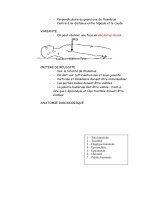
15.7.2 Pathophysiology of Trauma to External
Genitalia 261
15.7.2.1 Blunt Penile Trauma 261
15.7.2.2 Blunt Testicular Trauma 262
15.7.2.3 Blunt Vulvar Trauma 262
15.7.2.4 Penetrating Trauma of the External
Genitalia 262
Stab and Gunshot Genital Injuries 262
Genital Injuries Due to Bites 263
Straddle-Type Genital Injuries 263
Genital Mutilation 263
15.7.3 Diagnosis and Management of Genital
Trauma 264
15.7.4 Blunt Trauma of the Male Genitalia 264
15.7.4.1 Blunt Penile Trauma 264
15.7.4.2 Blunt Testicular Trauma 264
15.7.4.3 Blunt Female Trauma 265
15.7.4.4 Penetrating Trauma of the External
Genitalia 265
Penetrating Trauma in Men 265
Penetrating Women Trauma 265
15.7.5 Treatment of External Genital Trauma 265
15.7.5.1 Blunt Trauma 265
Blunt Penile Trauma 265
Blunt Testicular Trauma 266
Blunt Vulvar Trauma 266
15.7.5.2 Penetrating Trauma 266
Penetrating Penile Trauma 266
15.7.5.3 Penetrating Testicular Trauma 267
15.7.5.4 Penetrating Vulvar Trauma 267
References 267
15.7.1
Introduction
Traumatic injuries to the genitourinary tract are seen in
2.2%–10.3% of patients admitted to emergency units
(Brandes et al. 1995; Marekovic et al. 1997; Salvatierra et
al. 1969; Tucak et al. 1995; Archbold et al. 1981). Of these
injuries, between one-third and two-thirds are associat-
ed with injuries to the external genitalia (Brandes et al.
1995). Due to anatomy and prevalence of accidents, men
have a higher incidence of genital trauma than women,
since men have an increased exposure to violence, per-
formance of aggressive sports and motor vehicle acci-
dents. In addition, a worldwide increase in domestic vio-
lence has led to rising numbers of gunshot and stab
woundsoverthelastfewyears(Tiguertetal.2000;Cline
et al. 1998; Jolly et al. 1994; Bertini and Corriere 1988),
with as many as 35% of all gunshot wounds affecting also
the external genitalia (Monga and Hellstrom 1996).
Genitourinary trauma is seen in all age groups, most
frequently in males between 15 and 40 years of age.
However, 5% of trauma patients are less than 10 years
old, again undermining the broad spectrum of trau-
matic injuries requiring different specialists for man-
agement (Monga and Hellstrom 1996).
There are certain popular sports with an increased
risk for blunt and/or penetrating genital trauma, such
as off-road bicycling, horse-back riding, motorcycle ri-
ding, especially on bikes with a dominant gas tank (Lei-
bovitch and Mor 2005). In addition, blunt testicular
trauma has been reported in in-line hockey skating and
rugby players (Frauscher et al. 2001; de Peretti et al.
1993; Herrmann and Crawford 2002; Lawson et al.
1995; McAninch et al. 1984). Any type of full-contact
sport, without the use of necessary protective aids, may
be associated with genital trauma.
Besides these risk groups, severe trauma to the ex-
ternal genitalia is seen in female genital mutilation and
self-mutilation in psychotic patients and transsexuals
(McAninch et al. 1984).
Genitourinary trauma is commonly caused by blunt
injuries (80%), whereas 20% result from penetrating
lesions. For the above-mentioned reasons, blunt inju-
riestotheexternalgenitaliaaremorefrequentlyseenin
men than in women. Although the incidence of trau-
maticinjuriesishigherinmalesthanfemales,therisk
of associated injuries to neighboring organs (bladder,
urethra, vagina, and rectum) after blunt genital trauma
is higher in females than in males.
In men, blunt genital trauma frequently occurs uni-
laterally,withonly1%ofcasespresentingasbilateral
scrotal and/or testicular injuries (Monga and Hell-
strom 1996). However, penetrating scrotal injuries af-
fect both testes in 30% of cases (Monga and Hellstrom
1996; Cass et al. 1988). Besides locally extended lesions
associated with penetrating trauma, there is a 70% risk
of additional injuries in both genders.
15 Trauma
Table 15.7.1. American Association for the Surgery of Trauma
(AAST) organ injury severity scale for the vagina
Grade
a
Descriptionofinjury
I Contusion or hematoma
II Laceration, superficial (mucosa only)
III Laceration, deep into fat or muscle
IV Laceration, complex, into cervix or peritoneum
V Injury into adjacent organs (anus, rectum, urethra,
bladder)
a
AdvanceonegradeformultipleinjuriesuptogradeIII
Table 15.7.2. AASTorganinjuryseverityscaleforthevulva
Grade
a
Descriptionofinjury
I Contusion or hematoma
II Laceration, superficial (skin only)
III Laceration, deep into fat or muscle
IV Avulsion; skin, fat, or muscle
V Injury into adjacent organs (anus, rectum, urethra,
bladder)
a
AdvanceonegradeformultipleinjuriesuptogradeIII
Table 15.7.3. AASTorganinjuryseverityscaleforthetestis
Grade
a
Descriptionofinjury
I Contusion or hematoma
II Subclinical laceration of tunica albuginea
III Laceration of tunica albuginea with <50 % paren-
chymal loss
IV Major laceration of tunica albuginea with
50%
parenchymal loss
V Total testicular destruction or avulsion
a
AdvanceonegradeforbilaterallesionsuptogradeV
Table 15.7.4. AAST organ injury severity scale for the scrotum
Grade Descriptionofinjury
I Contusion
II Laceration <25% of scrotal diameter
III Laceration
25% of scrotal diameter
IV Avulsion <50%
V Avulsion
50%
Table 15.7.5. AAST organ injury severity scale for the penis
Grade Descriptionofinjury
I Cutaneous laceration/contusion
II
Buck’s fascia (cavernosum) laceration without tissue
loss
III Cutaneous avulsion/laceration through glans/mea-
tus/cavernosal or urethral defect <2 cm
IV Cavernosal or urethral defect
2 cm/partial penec-
tomy
V Tota l pen ectomy
Becauseofthishighincidenceofassociatedlesions,
accurate diagnosis and treatment of patients with pen-
etrating injuries are of utmost importance. The classifi-
cation of male and female genital trauma according to
the American Association for the Surgery of Trauma is
given in Tables 15.7.1–15.7.5.
Oneaspectthatmaynotbeforgottenintreating
trauma patients is the associated increased risk of infec-
tion of the emergency staff dealing with these patients,
especially hepatitis B and C. Recently, a 38% infection
rate with hepatitis B and/or C in males with penetrating
gunshot or stab wounds to the external genitalia was re-
ported (Cline et al. 1998). This incidence was signifi-
cantly higher compared with the normal population,
thus exposing emergency staff to an increased risk. It is
emphasized that standardized preventive procedures
mustbeinplaceandavailablefortheemergencystaff
not only to save the patient’s life but also to guarantee
co-workers’ health. Besides the risk of hepatitis infec-
tion, which is still higher than for HIV, the possible
transmission of HIV by trauma patients must be taken
into consideration. In a recent report by Xeroulis et al.,
a total of 287 consecutive trauma patients in Canada
were tested for Hep B/C and HIV infection (Xeroulis et
al. 2005). One patient was positive for hepatitis B, eight
for hepatitis C, and none for HIV. This revealed a three-
fold higher seroprevalence for hepatitis C compared
withthegeneralpopulation.Morethanhalfofthehepa-
titis C-positivepatients were men injured in a motor ve-
hicle crash with a mean Injury Severity Score of 19, de-
termining that hepatitis C poses the highest risk to the
trauma team. Although these numbers appear small,
there may be demographic differences at different cen-
ters, again emphasizing the importance of precautions
necessary for physicians and nursing staff.
15.7.2
Pathophysiology of Trauma to External
Genitalia
15.7.2.1
Blunt Penile Trauma
Blunt trauma to the flaccid penis may result in subcuta-
neous hematoma resulting from injury to the subcuta-
neous veins. Because the penile subcutaneous layers
(superficial, Colles fascia; deep, Buck’s fascia) meld into
lower abdominal fascial layers (superficial Camper’s
fascia, deep: Scarpa’s fascia), hematomas may spread to
the lower abdomen or to the penoscrotal base. De-
scending hematoma of the penile shaft can cause pre-
putial swelling that may cause obstructive voiding, re-
quiring transient catheterization.
Because of the thickness of the tunica albuginea in
theflaccidstate(approximately2mm),blunttraumato
the penis does not usually cause tearing of thetunica al-
15.7 Genital Trauma 261
buginea when there is no tumescence and rigidity. Dur-
ing erection, increasing rigidity and tumescence cause
a thinning of the tunica, reducing the thickness of the
tunica in the fully erect state. In these cases, a direct
blow to the erect penis may cause penile fracture, fre-
quently occurring during consensual intercourse,
which accounts for approximately 60% of penile frac-
tures (Haas et al. 1999). This usually occurs if the erect
penis slips out of the vagina and strikes against the
symphysis pubis or perineum, most frequently if the
womensitsontopoftheman.Penilefractureprimarily
affects the corporeal tunica by rupturing the tunica but
maybeassociatedwithlesionsofthecorpusspongio-
sum and urethra in 10%–22% (Nicolaisen et al. 1983;
Tsang and Demby 1992).
15.7.2.2
Blunt Testicular Trauma
Approximately 85% of testicular injuries result from
blunt trauma (Morey et al. 2004). Blunt trauma to the
scrotum can cause testicular dislocation, testicular
rupture, and/or subcutaneous scrotal hematoma.
Overall, traumatic dislocation of the testicle occurs
rarely,commonlyonlyunilaterallyandinvictimsofcar
or motorcycle accidents, or in pedestrians run over by
a vehicle (Lee et al. 1992; Shefi et al. 1999; Pollen and
Funckes 1982; Nagarajan et al. 1983). Bilateral disloca-
tion of the testes has been reported in up to 25% of
cases (Nagarajan et al. 1983). It can result in subcutane-
ous or internal dislocation of the testis. Subcutaneous
dislocation defines a subcutaneous epifascial displace-
ment of the testis, whereas during internal dislocation
of the testis it is positioned in the superficial external
inguinal ring, inguinal canal, or abdominal cavity.
Depending on the magnitude of blunt power acting
on the scrotum, testicular rupture may occur in ap-
proximately 50% of blunt scrotal traumas (Cass and
Luxenberg 1991). It can occur under intense, traumatic
compression of the testis against the inferior pubic ra-
mus or symphysis, resulting in a rupture of the tunica
albuginea of the testis. Wasko and Goldstein estimated
that a force of approximately 50 kg is necessary to cause
testicular rupture (Wasko and Goldstein 1996).
15.7.2.3
Blunt Vulvar Trauma
Blunt trauma to the vulva is rarely reported and may be
caused by obstetric, athletic, or sexual trauma or rarely
by car or bicycle accidents. The rich vulvar vascular
supply can be damaged by contusive frontal impacts,
whichcrushthevulvartissuesagainsttheosseous
planes (Virgili et al. 2000).
In obstetrics, incidence of traumatic vulvar hemato-
mas after vaginal deliveries was reported in only one
out of 310 deliveries (Sotto and Collins 1958). The fre-
quency in nonobstetric vulvar hematomas is even low-
er, with only several cases reported (Propst and Thorp
1998). Although the incidence of vulvar hematoma is
generally low, its presence indicates further investiga-
tions for associated lesions since vulvar hematoma is
closely related to an increased risk of vaginal, pelvic, or
abdominal injuries. Goldman et al. reported on the fre-
quency of blunt injuries of female external genitalia as-
sociated with pelvic trauma in 30%, consensual inter-
course in 25%, sexual assault in 20%, and other blunt
trauma in 15% (Goldman et al. 1998). Besides the pres-
ence of perforating associated lesions, blunt perineal
trauma may result in female sexual dysfunction classi-
fied as orgasmic disorders and/or hyposensitivity
(Munnarriz et al. 2002).
15.7.2.4
Penetrating Trauma of the External Genitalia
Penetrating trauma to the external genitalia is fre-
quently associated with complex injuries in other or-
gans. In children, penetrating injuries are most fre-
quently seen after straddle-type falls or laceration of
genital skin due to falls on sharp objects (Monga and
Hellstrom 1996; Okur et al. 1996). In any penetrating
trauma, the tetanus immunization status of the patient
has to be clarified. According to a recent review by Rhee
et al., tetanus toxoid booster was recommended in the
US for patients with the last immunization given more
than 10 years before. Since toxoid booster does not pro-
tect against the current injury, no urgency for the ad-
ministration of tetanus toxoid in the acute setting has
been suggested. This is divergent to suggestions by the
World Health Organization recommending tetanus
toxoid booster if tetanus immunization was received
more than 5 years before in patients with an open
wound(WorldHealthOrganization2000).Tetanusim-
munoglobulin should be reserved only for previously
nonimmunized injured patients (Rhee et al. 2005).
Stab and Gunshot Genital Injuries
Increasing worldwide domestic violence has led to a
rising incidence of stab and/or gunshot injuries associ-
ated with injuries of the genitourinary tract. The extent
of injuries associated with guns is related to the caliber
and velocity of the missile (Jolly et al. 1994). Handguns
or pistols range from 0.22 to 0.45 caliber, with a velocity
of 200–300 m/s. In addition, magnum handguns trans-
mit 20%–60% more energythan a standard handgun
to the tissue due to the higher velocity of the missile. In-
juries by rifles cause even more extensive lesions. Rifles
have a caliber ranging from 0.17 to 0.46 with a kinetic
energy transmission of up to 1,000 m/s.
262 15 Trauma
Missiles with a velocity of approximately 200–
300 m/s are considered as low velocity inducing a per-
manent cavity by entering the body. The energy along
the projectile path transmitted to the tissueis much less
than in high-velocity missiles, so that tissue destruc-
tion in low-velocity guns is less extensive (Jolly et al.
1994). On the contrary, high-velocity missiles (velocity
of 800–1,000 m/s) have an explosive effect with high-
energy transmission to the tissue causing a temporary
cavity. Due to the high-energy released, gaseous tissue
vaporization induces extensive damage, often associat-
ed with life-threatening injuries.
In relation to the weapon, caliber and configuration
of the missile, gunshot wounds are classified as pene-
trating, perforating, and avulsive.
a. Penetrating injuries with low-velocity missiles
often retain the projectile in the tissue, causing a
small, ragged entry wound.
b. Perforating gunshot wounds are frequently seen in
low- to high-velocity missiles. In these cases, the
missilepassesthroughthetissuewithasmallentry
wound, but larger exit wound.
c. Seriousinjuriesareassociatedwithavulsivegun-
shot wounds caused by high-velocity missiles, with
a small entry wound comparable to the caliber but
alargetissuedefectattheexitwound.
Genital Injuries Due to Bites
Although animal bites are common, bites involving in-
jury to the external genital are rare. Wounds are usually
minor but there is a potential risk of serious wound in-
fection.Thenatureoflocaltissuesandpolymicrobial
microbiology of bite wounds make genital bites a po-
tentially morbid event. Animal bites to external genita-
lia, especially to males, are rare. Of the affected pa-
tients, 60%–70% are boys aged under the age of
15 years (Gomes et al. 2000). Time to presentation since
trauma, severity of injury, and the type of management
have a direct influence on the outcome. A few small se-
ries (Gomes et al. 2001) and case reports (Kyriakidis et
al. 1979; Cummings and Boullier 2000) of genital bites
by different animals and humans have been reported.
But the lack of large retrospective or even prospective
trials make it difficult for a broad consensus on the
management of these injuries (Nabi and Mishriki
2005).
Approximately 30% of animal bite wounds already
present signs of infection within 48 h. The most com-
mon bacterial infection by a dog bite is Pasturella mul-
ticida, which accounts for up to 50% of infections (Do-
novan and Kaplan 1989). Other microorganisms com-
monly involved are Escherichia coli, Streptococcus viri-
dans, Staphylococ cus aureus, Bacteroides,andFusobac-
terium spp. (Donovan and Kaplan 1989; McAninch et
al. 1984). The first choice of antibiotics is penicillin fol-
lowed by cephalosporin or erythromycin. In addition
to antibiotics, proper wound management including
surgical exploration with debridement and daily
wound care are recommended (Kerins et al. 2004).
In animal bites, the possibility of rabies infection
must always be considered. In case of domestic pres-
ence of rabies infection in animals, vaccination must be
given to prevent life-threatening infections (Dreesen
and Hanlon 1998). The estimated worldwide number of
deaths due to rabies infection amounted to approxi-
mately 55,000 in 2004, most commonly in rural areas of
Africa and Asia. In addition to vaccination, local
wound management is an essential part of postexpo-
sure rabies prophylaxis. If rabies infection is suspected,
vaccination should be considered in relation to the ani-
mal involved, the specific nature of the wound and at-
tack (provoked/unprovoked), and the appearance of
the animal (aggressive, foam at the mouth). Presently,
vaccination with human rabies immunoglobulin and
human diploid cell vaccine is recommended (Dreesen
and Hanlon 1998; Anderson 1992).
Human bites to external genitalia include an even
broader range of possible infections with an additional
risk of sexually transmitted diseases, such as syphilis,
hepatitis, HIV, herpes, actinomycosis, or tuberculosis
(Franke et al. 1999).
Straddle-Type Genital Injuries
Straddle-type injuries may cause genitourinary trau-
ma, such as vaginal hematoma, vaginal contusion, pe-
nile laceration, or urethral injuries. In children, play-
ground equipment-specific injuries are attributed in
majority to monkey bars, jungle gyms, swings, and
slides (Waltzman et al. 1999).
Genital Mutilation
Female genital mutilation, often referred to as female
circumcision, comprises all procedures involving par-
tial or total removal of the external female genitalia (la-
bia majora/minora, clitoris) and/or other injuries to
the female genitalia (World Health Organization 2000).
It is still commonly performed in some parts of Africa
and the Middle East (Collinet et al. 2004). Some case re-
ports even reported genital mutilation performed in
Europe (Sheldon 2005; Holmgren et al. 2005; Turone
2004).
According to a recent report from southwestern Ni-
geria, the majority of genital mutilations were per-
formed by medically untrained personnel (89%) with a
complication rate up to 67% (Dare et al. 2004). The pro-
cedure is generally performed in young adrenarchal
women without anesthesia, with a high rate of hemor-
rhagic shock, urinary retention, and ulceration of the
genital region. Late complications include vulvar intro-
15.7 Genital Trauma 263
ital stenosis, HIV transmission, retention cysts and ab-
scesses, keloid scar formation, urinary incontinence,
dyspareunia, and sexual dysfunction, as well as diffi-
culties with childbirth (World Health Organization
2000).
15.7.3
Diagnosis and Management of Genital Trauma
Proper management of genital trauma requires a de-
tailed history, if possible, physical examination, and
imaging techniques. Especially in penetrating wounds,
information concerning the accident, possibly involved
persons, animals, vehicles, and weapons (knife, gun,
etc.) are important to estimate the extent of injury, the
potential risk of associated lesions, and subsequent in-
fections.
In addition to the history and physical examination,
a urine analysis is mandatory. Since an abusive assault
may be related to genital injuries, physicians must con-
sider the emotional difficulty for the patient as well as
their privacy in such examinations. This requires the
investigation of the patient alone without persons relat-
ed with the patient and may require short term anes-
thesia for physical examination. In case of suspicion,
takingswabsorvaginalsmearsfordetectionofsper-
matozoa is mandatory (Okur et al. 1996). Additionally,
other specialists may be requested (pediatrician, gyne-
cologist) for proper management of the patient. In or-
der to follow domestic rules and regulations, it is man-
datory to be aware of local guidelines such as the 2002
National Guidelines on the Management of Adult Vic-
tims of Sexual Assault (2002).
15.7.4
Blunt Trauma of the Male Genitalia
15.7.4.1
Blunt Penile Trauma
An essential part in the evaluation of blunt penile trau-
ma is the status of penile rigidity at injury. In case of a
flaccid penis at trauma, cavernosal and/or spongiosa
corporeal injuries are unlikely. Penile ultrasonography
with or without Duplex sonography and/or penile MRI
are not indicated.
Ifthepatientreportsonanerectionatinjury,diag-
nosisofpenilefracturecanbemadeafterathorough
history and examination in most cases. Patients most
commonly report a sudden cracking or popping sound
of the erect penis associated with moderate local pain
butimmediatepeniledetumescence.Asaresult,local
swellingofthepenileshaftdevelopswithprogressive
hematoma that may occur along fascial layers of the pe-
nile shaft extending to the lower abdominal wall in case
of rupture of Buck’s fascia. Depending on the extent of
the hematoma, rupture of the tunica may be palpated
(Morey et al. 2004).
In case of macro- or microhematuria, retrograde ur-
ethrography is mandatory to determine the presence of
urethral injury (Morey et al. 2004). Presence of micro-
hematuria without radiographic lesion of the urethra
requires no further intervention. In case of radiograph-
ic urethral lesion, a transurethral catheter can be
placed for bladder drainage.
Besides history and clinical examination, imaging
techniques may be performed by cavernosography and
magnetic resonance imaging (MRI) (Aboloyosr et al.
2005; Karadeniz et al. 1996; Pretorius et al. 2001). Both
techniques may identify laceration of the tunica albugi-
nea. Recent reports support the role of MRI as particu-
larly helpful in investigating the integrity of the tunica
albuginea, and presence of intracavernosal or extratu-
nical hematoma (Uder et al. 2002). Associated injuries
to adjacent structures (e.g., corpus spongiosum, ure-
thra) may also be found.
It remains uncertain whether the routine use of con-
trast material-enhanced MRI is justified in these cases
(Choi et al. 2000). Presently, cavernosography and/or
MRI are the most accurate imaging procedures in cases
wherepenilefractureissuspectedbuttheclinicalfind-
ings are unclear (Fedel et al. 1996).
15.7.4.2
Blunt Testicular Trauma
Patients report posttraumatic immediate scrotal pain,
nausea, vomiting, and sometimes they faint. They often
present with a tender, swollen scrotum and a impalpa-
ble testis. High-resolution, real-time ultrasonography
with a 7.5- to 10-MHz probe should be performed to
determine intra- and/or extratesticular bleeding, tes-
ticular contusion or rupture (Tsang and Demby 1992;
Pavlica and Barozzi 2001; Micallef et al. 2001; Patil and
Onuora 1994; Corrales et al. 1993; Mulhall et al. 1995;
Martinez-Pineiro et al. 1992; Fournier et al. 1989; Krat-
zik et al. 1989).
Controversial results have been presented regarding
the usefulness of ultrasonography in testicular trauma.
Some reported convincing results emphasizing the im-
portance of sonography with accuracy reaching 94%
(McAninch et al. 1984; Pavlica and Barozzi 2001; Marti-
nez-Pineiro et al. 1992; Fournier et al. 1989), whereas
others presented only low specificity (78%) and sensi-
tivity (28%) in determining testicular rupture (Cor-
rales et al. 1993). Some reported an overall accuracy of
scrotal ultrasound for testicular rupture of only 56%,
irrespective of the investigator (Corrales et al. 1993). So
far, it is the authors’ opinion that gray-scale ultrasonog-
raphy with 7.5- to 10-MHz remains a noninvasive tech-
nique with good reliability in experienced hands and
264 15 Trauma
should be performed in case of blunt testicular trauma.
Information may be increased by color Doppler duplex
ultrasonography to evaluate testicular perfusion. In
case of inconclusive scrotal sonography, testicular
computed tomography (CT) or MRI may be helpful in
elucidating scrotal dilemmas (Muglia et al. 2002). How-
ever, these techniques did not specifically increase the
detection of testicular rupture. The time delay associat-
ed with imaging studies has to be weighed against the
reliability of information in order to decide whether or
not surgical exploration is indicated. If imaging studies
cannot exclude testicular rupture, surgical exploration
should be initiated.
15.7.4.3
Blunt Female Trauma
In women, colposcopy and vulvovaginoscopy are a val-
id way of identifying genital injuries and are mandato-
ry if sexual assault is suspected (Mancino et al. 2003).
The presence of micro- or macrohematuria should not
be misinterpreted as menstrual bleeding. In women
with genital injuries and blood at the vaginal introitus,
it has been repeatedly emphasized that this may not on-
ly result from menstrual bleeding, but further investi-
gation is required to exclude vaginal injuries (Hussman
1998). As already mentioned, blunt genital trauma in
women seldom occurs, but if vulvar hematoma develop
there is a high chance of associated injuries. The per-
formance of flexible or rigid cystoscopy has been rec-
ommended to exclude urethral and bladder injury
(Goldman et al. 1998; Hussmann 1998). Complete vagi-
nal inspection with specula is mandatory and, because
of pain, should be carried out under sedation or gener-
al anesthesia in most cases. In case of suspected assault,
vaginalsmearsmustbetakenfordeterminationof
spermatozoa.
As blunt trauma to the vulva is often associated with
pelvic trauma, imaging studies of the pelvis with CT or
MRI should be performed to exclude intrapelvic pa-
thologies (Okur et al. 1996; Hussmann 1998).
15.7.4.4
Penetrating Trauma of the External Genitalia
As already mentioned in Sect. 15.7.2, “Pathophysiology
of Trauma to External Genitalia,” the importance of a
thorough history concerning the penetrating injury
must again be emphasized. Especially for gunshot
wounds, information concerning the type of weapons
used, the approximate distance of the missiles en-
trance,caliber,andsizeofthebulletishelpfulforfur-
ther treatment.
Penetrating T rauma in Men
Any kind of penetrating trauma of the external genital
requires urethrography irrespective of urine analysis to
exclude urethral lesion. Additionally, abdominal and a
pelvic CT scan, with or without cystography, may be
performed in those cases that do not require immediate
surgery.
Penetrating Women Trauma
Penetrating lesions of the external genitalia without le-
sions of adjacent organs are extremely rare, requiring
an abdominal and pelvic CT scan in any case. If the CT
scan cannot exclude associated bowel injuries or in-
traabdominal bleeding, exploratory laparoscopy has
been suggested in hemodynamically stable patients
prior to exploratory laparotomy (Okur et al. 1996). In
the hemodynamically unstable patient, exploratory
laparotomy is indicated.
15.7.5
Treatment of External Genital Trauma
15.7.5.1
Blunt Trauma
Blunt Penile Trauma
Blunt trauma to the flaccid penis usually develops only
subcutaneous hematoma requiring no surgical inter-
vention. The presence of subcutaneous hematoma,
without rupture of the cavernosal tunica albuginea and
no immediate detumescence of the erect penis, does
not require surgical intervention. In these cases, non-
steroidal analgetics and ice packs are recommended.
Preputial swelling and edema may require transient
catheterization with the need for percutaneous cystos-
tomy only in a few selected cases with an increased risk
of local inflammatory complications (i.e., necrotizing
fasciitis). In case of necrotizing fasciitis, rapid exten-
sive surgical debridement is very important in addition
to broad-spectrum antibiotic therapy.
Inthecaseofpenilefracture,immediatesurgicalin-
tervention with closure of the tunica albuginea is rec-
ommended. Closure of the tunica can be obtained by
using either absorbable or nonabsorbable sutures, with
good long-term outcome and protection of potency.
Postoperative complications were reported in 9 %, in-
cluding superficial wound infection and impotence in
1.3% (Haas et al. 1999; Orvis and McAninch 1989).
Conservative management of penile fracture is not rec-
ommended because of early and long-term complica-
tions, including penile abscess, missed partial urethral
disruption,penilecurvature,andpersistenthematoma
requiring delayed surgical intervention (Orvis and
McAninch 1989). In addition, fibrosis and penile angu-
15.7 Genital Trauma 265
lation were reported in 35% after conservative man-
agement of penile fracture (Haas et al. 1999; Orvis and
McAninch 1989).
Blunt Testicular Trauma
Blunt trauma to the scrotum can cause significant he-
matocele without testicular rupture. Conservative
management with ice packs, nonsteroidal analgetics,
and bed rest is recommended in hematoceles smaller
thanthreetimesthesizeofthecontralateraltestis(Ti-
guert et al. 2000). Several authors reported the risks of
conservative management in blunt scrotal trauma re-
quiring delayed interventions (>3 days) in many cases,
with a significantly higher rate of orchiectomy even in
the nonruptured testis (Monga and Hellstrom 1996;
Cass and Luxenberg 1988, 1991; McAninch et al. 1984;
Altarac 1994). The reasons for delayed interventions re-
quiring surgery were local infections and pain. It was
repeatedly reported that early surgical intervention,
i.e., within 72 h, resulted in more than 90% preserva-
tion of the testis, whereas delayed surgery necessitated
orchiectomy in 45%–55% (Cass and Luxenberg 1991).
If the integrity of testicular tunica albuginea cannot be
clearly visualized or duplex ultrasonography shows re-
duced perfusion in the injured testicles, scrotal explo-
ration is indicated.
Additionally, pain and duration of hospital stay may
be markedly reduced by early surgical intervention for
large hematoceles. Because of the long convalescence in
large hematoceles, surgical exploration is recommend-
ed, irrespective of testicle contusion or rupture. By
evacuation of the blood clot from the tunica vaginalis,
testicular pain is relieved and rehabilitation will be
more rapid (Altarac 1994).
In cases of testicular rupture, surgical exploration
with excision of necrotic testicular tubules, closure of
the tunica albuginea is mandatory and suction drainage
should be applied. By early intervention, 80% of injured
testicles can be saved (Fowler et al. 1992) and normal
testicular endocrine function can be maintained. By ap-
plying intravenous antibiotics and nonsteroidal anti-in-
flammatory drugs within 6 h after injury, a reduction in
infectious risk has been seen (Whelan et al. 2005).
Traumatic dislocation of the testis can be reposi-
tioned manually followed by delayed surgical orchido-
pexy. In cases of insufficient positioning of the dislo-
catedtestisposttraumatically,primaryorchidopexyis
indicated.
Blunt Vulvar Trauma
Blunt trauma to the vulva is rare and commonly pre-
sentsasextendedhematomas.Managementofvulvar
hematomas may range from conservative treatment to
surgical decompression. In most cases, vulvar hemato-
mas after blunt trauma do not require surgical inter-
vention, but they may cause significant blood loss re-
quiringtransfusion.Reporteddataarescarce,andrec-
ommendations for vulvar wound management are
based on empirical experience (Propst and Thorp 1998;
Goldman et al. 1998; Okur et al. 1996; Husmann 1998).
In hemodynamically stable women, nonsteroidal anti-
inflammatories and cold packs relieve pain, requiring
no surgical intervention in the majority of cases.
In extended vulvar hematoma or in unstable pa-
tients, hospitalization may be indicated for surgical in-
tervention, stabilization, and reduction of infectious
risks.Theadditionaluseofantibioticsisrecommended
in major vulvar trauma.
However, blunt trauma to the female external geni-
talia may be associated with voiding problems and/or
lesions to adjacent organs. Therefore, transurethral
catheterization for dip stick testing is indicated to ex-
clude hematuria requiring further investigations.
15.7.5.2
Penetrating Trauma
Penetrating trauma to the external genitalia require
surgical exploration in most cases, including debride-
ment and reconstruction in order to prevent late com-
plications such as urethral strictures, penile curvature
and erectile dysfunction, and testicular atrophy (Morey
et al. 2004). In complex wounds with persistent infec-
tion, negative-pressure wound therapy (vacuum de-
vices) complements surgical and medical intervention
(Whelan et al. 2005).
Penetrating Penile Trauma
Surgical exploration and conservative debridement of
necrotic tissue is recommended with primary closure
in most cases. Even in extended injuries of the penis or
complete dissection, primary repair should be tried
with only minor excision of necrotic tissue due to the
excellent blood supply of penile corpora. In complete
dissection of the penis, vascular and neuronal realign-
ment should be performed by a skilled microsurgeon in
addition to corporeal and urethral reconstruction
(McAninch et al. 1984; Van der Horst et al. 2004).
In extended loss of penile shaft skin, split-thickness
grafts can be utilized after infectious control. McA-
ninch et al. recommended the use of a skin graft thick-
ness of at least 0.001 inch in order to reduce the risk of
skin contractions restricting penile enlargement dur-
ing erection (McAninch et al. 1984). Additionally, dur-
ing reconstruction grafts should be placed circumfe-
rentially to the artificially erected penis to prevent con-
tracture, shortening, or deviation. In case of proper
surgicalmanagement,potencyratesofmorethan80%
can be achieved (Goldman et al. 1996). Excellent clini-
266 15 Trauma
cal results are also reported on the use of autologous
rectus fascia graft for coverage of a tunica or corporeal
defect (Pathak et al. 2005).
Besides postsurgical transient urethral stenting, a
suprapubic cystostomy may be placed in addition to
broad-spectrum antibiotics.
15.7.5.3
Penetrating Testicular Trauma
Penetrating injuries to the scrotum require surgical ex-
ploration with conservative debridement of nonviable
tissue. Primary realignment can be easily obtained, in
most cases. Only in severe infection or necrotizing fas-
ciitis would debridement with subcutaneous femoral
displacement of the testicles be required initially. After
proper wound granulation, reconstructive surgery ei-
ther by secondary closure of the scrotal skin and re-
placement of the testis can be obtained or split thick-
ness grafts may be used for scrotal reconstruction
(Rapp et al. 2005). In cases of high-velocity gunshot in-
juries, the testicle could not be saved in almost 90% of
the reported cases (Gomez et al. 1993).
Complete disruption of the spermatic cord occurs
and is treated with vascular realignment if possible. Mi-
crosurgical reconstruction of the vas deferens either by
vasovasostomy or tubulovasostomy should only be
performed in the hemodynamically stable patient or
secondarily after rehabilitation of the patient (Altarac
1993). If there is extensive destruction of the tunica al-
buginea, mobilization of a free tunica vaginalis flap can
be obtained for testicular closure. If the patient is un-
stable or reconstruction cannot be achieved, orchiecto-
my should be performed. If both testicles are severely
damaged, prior to surgery or even after orchiectomy,
testicular epididymal sperm extraction (TESE) map-
ping may be considered for future artifical reproduc-
tion (Baniel and Sella 2001; Negri et al. 2002).
Extended laceration of scrotal skin requires surgical
intervention for skin closure after removal of any for-
eign material. Due to the elasticity of the scrotum, most
defects can be primarily closed, even if the lacerated
skin is only minimally attached to the body (McAninch
et al. 1984). The recreative capacity of scrotal skin is
high, indicating conservative debridement and prima-
ry realignment in most cases. However, local wound
management with extensive rinsing of the wound is an
important fact for scrotal convalescence. Even in cases
of complete disruption of scrotal skin, it can be re-
aligned in most cases after debridement and washing.
In fact, there is an associated risk of harming the vascu-
lar plexus in the stratum reticulare of the skin, causing
partial necrosis of full-thickness skin grafts. This in
turn may require resection and staged closure with
split-thickness grafts or, depending upon the extent of
the defect, secondary granulation of the wound. It must
be noted that using thick skin flaps, or burying the tes-
ticle are not recommended for patients who wish to re-
main fertile, as the spermatogenesis deteriorates sub-
stantially after a period of 2 years (Wang et al. 2003).
Although the rehabilitative capacity of the scrotum
is very good, the use of antibiotics is indicated in any
case of penetrating trauma.
15.7.5.4
Penetrating Vulvar Trauma
Although penetrating vulvar trauma is rarely seen, it is
even more important to emphasize that vulvar hemato-
ma and/or blood at the vaginal introitus are an indica-
tion for vaginal exploration in order to identify possi-
ble associated vaginal and/or rectal injuries under se-
dation or general anesthesia (Husmann 1998). In case
of vulvar laceration, realignment after conservative de-
bridement is indicated. If there are associated injuries
to the vagina, these can be repaired immediately by pri-
mary suturing. Additional injuries to the bladder, rec-
tum, or bowel may require laparotomy for closure and,
in case of rectal injuries, may necessitate transient co-
lostomy.
References
Aboloyosr A, Moneim AE, Abdelatif AM, Abdalla MA, Imam HM
(2005) The management of penile fracture based on clinical
and magnetic resonance imaging findings. BJU Int 96:373
Altarac S (1993) A case of testicle replantation. J Urol 150:1507
Altarac S (1994) Management of 53 cases of testicular trauma.
Eur Urol 25:119
Anderson CR (1992) Animal bites. Guidelines to current man-
agement. Postgrad Med 92:134
Archbold JA, Barros d’sa AA, Morrison E (1981) Genito-uri-
nary tract injuries of civil hostilities. Br J Surg 68:625
Baniel J, Sella A (2001) Sperm extraction at orchiectomy for
testis cancer. Fertil Steril 75:260
Bertini JE Jr, Corriere JN Jr (1988) The etiology and manage-
ment of genital injuries. J Trauma 28:1278
Brandes SB, Buckman RF, Chelsky MJ, Hanno PM (1995) Ex-
ternal genitalia gunshot wounds: a ten-year experience with
fifty-six cases. J Trauma 39:266
Cass AS, Luxenberg M (1988) Value of early operation in blunt
testicular contusion with hematocele. J Urol 139:746
Cass AS, Luxenberg M (1991) Testicular injuries. Urology
37:528
Cass AS, Ferrara L, Wolpert J, Lee J (1988) Bilateral testicular
injury from external trauma. J Urol 140:1435
Choi MH, Kim B, Ryu JA, Lee SW, Lee KS (2000) MR imaging of
acute penile fracture. Radiographics 20:1397
Cline KJ, Mata JA, Venable DD, Eastham JA (1998) Penetrating
traumatothemaleexternalgenitalia.JTrauma44:492
C o llin e t P, S abb a n F, Lu c ot J P, B o uke r r ou M, S t i en L , L e roy JL
(2004) Management of type III female genital mutilation.
J Gynecol Obstet Biol Reprod 33:720
Corrales JG, Corbel L, Cipolla B, Staerman F, Darnault P, Guille
F, Lobel B (1993) Accuracy of ultrasound diagnosis after
blunt testicular trauma. J Urol 150:1834
References 267
Cummings JM, Boullier JA (2000) Scrotal dog bites. J Urol 164:57
DareFO,OboroVO,FadioraSO,OrjiEO,Sule-OduAO,Olabo-
de TO (2004) Female genital mutilation: an analysis of 522
cases in South-Western Nigeria. J Obstet Gynaecol. 24:281
De Peretti F, Cambas PM, Veneau B, Argenson C (1993) Fuel
tanks of motorcycles. Role in severe trauma of the pelvis.
Presse Med 22:61
Donovan JF, Kaplan WE (1989) The therapy of genital trauma
by dog bite. J Urol 141:1163
Dreesen DW, Hanlon CA (1998) Current recommendations for
the prophylaxis and treatment of rabies. Drugs 56:801
FedelM,VenzS,AndreessenR,SudhoffF,LoeningSA(1996)
Thevalueofmagneticresonanceimaginginthediagnosisof
suspected penile fracture with atypical clinical findings.
J Urol 155:1924
Fournier GR Jr, Laing FC, McAninch JW (1989) Scrotal ultraso-
nography and the management of testicular trauma. Urol
Clin North Am 16:377
Franke M, Lenk S, Petras T (1999) Genitalverletzung durch
Hundebisse. Extr Urol 22:15
Frauscher F, Klauser A, Stenzl A, Helweg G, Amort B, zur Ned-
den D (2001) US findings in the scrotum of extreme moun-
tain bikers. Radiology 219:427
Goldman HB, Dmochowski RR, Cox CE (1996) Penetrating
traumatothepenis:functionalresults.JUrol155:551
Goldman HB, Idom CB Jr, Dmochowski RR (1998) Traumatic
injuries of the female external genitalia and their associa-
tion with urological injuries. J Urol 159:956
Gomes CM, Ribeiro-Filho L, Giron AM, Mitre AI, Figueira ER,
Arap S (2000) Genital trauma due to animal bites. J Urol
165:80
Gomes CM, Ribeiro-Filho L, Giron AM, Mitre AI, Figueira ER,
Arap S (2001) Genital trauma due to animal bites. J Urol
165:80
Gomez RG, Castanheira ACC, McAninch JW (1993) Gunshot
wounds to the male genitalia. J Urol 150:1147
Haas CA, Brown SL, Spirnak JP (1999) Penile fracture and tes-
ticular rupture. World J Urol 17:101
Herrmann B, Crawford J (2002) Genital injuries in prepubertal
girls from inline skating accidents. Pediatrics 110:e16
Holmgren H, Almroth L, Berggren V, Bergstrom S (2005) Geni-
tal mutilation of children is an offence. Do health services
have a sufficient knowledge to see the problem? Lakartid-
ningen 102:1637
Husmann DA (1998) Editorial Comment. J Urol 159:959
Jolly BB, Sharma SK, Vaidyanathan S, Mandal AK (1994) Gun-
shot wounds of the male external genitalia. Urol Int 53:92
Karadeniz T, Topsakal M, Ariman A, Erton H, Basak D (1996)
Penile fracture: differential diagnosis, management and
outcome. Br J Urol 77:279
Kerins M, Greene S, O’Connor N (2004) A human bite to the
scrotum:acasereportandreviewoftheliterature.EurJ
Emerg Med 11:223
Kratzik C, Hainz A, Kuber W, Donner G, Lunglmayr G, Frick J,
Schmoller HJ (1989) Has ultrasound influenced the therapy
concept of blunt scrotal trauma? J Urol 142:1243
Kyriakidis A, Karydis G, Yannopoulos P (1979) An unusual
trauma of the glans penis. Br J Urol 51:161
Lawson JS, Rotem T, Wilson SF (1995) Catastrophic injuries to
the eyes and testicles in footballers. Med J Aust 163:242
Lee JY, Cass AS, Streitz JM (1992) Traumatic dislocation of tes-
tes and bladder rupture. Urology 40:506
Leibovitch I, Mor Y (2004) The vicious cycling: bicycling relat-
ed urogenital disorders. Eur Urol Mar 47:277
MancinoP,ParlavecchioE,MellusoJ,MontiM,RussoP(2003)
Introducing colposcopy and vulvovaginoscopy as routine
examinations for victims of sexual assault. Clin Exp Obstet
Gynecol 30:40
Marekovic Z, Derezic D, Krhen I, Kastelan Z (1997) Urogenital
war injuries. Mil Med 162:346
Martinez-Pineiro L Jr, Cerezo E, Cozar JM, Avellana JA, More-
no JA, Martinez-Pineiro JA (1992) Value of testicular ultra-
sound in the evaluation of blunt scrotal trauma without hae-
matocele. Br J Urol 69:286
McAninch JW, Kahn RI, Jeffrey RB, Laing FC, Krieger MJ
(1984) Major traumatic and septic genital injuries. J Trauma
24:291
MicallefM,AhmadI,RameshN,HurleyM,McInerneyD
(2001) Ultrasound features of blunt testicular injury. Injury
32:23
Monga M, Hellstrom WJ (2005) Testicular trauma. Adolesc
Med 1996 7:141
Morey AF, Metro MJ, Carney KJ, Miller KS, McAninch JW
(2004) Consensus on genitourinary trauma: external genita-
lia. Br J of Urol Int 94:507
Muglia V, Tucci S Jr, Elias J Jr, Trad CS, Bilbey J, Cooperberg PL
(2002) Magnetic resonance imaging of scrotal diseases:
when it makes the difference. Urology 59:419
Mulhall JP, Gabram SG, Jacobs LM (1995) Emergency manage-
ment of blunt testicular trauma. Acad Emerg Med 2:639
Munarriz R, Talakoub L, Somekh NN, Lehrfeld T, Chudnovsky
A, Flaherty E, Goldstein I (2002) Characteristics of female
patients with sexual dysfunction who also had a history of
blunt perineal trauma. J Sex Marital Ther 28 [Suppl 1]:175
Nabi G, Mishriki SF (2005) Bite wounds of genitalia: clinical
presentation and management. Hosp Med 66:112
Nagarajan VP, Pranikoff K, Imahori SC, Rabinowitz R (1983)
Traumatic dislocation of testis. Urology 22:521
National Guideline Clearinghouse (2002) 2002 National guide-
lines on the management of adult victims of sexual assault.
/>id=3050. Cited 5 December 2006
Negri L, Albani E, Di Rocco M, Levi-Setti PE (2002) Aspermia
and chronic testicular pain after imperforate anus correc-
tion. Cryopreservation of sperm cells extracted from whole
orchiectomized testis: case report. Hum Reprod 17:2935
Nicolaisen GS, Melamud A, Williams RD, McAninch JW (1983)
Rupture of the corpus cavernosum: surgical management.
J Urol 130:917
Okur H, Kucikaydin M, Kazez A, Turan C, Bozkurt A (1996)
Genitourinary tract injuries in girls. Br J Urol 78:446
Orvis BR, McAninch JW (1989) Penile rupture. Urol Clin
North Am 16:369
Pathak AS, Chang JH, Parekh AR, Aboseif SR (2005) Use of rec-
tus fascia graft for corporeal reconstruction during place-
ment of penile implant. Urology 65:1198
Patil MG, Onuora VC (1994) The value of ultrasound in the
evaluation of patients with blunt scrotal trauma. Injury
25:177
Pavlica P, Barozzi L (2001) Imaging of the acute scrotum. Eur
Radiol 11:220
Pollen JJ, Funckes C (1982) Traumatic dislocation of the testes.
J Trauma 22:247
Pretorius ES, Siegelman ES, Ramchandani P, Banner MP
(2001) MR imaging of the penis. Radiographics 21:S283; dis-
cussion S2989
Propst AM, Thorp JM Jr (1998) Traumatic vulvar hematomas:
conservative versus surgical management. South Med J
91:144
Rapp DE, Cohn AB, Gottlieb LJ, Lyon MB, Bales GT (2005) Use
of tissue expansion for scrotal sac reconstruction after scro-
tal skin loss. Urology 65:1216
Rhee P, Nunley MK, Demetriades D, Velmahos G, Doucet JJ
(2005) Tetanus and trauma: a review and recommendations.
J Trauma 58:1082
Salvatierra O Jr, Rigdon WO, Norris DM, Brady TW (1969)
268 15 Trauma
Vietnam experience with 252 urological war injuries. J Urol
101:615
Shefi S, Mor Y, Dotan ZA, Ramon J (1999) Traumatic testicular
dislocation: a case report and review of published reports.
Urology 54:744
Sheldon T (2005) Dutch doctors should tackle female genital
mutilation. BMJ 330:922
Sotto LSJ, Collins RJ (1958) Perigenital Hematomas. Obstet Gy-
necol 12:259
Tiguert R, Harb JF, Hurley PM, Gomes De Oliveira J, Castillo-
Frontera RJ, Triest JA, Gheiler EL (2000) Management of
shotgun injuries to the pelvis and lower genitourinary sys-
tem. Urology 55:193
Tsang T, Demby AM (1992) Penile fracture with urethral inju-
ry. J Urol 147:466
Tucak A, Lukacevic T, Kuvezdic H, Petek Z, Novak R (1995)
Urogenital wounds during the war in Croatia in 1991/1992.
J Urol 153:121
Turone F (2004) Controversy surrounds proposed Italian alter-
native to female genital mutilation. BMJ 328:247
Uder M, Gohl D, Takahashi M, Derouet H, Defreyne L, Kra-
mann B, Schneider G (2002) MRI of penile fracture: diagno-
sis and therapeutic follow-up. Eur Radiol 12:113
VanderHorstC,MartinezPortilloFJ,SeifC,GrothW,Jüne-
mann KP (2004) Male genital injury: diagnosis and treat-
ment. Br J Urol Int 93:927
Virgili A, Bianchi A, Mollica G, Corazza M (2000) Serious he-
matoma of the vulva from a bicycle accident. A case report.
J Reprod Med 45:662
Waltzman ML, Shannon M, Bowen AP, Bailey MC (1999) Mon-
keybar injuries: complications of play. Pediatrics 103:e58
Wang D, Zheng H, Deng F (2003) Spermatogenesis after scrotal
reconstruction. Br J Plast Surg 56:484
Wasko R, Goldstein AG (1966) Traumatic rupture of the testi-
cle. J Urol 95:721
Whelan C, Stewart J, Schwartz BF (2005) Mechanics of wound
healing and importance of vaccum assisted closure in urolo-
gy. J Urol May 173:1463
World Health Organization (2000) Fact sheet No. 241, June
2000
Pan American Health Organization (2000) Epidemiologic sur-
veillance and disease control. In: Natural disasters: protect-
ing the public health. World Health Organization, Pan
American Health Organization, Washington, DC, p 43
XeroulisG,InabaK,StewartTC,LanniganR,GrayD,Maltha-
ner R, Parry NG, Girotti M (2005) Human immunodeficien-
cy virus, hepatitis B, and hepatitis C seroprevalence in a Ca-
nadian trauma population. J Trauma 59:105
References 269
15.8 Management of Penile Amputation
G.H. Jordan
15.8.1 Introduction 270
15.8.2 History of Penile Replantation 271
15.8.3 Anatomy of the Penis 271
15.8.4 Penile Replantation 272
15.8.5 Summary 274
References 274
15.8.1
Introduction
When one reviews the literature surrounding penile
amputation, most of what is found is individual case re-
ports or reports of small series. Thus what is consid-
ered to be state-of-the-art management is gleaned from
literature review, and frankly reliant on expert opinion.
An exception to this statement is a series of penile am-
putation from Thailand published in 1983 (Bhangana-
da et al. 1983) in the American Journal of Surgery .That
report described the management of approximately
100 cases of penile amputation, many of which preced-
ed the description of microreplantation techniques and
validated much of what literature reviews have pro-
posed.
In Western culture, penile amputation injuries are
seen primarily as a result of felonious assault or self-
emasculation in the psychotic individual who is re-
sponding to command hallucinations. One will also
find descriptions of penile amputation as a conse-
quenceofcircumcision.Inmostcases,however,incir-
cumcision trauma, what is amputated is the penile skin
and/or only a portion of the glans (Neulander et al.
1996; Strimling 1996). True penile amputation is seen
in cultures that still perform ritual circumcision, but
again the literature reveals only sporadic reports
(Ameh et al. 1997; Ozkan and Gurpinar 1997; Hashem
et al. 1999; Silfen et al. 2000; Izzidien 1981).
In a review by Greilsheimer and Groves (1979), it
was found that patients who amputate or mutilate their
genitalia represent a heterogenous group. Eighty-seven
percent are believed or shown to be psychotic at the
time of the accident, with 51% in a decompensated
schizophrenic state. The other group represents indi-
viduals with severe character disorders or in some
cases gender identity problems. Those individuals are
often under the influence of drugs or alcohol at the time
of their genital amputation event. While many psycho-
tic individuals have a long history of mental illness,
usually the act of self-mutilation occurs during an
acute psychotic decompensation. There are some indi-
viduals who during their first psychotic break will at-
tempt the amputation of the penis or another body
part. In a paper by Hall et al. (1981), it was reported that
the psychotic individual often has a history of preexi-
sting conflicts about his role as a male; but with a psy-
chotic break, the individual comes under the effect of
hallucinations commanding him to amputate all of his
genitalia, or some other form of partial self-mutilation.
Blacker and Wong (1963) show that many of these
patients are born to a domineering older mother in the
home where there is no male influence. In many cases,
the families are impoverished, thus limiting the associ-
ation of the child with other adults and in particular
adult males. It was found that many of these individuals
were made to feel guilty or inadequate as males in their
childhood. Blacker and Wong have described self-mu-
tilation as a form of focal suicide. Dogma would say
that in the case of self-mutilation, replantation is con-
traindicated, as the patient, when capable, will “just
pull the replanted part off again.” This has not been the
experience of the author, and Stewart and Lowery
(1980) in their review state that self-inflicted injury is
not an absolute contraindication. The literature in fact
attests to a high degree of successful mental rehabilita-
tion in these patients. I believe that the dictum should
be replantation first and psychiatry second. Greilshei-
mer and Groves’ (1979) review of over 40 patients of pe-
nile amputation show that in that group there was only
one postoperative suicide and one repeat attempt at
genital self-mutilation. That said, however, when one is
confronted in the emergency room with a patient who
has undertaken genital self-mutilation, one must be
very careful to know the laws of the venue in which one
is “operating.” In some cases, court order is required, in
some states only the agreeing opinion of two practi-
tioners is required, and certainly many other variations
of this theme exist from state to state. Often times, get-
15 Trauma
ting consent from the patient is possible and where
possible should absolutely be done.
At our center, we have also become aware of another
interesting phenomenon, probably best described as
focal homicide by proxy. We have treated two cases in
whom a male child was the victim of penile amputation
by his mother. The motivation, however, for the attack
on the child was the behavior of the father. In one case,
the father was actively physically abusive of the woman;
and in another case the father had been discovered to
be having an affair. In neither case was replantation
possible, as the child’s mother took steps to ensure that
the amputated part was not available.
With regards to the patient with command halluci-
nations, the hallucinations not uncommonly involve
God or God’s representative telling the patient to muti-
late himself (Schweitzer 1990; Clark 1981; Waugh 1986;
Culliford 1987). Ames (1987) has suggested the eponym
of Klingsor syndrome for the phenomenon. In some
cases, the delusions involve the notion that there is
promise of great things currently denied the individual
because of sexual thoughts or sexual indiscretion.
Many of these patients find, initially, the hallucinations
to be troublesome. Many patients look to the Bible for
“confirmation of the will of God.” Examples of scrip-
ture which seem to support the notions of the halluci-
nations can be found in Matthew 5:9, Matthew 18:9,
and Mark 9:47. The delusional individual obviously
misrepresents, to himself, the intent of scripture, thus
interpreting the scripture as reaffirmation of the com-
mands. In talking with these patients, once the patient
has his affirmation, it is only a matter of time before he
proceeds with the act of genital mutilation.
The patient, often times, reports the commands to
doctorsorothermedicalpersonnel,butinvagueterms.
It is trite to say, however, that the best way to treat a pe-
nile amputation is to prevent it. Thus all primary care
practitioners, primary healthcare or not, must be alert
to the vagueness of these comments; and when they are
heard, they must be regarded as very serious and not
absurd and trifling, as in many cases they may seem.
15.8.2
History of Penile Replantation
In 1929, the first case of replantation of an amputated
penis was reported (Ehrich 1929). The patient had am-
putated his penis using a radial saw. During the trauma,
the patient’s penile skin was avulsed; the penis was re-
planted by macroscopic techniques and buried in the
patient’sscrotum.Twoyearslater,withliberationofthe
penis from the scrotum, the patient had a penis that
looked quite normal cosmetically and functioned very
normally. In 1976, two groups independently reported
the first successful microreplantation of an amputated
penis (Cohen et al. 1977; Tamai et al. 1977). Neither
group was aware of the other’s work, and since these
landmark reports, other cases using similar techniques
have been published with excellent, reproducible re-
sults. A review by Carroll and associates in 1985 (Car-
roll et al. 1985) proposed a logical sequence of care for
the patient with penile amputation. In that review, pa-
tients were reported to have excellent sensation; ability
to achieve intromission was not specifically addressed.
Using techniques that vary little from the initial reports
of 1977, microreplantation of the penis has been
changed from reportable to essentially nonreportable.
In 1968, McRoberts reported a case and review of the
literature(McRobertsetal.1968).Hethensummarized
the technique for replantation of the amputated penis
using macrotechniques. In that technique, all struc-
tures that could be coapted were coapted; this included
a repair of the urethra, coaptation of the erectile bod-
ies, and later it was proposed that anastomosis of the
dorsal vein was possible under loop magnification with
improved results. McRoberts had noted that using
these techniques, the skin of the penis if it was avulsed
atthetimeoftraumawasfrequentlysloughedduring
the postoperative period. He thus recommended de-
briding the skin of the penis to the coronal margin and
burying the penis in the scrotum. The penis could later
be liberated, and with the development of microsurgi-
cal techniques, nerve repair could later be undertaken.
In these cases, often times the glans will develop an es-
char; however, uniformly the spongy erectile tissue
seems to survive and will re-epithelialize. In the above-
mentioned series from Thailand, 18 of the 100 cases
were managed with microreplantation techniques,
many were managed by macroreplantation techniques,
and of course in some cases, the end of the penis of
someunfortunatepatientsdidnotmanagetomakeitto
the hospital with the patient.
15.8.3
Anatomy of the Penis
The deep vasculature of the penis is totally dependent
on branches of the deep internal pudendal arteries.
These are branches of the hypogastric artery. The pu-
dendalarterycoursestotheperineumviaAlcock’sca-
nal, and in the perineum gives off the posterior scrotal
arteries and the perineal arteries. The vessels then con-
tinue as the common penile artery (Fig. 15.8.1) where
the artery goes on to multiply bifurcate to provide vas-
culature to the corpus spongiosum and urethra, as well
as the corporal bodies and the glans penis (Kodos
1967). The skin of the penis is dependent on a fasciocu-
taneous blood supply based on the superficial external
pudendal artery (Fig. 15.8.2) (Quartey 1983). The ve-
nous drainage of the penis has likewise been nicely de-
15.8 Management of Penile Amputation 271
Fig. 15.8.1. Illustration of the common penile arterial system.
Thisisthevasculaturetothedeepstructuresofthepenis
Fig. 15.8.2. Illustration of the superficial external pudendal ar-
tery as described by the microinjection studies of Quartey
(Quartey 1983)
scribed. The venous system has been divided into three
systems: 1) the superficial dorsal system, 2) the deep
dorsal venous system, and 3) the crural vessels, which
depart from the corporal cavernosa at the crus of the
corpora and go on to drain into the periprostatic plex-
us, and the cavernosal venous system, which likewise
departs from the proximal crura and becomes part of
the dorsal vein to the penis and the periprostatic plexus
(Fig. 15.8.3) (Aboseif et al. 1993).
Fig. 15.8.3. Illustration of the venous drainage of the deep
structures of the penis (Aboseif 1983)
15.8.4
Penile Replantation
Whenoneisalerted,asasurgeon,thatapatientwith
penile amputation is being brought in, the initial atten-
tion must be directed to the preservation of the ampu-
tated portion of the penis. Hypothermia prolongs the
ischemic survival times of all tissues (Hayhurst et al.
1974). The amputated penis must be regarded as a free
flap. Literature that has examined the no-reflow phe-
nomenainarabbitflapsurvivalmodelshowsthatis-
chemia time clearly affects these phenomena (May et
al.1978).Thishasbeenshowninanumberofotherflap
models. In a study examining digital replantation, Hay-
hurst and his associates demonstrated that hypother-
miaprolongedtheischemiatimeorwascompatible
witheventualsurvivalfrom6to24h(Hayhurstetal.
1974). The precise response to hypothermia of the pe-
nis has not been studied; however, penile replantation
after16h,muchofwhichwasnormalthermicischemia
time, has been reported to be successful (Hashem et al.
1999; Mosahebi et al. 2001; Jezior et al. 2001). At our
center, a penis was successfully replanted after 18 h,
much of which was hypothermic ischemia time.
Thus I would recommend the following technique:
the penis should be placed in saline-soaked gauze and
then placed in a sterile plastic bag. The plastic bag can
then be placed in slush and the amputated part then
transported (Fig. 15.8.4).
The process that we have used in our patients begins
with obtaining approval to proceed with surgery. The
amputated organ remains in hypothermic preserva-
tion. It is essential that the patient be well hydrated, and
throughout the procedure, the patient’s body tempera-
272 15 Trauma
Fig. 15.8.4. Illustration of the technique of “cold ischemic”
preservation of organs. In the case of the penis, the amputated
part should be placed on a saline-soaked sponge and put into
a sterile (if none is available, clean) plastic bag. The bag is then
immersed in iced slush
ture should be kept normal. Thus we aggressively use
heat lamps and heating devices to keep the patient
warm and peripherally vasodilated.
Without question, microsurgical techniques have
been demonstrated to be superior and hence are the
preferred method of replantation whenever possible.
The technique has been well described. The structures
that must be anastomosed are the deep dorsal vein, the
dorsal arteries, and the dorsal nerves (Fig. 15.8.5). The
erectile bodies are coapted, and a two-layer spatulated
urethral reanastomosis is performed. Thus, minimal
debridement is required to expose these structures
(Fig. 15.8.5d). We proceed with the urethral recon-
struction first, and a Foley catheter is then plac-
ed through the urethra to stabilize the two parts
(Fig. 15.8.5d). The urethral epithelium is approximated
with small polyglycolic acid (PGA) sutures, and the
body of the corpus spongiosum reapproximated with a
small suture of poly diaxanone (PDS). While proximal-
ly the cavernosal arteries can be identified, it is not rec-
ommendedtotrytodoamicroanastomosisofthesear-
teries,asthetechniqueisdifficult,controloftheproxi-
mal arteries almost impossible, and nothing has shown
improved results with the attempts at coaptation. Next,
the corpora cavernosa are reapproximated. This is
done with small interrupted sutures of polydiaxanone
(PDS).
The dorsal neurovascular structures are then ad-
dressed (Fig. 15.8.5d). The vascular integrity of the cor-
pora cavernosa has been reestablished, and because the
corpus spongiosum has been reopposed, there will be
somevenousdrainageofthepenis.Hence,onecanpro-
ceed with the anastomosis of the dorsal arteries; 10-0 or
11-0 nylon suture is used for these anastomoses. The
dorsal vein is then reanastomosed and a 9-0 or 10-0 ny-
lon suture can be used. After the penis has been revas-
cularized,thesurgeoncanthendirecthisattentionto
the coaptation of the dorsal nerves. Proximally the epi-
neuriumcanbecoaptedusing9-0or10-0nylonsuture.
If the amputation is very distal, then the surgeon may
findthat,insomeareas,fascicularcoaptationmaybe
required. The Foley catheter isthen removed, a urethral
stentofsoftsiliconesilasticisplacedinthedistalure-
thra,toserveasadrain.Theurineisdivertedviaasu-
prapubiccystostomycatheter.Wehavekeptourpa-
Fig. 15.8.5a–d. Collage illustrating the technique of microre-
plantation of the penis. a The amputated part is placed on the
operating table. b Minimal dissection and debridement of
Buck’s fascia, the tunica albuginea in some cases, and the ure-
thral edges are required. The urethra is mobilized somewhat
both distally and proximally to allow for a spatulated anasto-
mosis. c The Foley catheter is placed through the urethra and a
two-layer spatulated anastomosis is performed. d The corpora
cavernosa have been reapproximated using an interrupted
long-acting absorbable suture. The microvasculature and
nerves are then anastomosed and coapted
15.8 Management of Penile Amputation 273
tients at bed rest for approximately 1 week and have
maintained their urinary diversion for 2–3 weeks, de-
pending on wound healing, i.e., the presence or lack of
presence of associated skin loss. We do not do pericath-
eter urethrograms, but rather at the time of the voiding
trial with contrast, the stent is removed, the patient’s
bladder is filled, and as mentioned a voiding film using
contrast is obtained. We do not routinely use anticoag-
ulation in these patients. As mentioned, during the
early postoperative period, the patients are kept in a
warm room, ostensibly keeping them peripherally di-
lated and somewhat hyperdynamic. They are kept well
hydrated, the hematocrit is kept at a level in the low 30s,
in other words, the vast majority of these patients do
not require transfusion. The patient is closely moni-
tored using Doppler.
If the patient is transferred to a facility without mi-
croreplantation capabilities or if the patient’s other
physical conditions would preclude the time required
for a microreplantation, then the technique described
by McRoberts and associates (McRoberts et al. 1968)
has also yielded surprisingly good and consistent re-
sults. Briefly, as already mentioned, they suggest that
thedistalpenileskinberemoved,andthereapproxi-
mated penile shaft be buried in the scrotum with the
glans protruding. The corpora cavernosa are coapted,
and the urethra is reconstructed; if possible the dorsal
veincanbecoapted,andonemustbecarefultoensure
that the proximal ends and the distal ends of the dorsal
arteries are ligated. A urethral stent is placed, a urethral
Foley catheter is not used, and a suprapubic cystostomy
is placed.
If the patient presents without the amputated end of
his penis, hemostasis must be obtained, and the issue
then is how to close the penis. In many cases, a great
deal of skin has been amputated, but not much of the
erectile bodies, and in these cases, primary grafting
with a split-thickness skin graft is acceptable. To avoid
subsequent meatal stenosis, the neomeatus must be
widely spatulated, no matter what technique of skin
coverage is employed.
15.8.5
Summary
The development of microsurgery techniques has dras-
tically modified the management of these injuries. The
results reported in the literature have been astonishing-
ly good (Stewart and Lowery 1980; Cohen et al. 1977;
Tamai et al. 1977; Aboseif et al. 1993; Jezior et al. 2001;
Yamano and Tanaka 1983; Wei et al. 1983; Tuerk and
Weir 1971; Strauch et al. 1983; Schulman 1973; Jordan
and Gilbert 1988; Heymann et al. 1977; Henriksson et
al. 1982; Evins et al. 1977; Einarsson et al. 1983; Gold-
stein 1978; Szasz et al. 1990; Peterson 1992; Zenn et al.
2000; Darewics et al. 2001; Yeniyol et al. 2002). After mi-
croreplantation, the patient can be expected to be left
with a penis that is cosmetically very normal in appear-
ance and function, with almost undetectable abnor-
malities, if any.
References
Aboseif S, Gomez R, McAninch JW (1983) Genital self-mutila-
tion. J Urol 150:1143
Ameh EA, Sabo SY, Muhammad I (1997) Amputation of the pe-
nis during traditional circumcision. Trop Doct 27:117
Ames D (1987) Autocastration and biblical delusions in schizo-
phrenia (letter) Br J Psychiatry 150:407
Bhanganada K, Chayavatana T, Pongnumkul C et al (1983) Sur-
gical management of an epidemic of penis amputations in
Siam. Am J Surg 146:376
Blacker KH, Wong N (1963) Four cases of autocastration. Arch
Gen Psychiatry 8:189
Carroll PR, Leu TF, Schmidt RA et al (1985) Penile replanta-
tion:currentconcepts.JUrol133:281
Clark RA (1981) Self-mutilation accompanying religious delu-
sion:acasereportandreview.JClinPsychiatry42:243
Cohen BE, May JW, Daly JS et al (1977) Successful clinical re-
plantation of an amputated penis by microneurovascular re-
pair. Plast Reconstr Surg 59:276
Culliford L (1987) Autocastration and biblical delusions in
schizophrenia (letter) Br J Psychiatry 150:407
DarewiczB,GalekL,DarewiczJ,KudelskiJ,MalczykE(2001)
Successful microsurgical replantation of an amputated pe-
nis. Int Urol Nephrol 33:385
Ehrich WS (1929) Two unusual penile injuries. J Urol 21:239
Einarsson G, Goldstein M, Laungani G (1983) Penile replanta-
tion. Urology 22:404
Evins SC, Whittle T, Rous SN (1977) Self-emasculation: review
of the literature, report of a case and outline of the objectives
of management. J Urol 188:775
Goldstein M (1978) Microsurgical reimplantation of amputat-
ed penis. Urol 12:237
Greilsheimer H, Groves JE (1979) Male genital self-mutilation.
Arch Gen Psychiatry 36:441
Hall DC, Lawson BX, Wilson LG (1981) Command hallucina-
tions and self-amputation of the penis and hand during a
first psychotic break. J Clin Psychiatry 42:322
Hashem FK, Ahmed S, al-Malaq AA, AbuDaia JM (1999) Suc-
cessful replantation of penile amputation (post-circumci-
sion) complicated by prolonged ischaemia. Br J Plast Surg.
52:308
Hayhurst JW, O’Brien BM, Ishida H et al (1974) Experimental
digital replantation after prolonged cooling. Hand 6:143
Henriksson TG, Hahne B, Hakelius S et al (1982) Microsurgical
replantation of an amputated penis. Scand J Plast Reconstr
Surg Suppl 19:75
Heymann AD, Bell-Thomson J, Rathod DM et al (1977) Suc-
cessful reimplantation of the penis using microvascular
techniques. J Urol 118:879
Izzidien AY (1981) Successful replantation of a traumatically
amputated penis in a neonate. J Pediatr Surg 16:202
Jezior JR, Brady JD, Schlossberg SM (2001) Management of pe-
nile amputation injuries. World J Surg 25:1602
Jordan GH, Gilbert DA (1988) Male genital trauma. Clin Plast
Surg 15:431
Kodos AB (1967) The vascular supply of the penis. Arkh Anat
Embriol 43:525
274 15 Trauma