báo cáo khoa học: "Spontaneous splenic rupture in Waldenstrom’s macroglobulinemia: a case report" doc
Bạn đang xem bản rút gọn của tài liệu. Xem và tải ngay bản đầy đủ của tài liệu tại đây (844.22 KB, 5 trang )
CAS E REP O R T Open Access
Spontaneous splenic rupture in Waldenstrom’s
macroglobulinemia: a case report
Michail Charakidis
*
, David Joseph Russell
Abstract
Introduction: We report the case of a patient with Waldenstrom’s macroglobulinemia complicated by
spontaneous splenic rupture.
Case presentation: A 49-year-old Caucasian woman was referred to our emergency department by her general
practitioner following a three-week history of malaise, night sweats, six kilograms of weight loss, intermittent
nausea and vomiting, progressive upper abdominal pain and easy bruising. On the fourth day following her
admission, she had a rapid clinical deterioration, with subsequent radiological investigations revealing a splenic
rupture. Her morphology, biochemistry, flow cytometry and histology were strongly suggestive of Waldenstrom’s
macroglobulinemia.
Conclusions: Spontaneous splenic rupture is not an expected complication of low-grad e lymphoplasmacytic
lymphomas, such as Waldenstrom’s macroglobulinemia. To the best of our knowledge, this is the only reported
case of early spontaneous splenic rupture due to Waldenstrom’s macroglobulinemia. Our case highlights that
despite the typical dis ease course of low-grade hematological malignancies, signs and symptoms of imminent
splenic rupture should be considered when formulating a clinical assessment.
Introduction
According to current World Health Organization
(WHO) consensus, Waldenstrom ’s macroglobulinemia
(WM) is defined as a lymphoplasmacytic lymphoma
(LPL) with bone marrow involvement and an IgM of
any concentration [1,2]. Although a familial c omponent
has been identified in up to 20 percent of patients, WM
is generally considered a sporadic disease [1,2]. The
most important risk factor is IgM monoclonal gammo-
pathy of u ndetermined significance ( MGUS). Another
implicated epidemiological factor in the development of
LPL is concomitant chronic hepatitis C viral (HCV)
infection, which is thought to potentially contribute to
the pathogenesis of the disease [3].
The presentation of WM is most commonly heralded
by the onset of non-specific symptoms, such as weak-
ness and fatigue. As the disease progresses, specific
symptoms such as cytopenias, visceral abdominal pain,
visual disturbances and peripheral neuropathies become
evident. These symptoms reflect tumour infiltration of
lymphoid tissues and bone marrow, increased serum
immunoglobulin, tissue deposition of IgM, and auto-
antibody activity of IgM [1].
Splenic rupture is a well-documented potential com-
plication of high-grade lymphomas and massive spleno-
megaly, but it is a rare pheno menon in low- grade
lymphomas, and previously has not been reported in
WM. We report the case of a patient with a sponta-
neous splenic rupture due to WM.
Case presentation
A 49-year-old Caucasian woman was referred to our
emergency department by her general practitioner
regarding a three-week history of generalised malaise,
night sweats, weight loss of 6 kg, intermittent nausea
and vomiting, progressive upper abdominal pain and
easy bruising.
Our observations revealed she had a low-grade fever,
blood pressure of 111/81 mmHg, a pulse rate of 80 bpm,
and SpO2 of 97 percent on room air. Examination
revealed a tender hepato-splenomegaly, with cervical,
axillary and inguinal lymphadenopathy.
* Correspondence:
Department of Haematology-Oncology, Royal Hobart Hospital, Tasmania,
7000, Australia
Charakidis and Russell Journal of Medical Case Reports 2010, 4:300
/>JOURNAL OF MEDICAL
CASE REPORTS
© 2010 Charakidis and Russell; licensee BioMed Central Ltd. This is an Open Access article distributed under the terms of the Creative
Commons Attribution License ( which permits unrestricted use, distribution, and
reproduction in any medium, provided the original work is properly cit ed.
Our initial investigations demonstrated a normo-chro-
mic-normo-cytic anemia with a hemoglobin of 103 g/L.
Leukocytosis of 12.9/nL; with a lymphocytosis of 9.9/nL.
Thrombocytopenia of 82/nL, and an elevated erythro-
cyte sedimentation rate of 31 mm. Her biochemistry
showed a total protein of 93 g/L, with a hypoalbumine-
mia of 29 g/L. A cholestatic picture was suggested on
liver function assay, with her alkaline phosphatase and
gamma-glutamyl transferase elevated at 142 units/L and
78 units/L respectively. Her lactate dehydrogenase was
elevated at 315 units/L and her international normalised
ratio was 1.2.
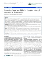
Her peripheral blood smear on medium-power field
revealed rouleaux formation, at ypical lymphocytes and
plasmacytoid cells (figure 1). High-power magnification
detected atypical B cells in her peripheral blood with
cytoplasmic expansion, coarse chromatin, multiple dis-
tinct nucleoli and peripheral vacuolation (figure 2).
Her bone marrow aspirate demonstrated a population
of small atypical lymphocytes admixed with normal cells
(figure 3). Interes tingly, a hematoxylin and eosin stain of
her bone marrow trephine identified large intratrabecular
lymphoid aggregates but the absence of CD10 (figure 4).
A further stain confirmed strong CD20 positivity of the
lymphoid aggregates (figure 5). The flow cytometry of
her bone marrow sample demonstrated t hat 77 perc ent
of the lymphoid cells were CD19, CD20, CD79b, and
cytoplasmic kappa posit ive. They were CD5, CD10,
CD38 and CD138 negative (figure 6).
Serum electrophoresis (table 1), appeared to show a
large IgM component - more than 30 g/L - in addition
to a mild increase in the serum IgG concentration.
Immunofixation revealed a monoclonal band that fixes
with anti-IgM and anti-kappa. Bence-Jones protein was
not detected. Her cytogenetics were 46 XX.
She progressed without event until the fourth day fol-
lowing her admission, when a medical emergency call
Figure 1 Medium-power field of peripheral blood smear
showing rouleaux formation, small atypical lymphocytes,
plasmacytoid cells and mild thrombocytopenia.
Figure 2 High-power field of peripheral blood smear revealing
a large, atypical B cell with mild cytoplasmic expansion, coarse
chromatin, multiple distinct nucleoli and peripheral
vacuolation.
Figure 3 Medium-power field of bone marrow aspirate
demonstrating a population of small atypical lymphocytes
admixed with normal cells of erythroid, myeloid and lymphoid
lineage.
Figure 4 Hematoxylin and eosin stain of bone marrow
trephine identifying large intratrabecular lymphoid aggregates
but CD10 negativity excluding follicular lymphoma.
Charakidis and Russell Journal of Medical Case Reports 2010, 4:300
/>Page 2 of 5
was initiated for an episode of worsening abdominal
pain and hypotension at 88/67 mmHg. Fluid resuscita-
tion was inadeq uate to maintain her systolic blood pres-
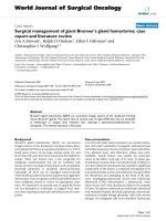
sure at greater than 100 mmHg. An urgent computed
tomography (CT) scan illustrated that her spleen was
enlarged with a craniocaudal extent of approximately 20
cm, with an extensive hemoperitoneum secondary to an
active hemorrhage from a laceration of the superior/
anterior pole of her spleen (Figure 7). Para-ao rtic, celiac
axis and porta hepaticus adenopathy was noted, with
the largest node measuring 19 mm.
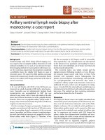
Splenic histology on low-power magnification dis-
played significant distortion of her splenic tissue and dif-
fuse infiltration by lympho id cells. There was also
expansion of the white pulp by this infiltrate (figure 8).
On high-power magnification, we saw infiltrates consist-
ing of small - and medi um-sized atypical lymphocytes,
which d isplayed dense chromatin clumping and promi-
nent nucleoli (figure 9).
Figure 5 CD20 stain of bone marrow trephine confirming the
strong CD20 positivity of the lymphoid aggregates.
Figure 6 Flow cytometry of bone marrow demonstrating that 77 percent of lymphoid cells are CD19, CD20, CD79b and cytoplasmic
kappa positive. They are CD5, CD10, CD38 and CD138 negative.
Table 1 Serum electrophoresis
Protein 94 g/L (60-80)g/L
IgA 7.98g/L (5.5-6.3)g/L
IgG 6.99g/L (0.65-4.21)g/L
IgM 30.93g/L (0.3-2.1)g/L
Large IgM component of more than 30 g/L and a mild increase in the serum
IgG
Charakidis and Russell Journal of Medical Case Reports 2010, 4:300
/>Page 3 of 5
Finally, in the splenic flow cytometry, the majority of
cells gated in the lymphoid region. Approximately 82
percent of lymphocytes were CD19, CD20, CD22 posi-
tive, Kappa positive B cells. A total of 25 percent were
CD38 and 21 percent CD138 positive.
Discussion
As mentioned previously an epidemiological link has
been established between HCV infection and non-Hodg-
kin’s lymphoma (NHL), (includin g LPLs), which is espe-
cially pronounced in Southern-European, Japanese, and
Brazilian populations, conferring an estimated relative
risk of 2.5 i n the development of any NHL. Marginal
zone lymphomas (MZL) are the most commonly-
encountered HCV-related lymphoma [3]. Despite this
association, the exact pathogenetic role of the virus is
not yet clearly established [3].
The 2008 WHO consensus on hematological malig-
nancies bases the diagnosis of WM on a number of
findings, including morphology, flow-cytometry, cytoge-
netics, and biochemistry. With regard to morphology,
the predominant features of WM are that of a diffuse
infiltration of the bone marrow by small lymphocytes,
plasma cells and plasmacytoid cells. Splenic architecture,
when availab le, demonstrates a lymphoplas macytic infil-
trate, composed predominantly o f small lymphocytes
that may form small nodules in the red pulp or appear
more diffusely infiltrated into the splenic parenchyma
[2]. Typically the immunophenotype of WM is CD19,
CD20, CD79a positivity, with light chain restriction.
This is identical to that of our patient. No cytogenetic
aberration is specific to WM and the karyotype is
usually normal, as opposed to B-cell lymphomas such as
splenic MZL and nodal MZL, which demonstrate immu-
noglobulin heavy and light chain aberrations in many
cases [2,4]. Serum electrophoresis demonstrates mark-
edly elevated levels of IgM protein; in addition, recipro-
cal depression of IgG and IgA can be seen in up to
25 percent of cases [5].
In our case report, an increase in IgG was seen.
Immunofixation showed that this was polyclonal and
therefore likely to be attributable to the underlying
HCVinfection.Itisofparticularnotethatantiviral
therapy directed against HCV infection in t he setting of
some LPLs has b een associated with a favourable prog-
nosis in terms of disease regression [4].
Clinicians are often faced with a diagnostic dilemma
when attempting to establish a definitive diagnosis o f
WM due to the overlap of clinicopathological features
with other B-cell lymphomas, including MZL (splenic
and nodal), mantle cell lymphoma, small cell lymphocytic
Figure 7 Abdominal computed tomography (CT) showing
significant hemoperitoneum, with extravasation of contrast
into the right flank/para-colic gutter. Hepatomegaly and
splenomegaly are clearly seen.
Figure 8 Low-power magnification of the splenic tissue.This
slide displays significant distortion and diffuse infiltration of the
splenic parenchyma by lymphoid cells. Of particular note is the
expansion of the white pulp by this infiltrate.
Figure 9 High-power mag nification o f splenic lymphoid
infiltrate. This slide demonstrates that the infiltrate consists of
small- and medium-sized atypical lymphocytes, which display dense
chromatin clumping and prominent nucleoli.
Charakidis and Russell Journal of Medical Case Reports 2010, 4:300
/>Page 4 of 5
lymphoma/chronic lymphocytic leukemia, and even dif-
fuse large B-cell lymphoma. As mentioned previously,
the diagnosis of B-cell lymphoma utilizes a combination
of both clinical features and a myriad of cellular para-
meters. In the absence of specific disease markers (such
as in WM), this combination of f actors is important in
attaining a ‘ pattern of best-fit’ when formulating a
diagnosis.
Splenic rupture is not an expected sequela o f WM.
Although infiltration of tissue parenchyma, venous c on-
gestion, and IgM-induced coagulopathies are features of
WM, they have not yet been reported to lead directly to
splenic rupture in the absence of a precipitating event. In
our case report, the absence of a previously abnormal
spleen, a precipitating traumatic event or a transformation
into a diffuse large B-cell lymphoma suggests that our case
may represent a more aggressive phenotype of WM.
Conclusions
Spontaneous splenic rupture is a complication of rapid
disease progression, and therefore is not an expected
complication of low-grade LPLs, such as WM. To the
best of our knowledge, this is the only reported case of
early spontaneous splenic rupture due to WM.
Our case report highlights that, despite the typical dis-
ease course of low-grade hematological malignancies,
signs and symptoms of imminent splenic rupture should
be considered when formulating a clinical assessment.
Consent
Written informed consent was obtained from the patient for publication of
this case report and any accompanying images. A copy of the written
consent is available for review by the Editor-in-Chief of this journal.
Competing interests
The authors declare that they have no competing interests.
Authors’ contributions
MC and DJR are the sole authors and contributed equally to the production
of this manuscript. All authors read and approved the final manuscript.
Acknowledgements
The authors thank Dr Roger Kimber for reviewing the manuscript and
providing valuable feedback.
Received: 25 September 2009 Accepted: 8 September 2010
Published: 8 September 2010
References
1. Vijay A, Gertz MA: Waldenström macroglobulinaemia. Blood 2007,
109:5096-5103.
2. WHO Classification of Tumours of Swerdlow SH, Campo E, Harris NL,
Jaffe ES, Pileri SA, Stein H, Thiele J, Vardiman JW: Haematopoietic and
Lymphoid Tissues. International Agency for Research on Cancer: Lyon, 4
2008.
3. Viswanatha DS, Dogan A: Hepatitis C virus and lymphoma. J Clin Pathol
2007, 60:1378-1383.
4. Fonseca R, Hayman S: Waldenström macroglobulinaemia. Br J Haematol
2007, 138:700-720.
5. Keren DF: Protein electrophoresis in clinical diagnosis Great Britain: Hodder
Arnold 2003, 72:145.
doi:10.1186/1752-1947-4-300
Cite this article as: Charakidis and Russell: Spontaneous splenic rupture
in Waldenstrom’s macroglobulinemia: a case report. Journal of Medical
Case Reports 2010 4:300.
Submit your next manuscript to BioMed Central
and take full advantage of:
• Convenient online submission
• Thorough peer review
• No space constraints or color figure charges
• Immediate publication on acceptance
• Inclusion in PubMed, CAS, Scopus and Google Scholar
• Research which is freely available for redistribution
Submit your manuscript at
www.biomedcentral.com/submit
Charakidis and Russell Journal of Medical Case Reports 2010, 4:300
/>Page 5 of 5