báo cáo khoa học: "Hashimoto’s encephalopathy presenting with neurocognitive symptoms: a case report" docx
Bạn đang xem bản rút gọn của tài liệu. Xem và tải ngay bản đầy đủ của tài liệu tại đây (939.32 KB, 4 trang )
CAS E REP O R T Open Access
Hashimoto’s encephalopathy presenting with
neurocognitive symptoms: a case report
Carlos Canelo-Aybar
*
, David Loja-Oropeza, Jose Cuadra-Urteaga, Franco Romani-Romani
Abstract
Introduction: Hashimoto’s encephalopathy is a neurological disorder of unknown cause associated with thyroid
autoimmunity. The disease occurs primarily in the fifth decade of life and may present in two types - a sudden
vasculitic type or a progressive subacute type associated to cognitive dysfunction, confusion and memory loss.
Case presentation: We report the case of a 62-year-old Hispanic woman, previously healthy, who developed a
subacute onset of declining upper brain function. Serologic studies demonstrated high levels of antithyroid
antibodies. Electroencephalographic and magnetic resonance image findings were consistent with Hashimoto’s
encephalopathy.
Conclusion: Hashimoto’s encephalopathy is a diagnosis of exclusion. This unusual disorder is often under-
recognized because of the multiple and protracted neurocognitive manifestations; therefore, it is important to be
aware of the clinical manifestations to make a correct diagnosis.
Introduction
Hashimoto’s encephalopathy (HE) is an uncommon neu-
rologic syndrome associated with Hashimoto’sthyroidi-
tis. It was initially described in 1966 [1], and it remains
a controversial disorde r. The cause of HE has been pro-
posed to be autoimmune because of its association with
other immunologic disorders (myasthenia gravis, glo-
merulonephritis, primary biliary cirrhosis, pernicious
anemia and rheumatoid arthritis), female predominance,
inflammatory findings in cerebrospinal fluid (CSF) and
response to treatment with steroids [1,2]. Other authors
suggest that HE may represent an autoimmu ne cerebral
vasculitis resulting from either endothel ial i nflamma tion
or immune complex deposition [1-3].
Clinical findings are variable and nonspecific. In this
case report, we present the case of a patient with suba-
cute onset of declining uppe r brain functions associated
with Hashimoto’s thyroiditis.
Case presentation
Over a five-month period, a 62-year-old Hispanic
woman who was previously healthy developed tremor in
the right arm, enuresis, slowness in performing her daily
activities, walking difficulties and trouble with getting
dressed. Additionally, her relatives observed transient
episodes of disorientation and inappropriate irritability.
Initially, the patient was a dmitted to another hospital,
where she was found to have apraxia, dysphasia, atten-
tion deficit and amnesic episodes. She had no sensory
or motor deficits.
Laboratory studies at that time revealed the presence
of antithyroid antibodies as well as slightly high serum
thyrotropin (TSH) concentrati on (Table 1). Examination
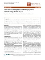
of the CSF was normal. Magnetic resonance images
(MRI) showed nodular focal subcortical lesions sugges-
tive of demyelination (Figure 1). A diagnosis of encepha-
litis and hypothyroidism was made, and th e patient
received levothyroxine.
Fifteen days later, the patient had two episodes of
inappropri ate behavior and transient anterograde amne-
sia. With these symptoms, she was admitted to our
hospital.
The laboratory examination showed no significant
change compared with the patient’s p revious laboratory
results except normalization of hemogram values. Addi-
tionally, antinuclear antibody titer, anti-double-stranded
DNA, anti-hepatitis B core antigen, hepatitis B surface
antigen, anti-hepatitis C virus, lupic antico agulant and
Venereal Disease Research Laboratory test results were
negative. Also, the anticardiolipin antibody IgG level
* Correspondence:
Department of Medicine, Arzobispo Loayza Hospital, Lima, Peru
Canelo-Aybar et al. Journal of Medical Case Reports 2010, 4:337
Http://www.jmedicalcasereports.com/content/4/1/337
JOURNAL OF MEDICAL
CASE REPORTS
© 2010 Canelo-Aybar et al; licensee Bi oMed Central Ltd. This is an Open Access article distributed under the terms of the Creative
Commons Attribution License (http://c reativecommons.org/licenses/by/2.0), which permits unrestricted use, di stribution, and
reproduction in any medium, provided the original work is properly cited.
was 10.8 U/GP L (reference range, <23 U/GPL), anticar-
diolipi n antibody Ig M was 5.9 U/MPL (reference range,
< 11 U/MPL), porphobilinogen deaminase level was 10.3
nmol/seg/L (reference range, 9.2-19.1 nmol/seg/L), 24-
hour urine porphobilinogen was 1.22 mg/24 h (reference
range, 0.2-2.00 mg/24 h), and 24-hour urine-delta-ami-
nolevulinic acid level was 2.46 mg/24 h (reference range,
0.1-4.5 mg/24 h).
Considering the clinical and laboratory findings, a
diagnosis of encephalopathy of undet ermined origin was
made. The electroencephalogram (EEG) showed a slow
background activity with theta waves and paroxysmal
activity at the hyperventilation maneuver (Figure 2). The
thyroid biopsy showed lympho cytic chronic thyroiditis,
and a diagnosis of HE was considered.
At discharge, the patient was treated with prednisone
at doses of 1 mg/kg body weight. Thirty days later, she
was experiencing a mild improvement in her sympt oms.
However, she never returned for her scheduled follow-
up medical appointments.
Discussion
HE is an unusual neurologic disorder whose etiology,
pathogenesis and histologic characteristics are unclear.
A systemat ic review published in 2003 [1] reported only
85 well-documented cases in the literature; however,
this syndrome may be underrecognized. A hospital-
based epidemiologic study of neurologic symptoms con-
sistent with HE estimated its prevalence to be about 2.1
per 100,000 [4]. The disorder occurs more frequently
between age 44 to 46 years, with a female-to-mal e ratio
of four to one [1,5].
The clinical manifestations usually include acute to
subacute onset of confusion with alteration of con-
sciousness. Two major patterns of presentation were
described: (1) 25% of patients follow a stroke-like pat-
tern of multiple recurrent episodes of focal neurologic
deficits with a variable degree of cognitive dysfunction
and consciousness impairment [1,2], and (2) the remain-
ing 75% present with a diffuse progressive pattern of
slow cognitive decline with dementia, confusion and hal-
lucinations [1,2]. These two clinical patterns may over-
lap over the course of the disease. In this case report,
our patient’s clinical manifestations are more consistent
with the second form of presentation, which is more
common.
Two-thirds of p atients may experience focal or gener-
alized tonic-clonic seizures, and 12% may present with
status epilepticus. Also, myoclonus or tremor is seen in
up to 38% of patients; hyperreflexia and other pyramidal
tract signs in 85% of patients; and psychosis, visual
Table 1 Laboratory studies prior admission on Arzobispo
Loayza Hospital
Studies Value Normal Range
Hemoglobin 11.7 g/dL 12-16 g/dL
Leukocyte count 3,000 cells/μL 4,500-10,000 cells/μL
Platelet count 800,000 cells/μL 150,000-400,000 cells/μL
INR* 1.7 1.0
Aspartate aminotransferase 39 U/L 0-37 U/L
Alanine aminotransferase 72 U/L 0-34 U/L
Albumin 3.7 g/dL 3.5-5.2 g/dL
Globulin 3.5 g/dL 2.5-3.0 g/dL
Thyroid-stimulating hormone 7.7 μU/mL 2.3-4.0 μU/mL
Free thyroxine (T4) 0.9 ng/mL 1.0-2.0 ng/dL
Antithyroglobulin antibody 135 IU/mL <10.0 IU/mL
Antithyroid peroxidase 715 IU/mL <10.0 IU/mL
*INR: International Normalizated Ratio of prothrombin time.
Figure 1 Axial magnetic resonance images (MRI) of the brain demonstrating nodular subcortical lesions suggestive of demyelination
in frontal and parietal lobes . A) T1-weighted MRI. B) T2-weighted MRI.
Canelo-Aybar et al. Journal of Medical Case Reports 2010, 4:337
Http://www.jmedicalcasereports.com/content/4/1/337
Page 2 of 4
hallucinations and paranoid delusions have been
reported in 25% to 36% of patients [1,2,5].
The mechanism of HE does not appear to be related
to the thyroid status, which can vary greatly in patients
with HE. In two recent reviews, 23% to 35% of patients
had subclinical hypothyroidism, 17% to 20% had
hypothyroidism, 7% had hyperthyroidism and 18% to
45% were euthyroid [1,5]. The development of neurolo-
gic symptoms may occur up to three years before the
onset of autoimmune thyroiditis [6].
The p resence of elevated serums levels of a ntithyroid
antibodies remains an essen tial characteristic of HE
diagnosis, and suggest the pres ence of thyroid autoim-
munity [1,5]. Although in some cases, the diagnosis is
supported by the association with Hashimoto’sthyroi-
ditis, it is possible that some patients develop HE with-
out a concomitant clinical thyroid disease because
asymptomatic thyroid autoimmunity is frequent in
these pat ients [1,5].
Thepathogenicroleofthyroidantibodiesremains
unknown, there is no evidence that any antithyroid anti-
body reacts with brain tissue or affects nerve function,
and there is no clear correlation between the severity of
the neurolog ic symptoms and the concentration of these
antibodies [1,4].
Antithyroid antibodies have also been related to other
autoimmune conditions such as myopathy, d epression,
bipolar disease and dementia, but the prevalence of
these antibodies in the general population (ranging from
2%-20%) make it difficult to establish whether a real
association exists [7].
Infrequently, the titers of antithyro id antibodies
(TPOAb and TgAb) are measured in the CSF. In one
case series, nine of 12 patients with encephalopathy and
elevated serum antithyroid antibodies had elevated CSF
autoantibody titers [4]. A systematic review found that
13% of publis hed cases of HE reported antithyroid anti-
bodies in the CSF [5]. However, the titers of antithyroid
antibodies in the CSF do not co rrelate with the clinical
stage of the dis ease, and the sensitivity and specificity of
this finding remain unclear [4,5].
An autoantibody against the amino terminal end of
the enzyme a-enolase, an antigen of the thyroid and the
brain, has been identified as a potential biomarker of
HE [5,8]. A study found serum autoantibody reactivity
in five of six patients with HE compared with two of 17
patients with Hashimoto’ s thyroiditis but no HE and in
none of 25 healthy control subjects [8]. This antigen is
also found in endothelial cells, suggesting an autoim-
mune vasculitic mechanism; however, this has not been
confirmed by neuroimaging techniques [5].
In some patients, C-reactive protein and the erythro-
cyte sedimentati on rate are elevated [9], and in one ser-
ies, mild elevation of liver enzymes was found in 12 of
20 patients [9)], which is concordant with the mild ele-
vation observed in our patient.
Although the CSF analy sis results were normal in our
patient, a lymphocytic pleocytosis has been foun d in
14% of reported patients; in 4% of patients, it may con-
tain more than 100 cells/mm
3
. An elevated protein con-
centration occurs in 78% of patients, and in 20% of
patients, it may be greater than 100 mg/dL. The blood
glucose concentration is usually normal [1,2].
Nonspecific EEG abnormalities are seen in 90% to
98% of patients, which is usually a nonspecific slow
background activity. The same pattern was observed in
our patient. Focal spikes or sharp waves and transient
epileptic activity are less common [2,10].
Figure 2 An electroencephalogram showing a slowing background activity with theta waves and paroxysmal activity at
hyperventilation maneuver .
Canelo-Aybar et al. Journal of Medical Case Reports 2010, 4:337
Http://www.jmedicalcasereports.com/content/4/1/337
Page 3 of 4
In a review of 82 pat ients with HE, brain computed
tomography or MRI showed abnormalities in 49% such
as cerebral atrophy, focal cortical abnormality, diffuse
subcortical abnormality and nonspecific subcortical focal
white matter abnormality . The latter was observed in
our patient as subcortical foci of demyelination [1].
The differential diagnosis of HE must consider any
condition a ssociated with delirium, rapidly progressive
dementia, seizures or focal neurologic deficits [5]. Th us,
the list of diseases that can be confused with HE is vast,
including stroke or transient ischemic attack, cerebral
vasculitis, carcinomatous meningitis, toxic metabolic
encephalopathies, paraneoplastic syndromes, Creutz-
feldt-Jakob disease, degenerative dementia and psychia-
tric diseases [1,5].
The long-term prognosis is variable, although a h igh
percentage of patients respond to treatment; others
could have a progressive or a relapsing course [1,5]. The
symptoms usually improve with glucocorticoid therapy;
however, it is not necessary because of treatment. A sys-
tematic review of 85 cases published of HE found clini-
cal response in 98% o f patients treated with
glucocorticoids, 92% of patients treated with glucocorti-
coids and levothyroxine and 67% of patients treated
with levothyroxine only [1].
Although our patient had a mild improvement of her
symptoms, the long-term effect of the therapy could not
be assessed because the patient did not return for her
follow-up medical appointments.
Conclusion
HE frequently presents with a myriad of neurocognitive
symptoms and normal findings in several different
examinations. This syndrome may go unrecognized for
a l ong time; therefore, it should be kept in mind when
evaluating a patient with cognitive dysfunction and high
titers of antithyroid antibodies.
Consent
Written consent was obtained from th e patient for pub-
lication of the case report and any accompanying
images. A copy of the written consent is available for
review by the Editor-in-Chief of the journal.
Authors’ contributions
CCA contributed to patient care, drafting the manuscript and literature
review. JCU contributed to interpretation of data and drafting of the
manuscript. FRR contributed to data collection and literature search for the
manuscript. DLO contributed to patient care, drafting the manuscr ipt,
revision and approval of the manuscript. All authors read and approved the
final manuscript.
Competing interests
The authors declare that they have no competing interests.
Received: 14 October 2009 Accepted: 25 October 2010
Published: 25 October 2010
References
1. Chong J, Rowland L, Utiger R: Hashimoto encephalopathy: syndrome or
myth? Arch Neurol 2003, 60:164-71.
2. Kothbauer-Margreiter I, Sturzenegger M, Komor J, Baumgartner R, Hess C:
Encephalopathy associated with Hashimoto thyroiditis: diagnosis and
treatment. J Neurol 1996, 243:585-593.
3. Forchetti C, Katsamakis G, Garron D: Autoimmune thyroiditis and a rapidly
progressive dementia: global hypoperfusion on SPECT scanning suggest
a possible mechanism. Neurology 1997, 49:623-626.
4. Ferracci F, Bertiato G, Moretto G: Hashimoto’s encephalopathy:
epidemiologic data and pathogenetic considerations. J Neurol Sci 2004,
217:165-168.
5. Ferracci F, Carnevale A: The neurological disorder associated with thyroid
autoimmunity. J Neurol 2006, 253:975-984.
6. Peschen-Rosin R, Schabet M, Dichgans J: Manifestation of Hashimoto’s
encephalopathy years before onset of thyroid disease. Eur Neurol 1999,
41:79-84.
7. Vanderpump M, Tunbridge W, French J, Appleton D, Brewis M, Clark F, et al:
The incidence of thyroid disorders in the community: a twenty-year
follow-up of the Whickham Survey. Clin Endocrinol 1995, 43:55-68.
8. Fujii A, Yoneda M, Ito T, Yamamura O, Satomi S, Higa H, et al:
Autoantibodies against the amino terminal of alpha-enolasa are useful
diagnostic marker of Hashimoto’s encephalopathy. J Neuroimmunol 2005,
162:130-136.
9. Castillo P, Woodruff B, Caselli R, Vernino S, Lucchinetti C, Swanson J, et al:
Steroid-responsive encephalopathy associated with autoimmune
thyroiditis. Arch Neurol 2006, 63:197-202.
10. Rodrigez A, Jicha G, Steeves T, Benarroch E, Westmoreland B: EEG changes
in a patient with steroid responsive encephalopathy associated with
antibodies to thyroperoxidase (SREAT, Hashimoto’s encephalopathy). J
Clin Neurophysiol 2006, 23:371-373.
doi:10.1186/1752-1947-4-337
Cite this article as: Canelo-Aybar et al.: Hashimoto’s encephalopathy
presenting with neurocognitive symptoms: a case report. Journal of
Medical Case Reports 2010 4:337.
Submit your next manuscript to BioMed Central
and take full advantage of:
• Convenient online submission
• Thorough peer review
• No space constraints or color figure charges
• Immediate publication on acceptance
• Inclusion in PubMed, CAS, Scopus and Google Scholar
• Research which is freely available for redistribution
Submit your manuscript at
www.biomedcentral.com/submit
Canelo-Aybar et al. Journal of Medical Case Reports 2010, 4:337
Http://www.jmedicalcasereports.com/content/4/1/337
Page 4 of 4