báo cáo khoa học: "Takayasu’s disease presenting as convulsive syncope which had been misinterpreted as epilepsy: a case report" doc
Bạn đang xem bản rút gọn của tài liệu. Xem và tải ngay bản đầy đủ của tài liệu tại đây (658.08 KB, 4 trang )
CAS E REP O R T Open Access
Takayasu’s disease presenting as convulsive
syncope which had been misinterpreted as
epilepsy: a case report
Bindu Menon
*
, A Himabindu
Abstract
Introduction: Takayasu’s arteritis is a chronic vasculitis mainly involving the aorta and its main branches. The
disease has protean clinical manifestation ranging from asymptomatic to catastrophic illness.
Case presentation: A 19-year-old woman of Asian origin was referred to our neurology out-patient department
for the management of refractory seizures. She reported several episodes of a loss of consciousness with tonic
posturing when she assumed an upright position, which was accompanied by constitutional symptoms. A clinical
examination showed orthostatic hypotension and an investigation confirmed the diagnosis of Takayasu’s disease
with presentation as convulsive syncope.
Conclusion: Our case highlights the importance of a thorough clinical history and physical examination in order to
distinguish events mimicking epileptic seizure. We also describe an unusual presentation of Takayasu’s disease with
convulsive syncope and systemic constitutional symptoms.
Introduction
Takayasu’s arteritis (TA) is an inflammatory and stenotic
disease of medium-sized and large-sized arteries character-
ized by a str ong predilection for the aortic arch and its
branches. Symptoms of the disease start with non-specific
constitutional symptoms in the first stage to organ specific
ischemic symptoms in the second stage. Occasionally
there is no clear demarcation between stages, and diagno-
sis can be difficult in such a setting. We report the case of
a 19-year-old woman who was referred to our neurology
out-patient department for recurrent episodes of loss of
consciousness and systemic constitutional symptoms.
Case presentation
A 19-year-old Asian woman was referred to the depart-
ment of neurology for the management of recurrent sei-
zures. She had been having recurrent episodes of a loss of
consciousness. On detailed enquiry she revealed that these
episodes were present immediately on changing to a n
upright posture from a lying position. She would lose con-
sciousness, become pale, and fall to the ground. She would
then exper ience tonic posturing of the limbs lasting for a
few seconds. She would immediately regain consciousness
in the supine position, with no postictal drowsiness. There
were several similar episodes a day, and this had persisted
for the last three months. Her symptoms persisted even
after t reatment with phenytoin, phenobarbit one, and
sodium valproate, with no decrease in the frequency of
episodes. She also complained of fatigue, malaise, myalgia,
giddiness, fever, decreased appetite and weight loss over
the preceding three months. Her medical history was not
significant. There was no prev ious or family history of
tuberculosis.
Physical examination revealed a thin built, frail and
anxious patient. She was afebrile . Her radial pulses were
very feeble while lower limb pulses felt normal. Her
bilateral carotid pulses were also feeble. Upper limb
blood pressure (BP) was measured (with difficulty) as
being72/58mmHginbothupperlimbsinasupine
position. Standing BP was 50/48 mmHg in the upper
limbs, suggestive of orthostatic hypotension. Lower limb
blood pressures were130/70 mmHg bilaterally. The
results of cardiac examination were normal. Her higher
mental functions and fundi were normal. There was no
cranial nerve, motor or sensory deficit.
* Correspondence:
Department of Neurology, Narayana Medical College and Superspeciality
Hospital, Chintareddypalem, Nellore-2 AP, India
Menon and Himabindu Journal of Medical Case Reports 2010, 4:352
/>JOURNAL OF MEDICAL
CASE REPORTS
© 2010 Menon and Himabindu; licensee BioMed C entral Ltd. This is an Open Access article distributed under the terms of the Creative
Commons Attribution License ( which permits unrest ricted us e, distribution, a nd
reproduction in any medium, provided the original work i s properly cited.
Blood test results indicate d anemia with hemoglobin of
9 gm/dlL, an elevated erythrocyte sedimentation rate
(ESR) of 42 mm/hour and a total leukocyte count of
13,400 cells/mm
3
. Her C-reactive protein level was ele-
vated at 26 mg/L. Tests for plasma sugars, liver function
tests, renal function tests, HIV/hepatitis B virus surface
antigen (HBsAg) antibodies, hepati tis C virus antibo dies,
and Venereal Disease Research Laboratory test results
(protoplasmic-staining anti-neutrophil cytoplasmic antibo-
dies (p-ANCA), cytopla smic staining ANCA (c-ANCA)
and Hbs Ag) were all negative. Her chest X-ray, electro-
cardiography, echocardiography and electroencephalogra-
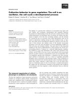
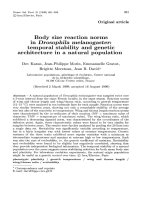
phy findings were normal. A color Doppler of the carotid
vessels showed right (Figure 1A) and left (Figure 1B) com-
mon carotid arteries showing diffuse wall thickening and
streaky flow in the lumen. The left subclavian origin
showed no flow (Figure 1C). Bilateral brachia l, radial and
ulnar arteries showed low velocity monophasic flow. Renal
Doppler results showed normal renal vessels. The abdom-
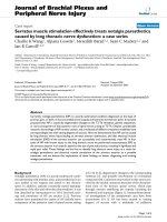
inal aorta was not imaged. A magnetic resonance angio-
graphy (MRA) study of the carotid and vertebral vessels
was performed with a 0.35 Tesla Siemens Magnetom C
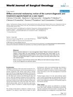
device (Siemens, Mumbai, India). Maximum intensity pro-
jection (Figure 2A) a nd coronal reconstruction images
(Figure 2B) from two-dimensional ‘time of flight’ MRA
showed no flow in the brachiocephalic artery beyond the
origin. Origin of left subclavian artery did not show a flow
signal. The left common carotid artery showed streaky
flo w. Multiple collateral vessels were seen. The vertebral
arteries were normally visualized.
An axial reconstruction image (Figure 2C) showed dif-
fuse wall thickening of arteries with luminal narrowing.
Our patient was diagnosed as having TA, fulfilling
four of the six criteria for diagnosis: development of
symptoms or findings related to TA at age ≤40 years,
decreased pulsation of one or both brachial arteries,
arteriographic narrowing or occlusion of the primary
branches of the aorta, and a difference of >10 mmHg in
systolic blood pressure between arms [1]. The results of
laboratory investigations for anemia, elevated ESR and
C-reactiv e prot ein were suggestive of active disease. She
was hospitalized a nd prescribed complete bed rest. Pre-
dnisolone (40 mg/day) along with antacids were given.
Our patient started feeling better after a week and was
therefore gradually mobilized. She did have episodes of
giddiness, but there was no loss of consciousness or
tonic posturing of limbs. She was discharged after two
weeks. After a year, at follow-up she had made a clinical
Figure 1 Color Doppler images of right (A) and left (B) common carotid arteries (*) show diffuse wall thickening and a streaky flow in
the lumen. The origin of left subclavian artery (C) shows no flow.
Menon and Himabindu Journal of Medical Case Reports 2010, 4:352
/>Page 2 of 4
improvement with no constitutional symptoms or syn-
copal seizures but her radial pulses remained feeble.
There was no evidence of postural hypotension.
Discussion
TA is a chronic vasculitis that mainly involves the aorta
and its main branches (the brachiocephalic, carotid, sub-
clavian, vertebral and renal arteries), as well as the cor-
onary and pulmonary arteries. The disease is most
commonly seen in Asian and Latin American countries
[2]. The incidence of TA is about 2 in 10,000 person-
years [3]. The female/male ratio varies from 9:1 in
reports fro m Japan to 1.3:1 in India [4]. The underlying
pathology is inflammation leading to stenosis, blockage
or aneurysm formation. The disease is also called the
pulseless disease because of the difficulty in detecting
the peripheral pulses.
The etiology of the disease is still poorly understoo d.
The clinical presentation is widely heterogeneous
depending on the involvement of the vessels. The disease
has three stages, in which there is an initial pre-pulseless
stage predominated by systemic constitutional symptoms.
As a result of the non-specificity of the symptoms the
diagnosis is often delayed until the next stage of vascular
insufficiency. The second stage may be devoid of any
signs of inflammation. Hypertension due to renal artery
stenosis, retinopathy, aortic regurgitation, aneurismal
enlargement of aorta, congestive cardi ac fail ure, postural
dizziness, amaurosis, transient ischemic attacks and
stroke are some of the presenting features in the second
stage. The third stage is the stage of quiescence. Collat-
eral circulation develops because of the chronic nature of
the illness. Neurological manifestation is seen in about
20% of cases. The initial manifestations are predomi-
nantly ischemic in nature because of the stenosis of the
vessels. Seizures as an initial manifestation have been
reported but are rare [5]. Syncopal seizures have not
been reported. A MeSH database searches with the head-
ings ‘Syncope’, ‘convulsion’ and ‘Takayasu’sarteritis’ did
produce any reported studies.
Our patient had stenosis of the major branches of the
aorta, brachiocephalic, subclavian and left common carotid
artery. The cerebral perfusion was being maintained by the
collateral circulation. The differential diagnosis of episodic
neurological dysfunction included a vast range of disor-
ders. The loss of consciousness, with tonic posturing, led
to the misdiagnosis of epilepsy. However, these symptoms
were suggestive of convulsive syncope because: all epi-
sodes had a postural component, episo des were only for
few seconds and there was no postictal drowsiness, our
patient immediately regained consciousness in the recum-
bent position and documented orthostatic hypotension
Figure 2 Maximum intensity projection (A) and thin coronal reconstruction images (B) from two-dimensional ‘time of flight’ magnetic
resonance angiography show no flow in the brachiocephalic artery (a) beyond the origin. The origin of left subclavian artery (b) does not
show a flow signal. The left common carotid artery (c) shows a streaky flow. The image in (A) shows multiple collateral vessels (d) along with
normal flow in bilateral vertebral arteries (e). Axial reconstruction image (C) shows diffuse wall thickening of arteries with luminal narrowing.
Menon and Himabindu Journal of Medical Case Reports 2010, 4:352
/>Page 3 of 4
was found on examination. Syncope is transient loss of
consciousness due to cerebral hypoxia. This may be
accompanied with convulsive episodes. Convulsive syn-
cope merely represents a variant of syncope, which is
accompanied by tonic or myoclonic activity.
The differential diagnoses of TA include tuberculosis,
temporal arteritis, atherosclerosis, fibro muscular dyspla-
sia and syphilitic aortitis. Apart from syphilitic aortitis,
which affects the aorta with calcification, all the other
differentials have a predilection for other vessels. The
gold standard of investigation was considered to be
angiography, but non-invasive Doppler and MRA can
provide equally good results [1].
In the active stage of the disease, judged by systemic
symptoms, glucocorticoids are the mainstay of treat-
ment. This is thought to halt the inflammation and
further stenosis in vessels. Serological tests have not so
far proved helpful in differentiating the active from the
inactive stage [6]. Surgery is indicated in certain patients
with inactive disease.
Conclusion
Misdiagnosis of epilepsy remains a majo r clinical pro-
blem. We presented this case in order to highlight two
findings: the importance of a detailed clinical history
and examination, which will help to determine whether
or not an epileptic seizure actually occurred in a patient
and to differentiate seizure mimics and the absence of
stage-wise progression of the symptoms in TA which
can create a diagnostic dilemma.
Consent
Written informed consent was obtained from the patient
for publication of this case report and accompanying
images. A copy of the written consent is available for
review by the Editor-in-Chief of this journal.
Authors’ contributions
BM diagnosed the case of TA, collected the requisite literature and analyzed
and interpreted the data from our patient. AB performed and interpreted
the radiological examination. Both authors read and approved the final
manuscript.
Competing interests
The authors declare that they have no competing interests.
Received: 23 October 2009 Accepted: 2 November 2010
Published: 2 November 2010
References
1. Arend WP, Michel BA, Bloch DA, Hunder GG, Calabrese LH, Edworthy SM,
et al: The American College of Rheumatology 1990 criteria for the
classification of Takayasu arteritis. Arthritis Rheum 1990, 33:1129-1134.
2. Nagasawa T: Current status of large and small vessel vasculitis in Japan.
Int J Cardiol 1998, 54:S98.
3. Smeeth L, Cook C, Hall AJ: Incidence of diagnosed polymyalgia
rheumatica and temporal arteritis in the United Kingdom, 1990 to 2001.
Ann Rheum Dis 2006, 65:1093-1098.
4. Johnston SL, Lock RJ, Gompels MM: Takayasu arteritis: a review. J Clin
Pathol 2002, 55:481-486.
5. Ioannides MA, Eftychiou C, Georgiou GM, Nicolaides E: Takayasu arteritis
presenting as epileptic seizures: a case report and brief review of the
literature. Rheumatol Int 2009, 29:703-705.
6. Tann OR, Tulloh RM, Hamilton MC: Takayasu’s disease: a review. Cardiol
Young 2008, 18:250-259.
doi:10.1186/1752-1947-4-352
Cite this article as: Menon and Himabindu: Takayasu’s disease
presenting as convulsive syncope which had been misinterpreted as
epilepsy: a case report. Journal of Medical Case Reports 2010 4:352.
Submit your next manuscript to BioMed Central
and take full advantage of:
• Convenient online submission
• Thorough peer review
• No space constraints or color figure charges
• Immediate publication on acceptance
• Inclusion in PubMed, CAS, Scopus and Google Scholar
• Research which is freely available for redistribution
Submit your manuscript at
www.biomedcentral.com/submit
Menon and Himabindu Journal of Medical Case Reports 2010, 4:352
/>Page 4 of 4