báo cáo khoa học: "Omental infarction in the postpartum period: a case report and a review of the literature" docx
Bạn đang xem bản rút gọn của tài liệu. Xem và tải ngay bản đầy đủ của tài liệu tại đây (772.85 KB, 3 trang )
CAS E REP O R T Open Access
Omental infarction in the postpartum period:
a case report and a review of the literature
Michael Tachezy
1*
, Rainer Grotelüschen
1
, Florian Gebauer
1
, Andreas H Marx
2
, Jakob R Izbicki
1
, Jussuf T Kaifi
1
Abstract
Introduction: Omental infarction is a rare and often misdiagnosed clinical event with unspecific symptoms. It
affects predominantly young and middle aged women.
Case presentation: This is a case report of a 26-year-old Caucasian woman with spontaneous omental infarction
two weeks after normal vaginal delivery.
Conclusion: Omental infarction is a differential diagnosis in the postpartum acute abdomen. As some cases of
omental infarction, which are caused by torsion, can be adequately diagnosed via computed tomography, a
conservative treatment strategy for patients without complications should be considered in order to avoid any
unnecessary surgical intervention.
Introduction
Omental infarction is a rare clinical event that affects
predominantly young and middle aged women [1]. It is
usually caused by omental torsion, but the reasons for
this remains poorly understood. Omental infar ction was
first reported in 1882 by Oberst [2]. Patients present
symptoms of an acute abdomen. T he clinical findings
are very unspecific and, therefore, in most cases it is
surgical exploration that leads to the diagnosis.
This report highlights the case of a spontaneous
omental in farction in a young woman in the postpartum
period.
Case presentation
A 26-year-old Caucasian woman presented with a five
day history of increasing epigastric pain and nausea two
weeks after the vaginal delivery of a healthy child of
normal weight and size.
Physical examination revealed a normal pe ristalsis and
supraumbilical tenderness. A small umbilical hernia
(<1 cm diameter), wi th no signs o f incarceration, was
described b y the initial examining physician. Pulse and
blood pressure were normal (85 beats/min, 123/83
mmHg). She was apyrexial but adynamic, with pale and
clammy skin. In summary, the general status of the
patient was impaired on admission (American Society of
Anesthesiologists score 2-3).
Blood tests revealed an elevated white blood cell count
(14.7/nL) and serum C-reactive protein (120 mg/dL).
A coagulation study (international normalised ratio, par-
tial thromboplastin time, fibrinogen and platelet count)
revealed no abnormalities.
Abdominal ultrasound showed no specific pathological
findings and, for further clarification, a contrast-enhanced
abdominal computed tomography (CT) was performed.
The morphologic findings of the CT were interpreted as
an incarcerated umbilical hernia by the radiologist. How-
ever, due to the clinical presentation of an acute abdomen
and the elevated inflammatory blood parameters, the
patient was asked to consent to an exploratory laparot-
omy. A small laparotomy (5 cm long midline incision
around the umbilicus) was performed. Contrary to the CT
findings, and in accordance to the clinical examination, no
umbilical hernia could be detected intraoperatively. Sur-
prisingly, a hemorrhagic greater omentum measuring 11 ×
7.5 × 2.5 cm was discovered and resected. A small amount
of sanguinous ascites was also found. On further explora-
tion we found no adhesions or other underlying causes for
the infarction, such as an exte rnal or internal hernia or a
vascular pedicle.
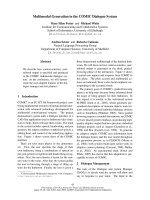
In a retrospective repeat analysis of the CT scan, a
hypoperfused mass of fatty appearance in the anterior
* Correspondence:
1
Department of General, Visceral and Thoracic Surgery, University Medical
Center Hamburg-Eppendorf, Martinistraße 52, 20246 Hamburg
Full list of author information is available at the end of the article
Tachezy et al. Journal of Medical Case Reports 2010, 4:368
/>JOURNAL OF MEDICAL
CASE REPORTS
© 2010 Tachezy et al; licensee BioMed Central Ltd. This is an Open Access article distributed under the terms of the Creative Commons
Attribution Lice nse ( which permits unrestricted use, distribution, and reprodu ction in
any medium, provided the original work is properly cited.
portion of the midabdomen and small amounts of free
fluid surrounding the liver were observed (Figure 1).
Histopathological findings of the resected omental
specimen confirmed fresh hemorrhagic infiltrations o f
the tis sue, partial thrombosis of the small vessels and, in
some parts, necrotic fatty tissues with an acute inflam-
matory cellular infiltrate (Figure 2). Further laboratory
testing exclu ded potentially underlying coagulopathy or
rheumatic disease.
The patient was discharged after an uneventful recov-
ery three days after surgery.
Discussion
Omental infarction was first described in the late 19th
century and, since then, only a f ew hundred cases have
been published in the English literature [3]. This is one
of the first cases showing spontaneous omental infarc-
tion in the puerperium after a vaginal birth. Two pre-
viously published cases describe omental infarction in
the postpartum period - one af ter caesarean section and
another after vaginal delivery [4,5]. Torsion of the
omentum is the main reason for infarction and two dif-
ferent forms have been described: primary torsio ns
(without other pathologic intraabdominal findings) and
secondary torsions (tumors, cysts, inflammatory changes,
adhesions, hernias). Predisposing factors for torsion are
anomalies of the omentum, such as a small root, irregu-
lar vascular anatomy, abdominal trauma, cough and
physical strain [2].
The etiology of omental infarction without torsion
remains uncertain but s everal mechanisms have been
proposed, such as an anomaly of venous vessels [6].
Other possible causes for primary infarctions could be
disorders of hemostasis or vascular diseases. It is known
that hematologic changes occur during pregnancy and
the puerperium and that hypercoagulability leads to an
increased risk of thromboembolic events [7]. The exact
mechanism leading to infarction in this case remains
unclear. Possible changes during the return of the
mother’s body to the pre-pregnancy physiological condi-
tion may have provoked the infarction. Usually the clini-
cal symptoms o f an infarction of the omentum are
localized peritoneal irritation on the right side of the
abdomen, sometimes associated with low-grade fever.
As in the present case, the C-reactive protein an d white
blood count may be elevated. The clinical picture
often misleads physicians to a ssume an incorrect preo-
perative diagnosis such as acute cholecystitis, appendici-
tis, diverticulitis, appendicitis epiploica or umbilical
hernia [3,8,9].
As most patients show symptoms of an acute abdo-
men, CT of the abdomen and pelvis should be the diag-
nostic imaging of choice [10]. If omental infarction is
caused by torsion, characteristic CT-findings might be
detectable. The torsion leads to the presence of con-
centric linear strands in the fatty mass, a so-called ‘fat
spiral patte rn’ [11]. In our case no omental torsion was
present and, consequently, the radiologist was unable to
identi fy this diagnostic radiologic sign. Therefore, differ-
entiating the omental infarction from other abdominal
or ome ntal diseases was challenging and the radiological
findings were misinterpreted as a small incarcerated
umbilical hernia.
Diagnosis of an omental infarction has traditionally
been made intraoperatively during an explor atory lapar-
otomy or laparoscopy and the treatment has been partial
or total omentectomy. Recent r eports highlight cases of
patients with CT diagnosed omental torsions who have
been successfully treated conservatively without any
Figure 1 Computed tomography scan of the abdomen
showing a hypoperfused mass in the anterior portion of the
median epigastrium with fatty density (®) and a thin layer of
free fluid surrounding the liver.
Figure 2 Histological findings o f omentum majus show fresh
hemorrhagic circulation disorders (arrows), partial necrosis of
fatty tissue with acute inflammatory cell infiltrate (hematoxylin
staining, original magnification × 100).
Tachezy et al. Journal of Medical Case Reports 2010, 4:368
/>Page 2 of 3
other complications (such as bac terial superinfections)
[12-15]. Whenever conservative treatment fails, or the
clinical status of the patient worsens, a surgical interven-
tion should be quickly implemented.
Conclusion
Omental infarctions are often not initially considered in
the d ifferential diagnosis of a post partum acute abdo-
men. When omental infarction is caused by torsion, a
correct preoperative diagnosis by contrast-enhanced CT
scanning can avoid surgery. Recently published case ser-
ies have reported successful conservative management.
Consent
Written informed consent was obtained from the patient
for publication of this case report and any accompany-
ing images. A copy of the written consent is available
for review by the Editor-in-Chief of this journal.
Acknowledgements
The authors would like to thank Shazia Hussain and Katharina Tornow for
their help in proofreading and editing the manuscript.
Author details
1
Department of General, Visceral and Thoracic Surgery, University Medical
Center Hamburg-Eppendorf, Martinistraße 52, 20246 Hamburg.
2
Institute of
Pathology, University Medical Center Hamburg-Eppendorf, Martinistraße 52,
20246 Hamburg, Germany.
Authors’ contributions
MT, RG and JTK managed the patient and reviewed the literature. MT and
RG were the main authors of the manuscript. AHM analyzed the
histopathological specimen. FG, JTK and JRI made modifications to the
manuscript. All authors read and approved the final manuscript.
Competing interests
The authors declare that they have no competing interests.
Received: 30 November 2009 Accepted: 17 November 2010
Published: 17 November 2010
References
1. Kimber CP, Westmore P, Hutson JM, Kelly JH: Primary omental torsion in
children. J Paediatr Child Health 1996, 32:22-24.
2. Knoop M, Vorwerk T: [Inflammatory alterations of the greater omentum–
a difficult preoperative diagnosis]. Zentralbl Chir 2002, 127:626-628.
3. Leung R, Kreis DJ Jr: Infarction of the omentum in pregnancy. South Med
J 1986, 79:1597.
4. Guerquin B, Pannequin L, Gregoire J, Legoulme C: [Tumor syndrome of
omental origin in the post-partum period]. J Gynecol Obstet Biol Reprod
(Paris) 1994, 23:96-98.
5. Phillips RW, Peterson CM: Infarction of the omentum after cesarean
section. A case report. J Reprod Med 1988, 33:382-384.
6. Maternini M, Pezzetta E, Martinet O: Laparoscopic approach for idiopathic
segmental infarction of the greater omentum. Minerva Chir 2009,
64:225-227.
7. James AH: Pregnancy-associated thrombosis. Hematology Am Soc Hematol
Educ Program 2009, 277-285.
8. Tompkins RK, Sparks FC: Primary torsion of the omentum - mimic of
appendicitis: review of six cases. Am Surg 1966, 32:399-402.
9. Basson SE, Jones PA: Primary torsion of the omentum. Ann R Coll Surg
Engl 1981, 63:132-134.
10. Naffaa LN, Shabb NS, Haddad MC: CT findings of omental torsion and
infarction: case report and review of the literature. Clin Imaging 2003,
27:116-118.
11. Ceuterick L, Baert AL, Marchal G, Kerremans R, Geboes K: CT diagnosis of
primary torsion of greater omentum. J Comput Assist Tomogr 1987,
11:1083-1084.
12. Puylaert JB: Right-sided segmental infarction of the omentum: clinical,
US and CT findings. Radiology 1992, 185:169-172.
13. Coulier B, Pringot J: [Pictorial essay. Infarction of the greater omentum:
can US and CT findings help to avoid surgery?]. JBR-BTR 2002, 85:193-199.
14. van Breda Vriesman AC, Lohle PN, Coerkamp EG, Puylaert JB: Infarction of
omentum and epiploic appendage: diagnosis, epidemiology and natural
history. Eur Radiol 1999, 9:1886-1892.
15. Balthazar EJ, Lefkowitz RA: Left-sided omental infarction with associated
omental abscess: CT diagnosis. J Comput Assist Tomogr 1993, 17:379-381.
doi:10.1186/1752-1947-4-368
Cite this article as: Tachezy et al.: Omental infarction in the postpartum
period: a case report and a review of the literature. Journal of Medical
Case Reports 2010 4:368.
Submit your next manuscript to BioMed Central
and take full advantage of:
• Convenient online submission
• Thorough peer review
• No space constraints or color figure charges
• Immediate publication on acceptance
• Inclusion in PubMed, CAS, Scopus and Google Scholar
• Research which is freely available for redistribution
Submit your manuscript at
www.biomedcentral.com/submit
Tachezy et al. Journal of Medical Case Reports 2010, 4:368
/>Page 3 of 3