báo cáo khoa học: "Living donor liver transplantation for neonatal hemochromatosis using non-anatomically resected segments II and III: a case report" doc
Bạn đang xem bản rút gọn của tài liệu. Xem và tải ngay bản đầy đủ của tài liệu tại đây (681.47 KB, 4 trang )
CAS E REP O R T Open Access
Living donor liver transplantation for neonatal
hemochromatosis using non-anatomically
resected segments II and III: a case report
Amit Sharma
*
, Adrian H Cotterell, Daniel G Maluf, Marc P Posner, Robert A Fisher
*
Abstract
Introduction: Neonatal hemochromatosis is the most common cause of liver failure and liver transplantation in
the newborn. The size of the infant determines the liver volume that can be transplanted safely without incurring
complications arising from a large graft. Transplantation of monosegments II or III is a standard method for the
newborns with liver failure.
Case presentation: A three-week old African-American male neonate was diagnosed with acute liver failure
secondary to neonatal hemochromatosis. Living-related liver transplantation was considered after the failure of
intensive medical therapy. Intra-operatively a non-anatomical resection and transplantation of segments II and III
was performed successfully. The boy is growing normally two years after the transplantation.
Conclusion: Non-anatomical resection and transplantation of liver segments II and III is preferred to the
transplantation of anatomically resected monosegements, especially when the left lobe is thin and flat. It allows
the use of a reduced-size donor liver with intact hilar structures and outflow veins. In an emergency, living-related
liver transplantation should be offered to infants with liver failure secondary to neonatal hemochromatosis who fail
to respond to medical treatment.
Introduction
Neonatal hemochromatosis (NH), although rare, is the
most common cause of liver failure and liver transplan-
tation in neonates. Liv er transplantation is the main
therapy for infants who fail to respond to medical treat-
ment [1]. Liver t ransplantation using either mono-
segment II or III [2,3] is a technically challenging option
that is especially beneficial for small infants in whom a
left lateral segment [4] is large-for-size. We report that
non-anatomical resection and transplantation of seg-
ments II and III may be a simpler, yet effective, surgical
option for neonates with liver failure.
Case presentation
A three-week old African-A merican male newborn,
weighing 2.5 kg, was admitted to our unit with jaun dice,
abdo minal distension and hepatomegaly. The pregnancy
had been uncomplicated and there was no family history
of metabolic or liver disease.
Laboratory studies for liver failure revealed a total
serum bilirubin of 22.5 mg/dL, an international normal-
ized ratio 4.9, aspartate aminotransferase 45 U/L and
alanine aminotransferase 23 U /L. The boy had a serum
iron of 157 μg/dL (normal 30-165 μg/dL), serum ferritin
994 ng/mL (normal 30-330 ng/mL), serum transferrin
103 mg/dL (normal 215-380 mg/dL) and transferrin
saturation 109% (range 16%-60%). He had an elevated
alpha-fetoprotein level (3289 ng/mL). Investigations for
infectious and inherited metabolic pathologies were
negative. Magnetic r esonance imaging (MRI) of the
abdomen was su ggesti ve of iron deposition in the liver,
with conventional, patent arterial and venous anatomy.
NH was suspected and confirmed by minor salivary
gland biopsy from the lower lip.
Medical therapy consisting of anti-oxidants and chel a-
tion with desferroxamine was initiated. As there was a
progressive worsening of the boy’s condition on medical
therapy, his mother vol unteered to be a living li ver
* Correspondence: ;
Department of Surgery, Hume-Lee Transplant Center, Virginia
Commonwealth University, PO Box 980057, Richmond, Virginia 23298-0057 ,
USA
Sharma et al. Journal of Medical Case Reports 2010, 4:372
/>JOURNAL OF MEDICAL
CASE REPORTS
© 2010 Sharma et al; licensee BioMed Centra l Ltd. This is an Open Access article distributed under the terms of th e Creative Commons
Attribu tion License (h ttp://creativecommons.org/licenses/by/2.0), which permits unrestricted use, distribution, and reproduction in
any medium, provided the original work is prope rly cite d.
donor. After a standard expedited two-day donor-
workup, the related living donor liver transplantation
was planned. Pre-operative MRI showed that the
mother’s left lateral segme nt volume was approximately
200 cc
3
. We therefore decided to do a left lateral resec-
tion with back-table monosegmentectomy followed by
transplantation.
Intra-operatively, after isolation of the mother’ sleft
hepatic artery, hepatic duct and portal branch, the hepa-
tic parenchyma of segment IV wa s transected 5 mm to
the right of the falciform ligament without blood inflow
occlusion or graft manipulation. The segment II and III
ducts united, just lateral to the umbilical portion of the
left portal vein and the segment IV duct, then joined
medial to umbilical portion. This confluence of segment
II and III ducts was divided and used for anastomosis in
the recipient. This was a thin ‘pancake’ left lateral seg-
ment that was transected using ultrasound guidance. On
the back-table, a 2.5 mm endostapler with two, triple-
staggered rows of titanium staples (Autosuture™GIA™ U-
NIVERSAL stapler, US Surgical, Division of Tyco
Healthcare Group LP, C T, USA) was used to staple and
divide across the mid-portion of the left lateral segment
just to the left of the secondary portal vasculature
branching (Figure 1). The final graft consisted of the
confluence of segment II and III bile ducts, left portal
vein, left hepatic artery arising from left gastric artery
and two in-proximity left hepatic veins, joined as one.
This non-anatomically resected portion of the lateral
segment was used for transplantation in standard piggy-
back fashion with Roux-en-Y jejuno-biliary anastomosis.
The discarded part was used for hepatocyte isolation
[5]. Heparin and aspirin were used in the first post-
operative week to prevent vascular thrombosis. The
patient was re-explored in the first week for clot evacua-
tion around the transplanted liver segments, with no
active bleeding on the cut surface or the stapled edge.
Patient was discharg ed home after three weeks and con-
tinues to do well two years post-transplant.
Discussion
Neonatal hemochromato sis is a syndrome with an
aggressive course and a poor prognosis. The etiopatho-
genesis is not very clear although siderosis resulting from
infections, genetics a nd auto-immunity may play a role
[6]. The pregnancy may be complicated by intrauterine
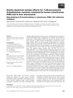
Figure 1 Liver segments II and III with reconstructed hilar structures just before non-anatomical resection. The donor had a left hepatic
arterial branch arising from the left gastric artery and two left hepatic vein tributaries draining into the supra-hepatic vena cava. The final
arrangement of the left portal vein, the reconstructed hepatic veins, replaced left hepatic artery arising from the left gastric artery and the bile
duct is demonstrated here. On the back-table, transection was carried through segments II and III along a non-anatomical plane (thick black line)
using an endovascular stapling device.
Sharma et al. Journal of Medical Case Reports 2010, 4:372
/>Page 2 of 4
growth restriction, oligohydramnios or still birth. The
neonate may present with signs of hepatic insufficiency
within hours of birth. Abnormal laboratory parameters
include: decreased transferri n; ceruloplasmin; increased
ferritin (non-specific, > 800 ng/mL); mixed hyperbilirubi-
naemia; low aminotransferases; low factors V and VII
(< 10% of normal); thrombocytopenia, anaemia and
increased alpha-fetoprotein (> 200 ng/mL). Hepatic and
extra-hepatic siderosis with reticuloendothelum sparing
is diagnostic of NH. Lower lip biopsy is safe and conveni-
ent for documenting siderosis in minor salivary glands.
MRI is used to support the diagnosis of NH and is char-
acterised by low signal intensity on T2 weighted liver
images [7]. Since NH recurs in 75-80% of siblings, the
parents should be discouraged from having any further
pregnancies. Gestational high dose intravenous immuno-
globulin administered to the mother, from 18 weeks to
birth, appears to decrease the lethality of recurrent neo-
natal hemochromatosis [8]. Medical therapy with desfer-
rioxamine and antioxidant cocktai l (N-acetylcysteine,
vitamin E, prostaglandin E1 and selenium), although not
highly successful, are still used to treat neonates. Liver
transplantation is considered to be the treatment of
choice for infants not responding to medica l therapy.
Early medical therapy results in a 10 %-20% survival rate
while long-term survival after liver transplantation m ay
range from 50% to 66% [1].
Liver transplantation using either monosegment II or III
is a useful option for small infants in whom a whole left
lateral se gment is large-for-size [4]. Monosegment trans-
plantation is mostly used for infants with a calculated
graft-to-recipient weight ratio of less than or equal to 4.0%
when using the left lateral segment. Splitting of the left lat-
eral segment can be been done either in situ in the donor
or on the back-table. Despite these surgical innovations,
neonatal liver transplantation still poses challenges
because of the size of the recipients who usually weigh less
than 10 kg [9]. De pending on the donor size, even t he
transplanted monosegment may be large-for-size and
make graft placement technically difficult and may lead to
post-operative complications [10]. More importantly, the
use of a segment II or III may result in a smaller diameter
bile duct t hat may be more prone t o strictures and leaks
as the liver regenerates [1]. In our report, the left lateral
segment was split along a non-anatomical plane and the
confluence of segments II and III bile ducts provided us
with a larger caliber (4 mm) duct in the donor segments.
The caliber of the hepatic and the portal veins u sed for
anastomosis were the same as when using a complete left
lateral segment. The use of a stapling device made this
division technically easier and more efficient. However,
the stapled edge may be prone to bleeding after reperfu-
sion. This can be minimized by selecting thin and flat left
lateral segments for stapling. This case also demonstrate s
that emergent living-related liver transplantation is a viable
opt ion for neonates with acute liver fai lure who may not
survive the time spent on the waiting list for a whole or a
split-liver from a deceased donor.
Conclusion
Urgent living-related liver transplantation should be
offered to infants with acute liver failure secondary to
neonatal hemochromatosis who are non-responsive to
medical therapy. The left lateral segment can be reduced
in size, especially when it is flat (like a pancake), by
splitting it along a non-anatomical plane. This simple
technique allows the use of the confluence of donor seg-
ment II and III bile ducts that are less prone to strictur-
ing due to the ir larger ca liber. However, t his advantage
may be lost in cases where the segment II and III bile
ducts join separately, medial to the umbilical portion of
the portal vein.
Consent
Written informed consent was obtained from the
patient’s mother for publication of this case report and
any accompanying images. A copy of the written con-
sent is available for review by the Editor-in-Chief of this
journal.
Abbreviations
IUGR: intrauterine growth restriction; MRI: magnetic resonance imaging; NH:
neonatal hemochromatosis.
Acknowledgements
We would like to thank Mr Jose Rodriguez for his technical help in the
drafting of this manuscript.
Authors’ contributions
AS collected data, designed and wrote the manuscript. AHC was a major
contributor to the manuscript. DGM assisted in the critical revisions of the
manuscript. MPP reanalyzed the surgical facts and provided comments on
the critical intellectual content of the manuscript. RAF helped to conceive,
critically revise and write the manuscript. All authors read and approved the
final manuscript.
Competing interests
The authors declare that they have no competing interests.
Received: 25 March 2010 Accepted: 19 November 2010
Published: 19 November 2010
References
1. Rodrigues F, Kallas M, Nash R, Cheeseman P, D’Antiga L, Rela M,
Heaton ND, Mieli-Vergani G: Neonatal hemochromatosis-medical
treatment vs transplantation: the king’s experience. Liver Transpl 2005,
11:1417-1424.
2. de Santibañes E, McCormack L, Mattera J, Pekolj J, Sívori J, Beskow A,
D’Agostino D, Ciardullo M: Partial left lateral segment transplant from a
living donor. Liver Transpl 2000, 6:108-112.
3. Srinivasan P, Vilca-Melendez H, Muiesan P, Prachalias A, Heaton ND, Rela M:
Liver transplantation with monosegments. Surgery 1999, 126:10-12.
4. Broelsch CE, Whitington PF, Emond JC, Heffron TG, Thistlethwaite JR,
Stevens L, Piper J, Whitington SH, Lichtor JL: Liver transplantation in
children from living related donors. Surgical techniques and results. Ann
Surg 1991, 214:428-437.
Sharma et al. Journal of Medical Case Reports 2010, 4:372
/>Page 3 of 4
5. Fisher RA, Strom SC: Human hepatocyte transplantation: worldwide
results. Transplantation 2006, 82:441-449.
6. Sigurdsson L, Reyes J, Kocoshis SA, Hansen TW, Rosh J, Knisely AS: Neonatal
hemochromatosis: outcomes of pharmacologic and surgical therapies.
J Pediatr Gastroenterol Nutr 1998, 26:85-89.
7. Udell IW, Barshes NR, Voloyiannis T, Lee TC, Karpen SJ, Carter BA,
Finegold M, Goss JA: Neonatal hemochromatosis: radiographical and
histological signs. Liver Transpl 2005, 11:998-1000.
8. Whitington PF, Hibbard JU: High-dose immunoglobulin during pregnancy
for recurrent neonatal haemochromatosis. Lancet 2004, 364:1690-1698.
9. Enne M, Pacheco-Moreira L, Balbi E, Cerqueira A, Santalucia G, Martinho JM:
Liver transplantation with monosegments. Technical aspects and
outcome: a meta-analysis. Liver Transpl 2005, 11:564-569.
10. Ogawa K, Kasahara M, Sakamoto S, Ito T, Taira K, Oike F, Ueda M, Egawa H,
Takada Y, Uemoto S: Living donor liver transplantation with reduced
monosegments for neonates and small infants. Transplantation 2007,
83:1337-1340.
doi:10.1186/1752-1947-4-372
Cite this article as: Sharma et al.: Living donor liver transplantation for
neonatal hemochromatosis using non-anatomically resected segments
II and III: a case report. Journal of Medical Case Reports 2010 4:372.
Submit your next manuscript to BioMed Central
and take full advantage of:
• Convenient online submission
• Thorough peer review
• No space constraints or color figure charges
• Immediate publication on acceptance
• Inclusion in PubMed, CAS, Scopus and Google Scholar
• Research which is freely available for redistribution
Submit your manuscript at
www.biomedcentral.com/submit
Sharma et al. Journal of Medical Case Reports 2010, 4:372
/>Page 4 of 4