báo cáo khoa học: " A rare cause of chronic mesenteric ischemia from fibromuscular dysplasia: a case report" potx
Bạn đang xem bản rút gọn của tài liệu. Xem và tải ngay bản đầy đủ của tài liệu tại đây (2.8 MB, 9 trang )
CAS E REP O R T Open Access
A rare cause of chronic mesenteric ischemia from
fibromuscular dysplasia: a case report
Viplove Senadhi
Abstract
Introduction: Chronic mesenteric ischemia is a condition that is classically associated with significant
atherosclerosis of the abdominal arteries, causing postprandial abdominal pain out of proportion to physical
examination. The abdominal pain is exacerbated after meals due to the shunting of blood away from the
intestines to the stomach, causing relative ischemia. More than 95% of chronic mesenteric ischemia cases are due
to atherosclerosis. We report the first known case of chronic mesenteric ischemia from fibromuscular dysplasia. To
the best of our knowledge, this is also the first known case in the literature where postprandial abdominal pain
was the presenting symptom of fibromuscular dysplasia.
Case presentation: A 44-year-old Caucasian woman with a history of hypertension and preeclampsia, who had
taken oral contraceptive pills for 15 years, presented with an intractable, colicky abdominal pain of two weeks
duration. This abdominal pain worsened with oral intake. It was also associated with diarrhea and vomiting.
Physical examination revealed stage III hypertension o ut of proportion to her risk factors and diffuse abdominal
pain without peritoneal signs. An a bdominal computed tomography scan, completed in the emergen cy room,
revealed nonspecific colitis. Laboratory work revealed leukocytosis with a left shift, an erythrocyte sedimentation
rate of 79 and a C-reactive protein level of 100. She was started on intravenous flagyl and intravenous
ciprofloxacin. However, all m icrobial cultures were negative including three cultures for clostridium difficile.
Urine analysis revealed nephritic range proteinuria. The laboratory profile wa s within normal limits for
perinuclear-anti-neutrophil cytoplasmic antibody, cytoplasmic-anti-neutrophil cytoplasmic antibody, anti-
saccharomyces cerevisiae antibody, antinuclear antibody test, celiac profile, lactate, carbohydrate antigen-125
and thyroid stimulating hormone. A colonoscopy was completed, which revealed diffuse colonic lymphoid
reactive hyperplasia. A small bowel series was negative for any inflammation. An indium scan, pan-computed
tomography scan and tran svaginal ultrasound were also negative. Magnetic resonance angiograp hy of her
abdomen revealed proximal superior mesenteric artery stenosis, which was confirmed by computed
tomography angiogram findings of severe proximal and distal superior mesenteric artery stenosis, consistent
with the appe arance of fibromuscular dysplasia on angiography in the absence of vasculitis or atherosclerotic
disease. The patient’ s superior mesenteric artery stenosis was subsequently angioplastied sub optimally and had
to be stented with an Angioplus stent. One month after she was admitted, her abdominal pain and tolerance
to oral feeds improved tremendously.
Conclusion: Fibromuscular dysplasia most commonly presents with renal artery stenosis, which rarely causes
abdominal pain. This case illustrates how fibromuscular dysplasia can present as a rare cause of chronic mesenteric
ischemia, similar to chronic mesenteric ischemia from atherosclerosis.
Correspondence:
Johns Hopkins University/Sinai Hospital Program in Internal Medicine,
Department of Internal Medicine, Sinai Hospital, Baltimore, MD, USA
Senadhi Journal of Medical Case Reports 2010, 4:373
/>JOURNAL OF MEDICAL
CASE REPORTS
© 2010 Senadhi; licensee BioMed Central Ltd. This is an Open Access article distributed under the terms of the Creat ive Commons
Attribution License ( which permits unrestricted use, distribution, and reproduction in
any medium, provided the original work is properly cited.
Introduction
Chronic mesenteric ischemia is a condition classically
associated with significant atherosclerosis of the abdom-
inal arteries causing postprandial abdominal pain out of
proportion to physical examination [1]. The abdominal
pain is exacerbated after meals due to the shunting of
blood away fro m the intestines to the stomach causing
relative ischemia. More than 95% of chronic mesenteric
ischemia cases are due to atheroscler osis [1]. We report
the first known case of chronic mesenteric ischemia
from fibromuscular dysplasia. This is the first known
case in the literature where the presenting symptom of
fibromuscular dysplasia was postprandial abdominal
pain.
Case presentation
A 44-year-old Caucasian woman with a history of
hypertension and preeclampsia who had taken o ral con-
traceptive pills (OCPs) for 15 years, presented with
intractable, colicky abdominal pain of two weeks dura-
tion. This abdominal pain worsened with oral intake. It
was also associated with diarrhea and vomiting. The
diarrhea was watery at times a nd was confirmed to be
hemoccult positive. Her vomiting was non-bilious and
was consistent with gastric contents, with a negative
gastroccult test. No abnormal findings were found on
physical examination except for stage III hypertension
out of proportion to her risk factors and diffuse abdom-
inal pain, which was the most prominent in the perium-
bilical area.
Routine laboratory work revealed leukocytosis (white
blood count = 16.1) with a left shift (91% polymorpho-
nuclear neutrophils), an erythrocyte sedimentation rate
(ESR) of 79, and a C-reactive protein level (CRP) of 100.
An abdominal computed tomography (CT) scan com-
pleted in the emergency room revealed nonspecific coli-
tis . It was thought that she had infect iou s col itis and so
she was started on i ntravenous (IV) flagyl and IV cipro-
floxacin. However, all microbial cultures were negative,
including three cultures for clostridium difficile. A uri-
nalysis (UA) revealed nephritic range proteinuria. At
this point, there was a concern for vasculitis, hypothyr-
oidism, and inflammatory bowel disease (IBD). The
laboratory profile was negative for perinuclear anti-neu-
trophil cytoplasmic antibodies (P-ANCA), cytoplasmic
anti-neutrophil c ytoplasmic antibodies (C-ANCA), anti-
saccharomyces antibody (ASCA), antinuclear antibody
(ANA), celiac profile, lactate, carbohydrate antigen 125
(CA-125), and a thyroid stimulating hormone (TSH). A
colonoscopy was completed in order to rule out micro-
scopic colitis and occult inflammatory bowel disease
(IBD), but revealed a nonspecific finding of diffuse colo-
nic lymphoid reactive hyperplasia. A small bowel series
was negative for any inflammation and the possibility of
IBD was negated. An indium scan, looking for occult
infection, was also negative. A pan-CT scan with con-
trast looking for malignancy of the abdomen, pelvis,
chest and head were all ne gative. A transvaginal ultra-
sound and pelvic examination ruled out an occult early
ovarian cancer, endometrial or cervical cancer.
Although the patient was n eve r hypotensive, nor had
any hypercoagulable conditions in the past, the diagnosis
of mesenteric ischemia was pursued based on her expo-
sure to OCP. Her OCPs had been discontinued at the
beginning of her hospitalization, which was more than a
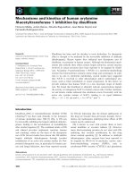
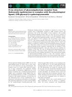
month prior to this point in the workup. A magnetic
resonance angiography (MRA) of the abdomen revealed
proximal superior mesenteric artery (SMA) stenosis
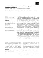
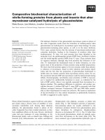
which was confirmed by CT angiogram findings of
severe proximal and distal SMA stenosis (Figures 1 and
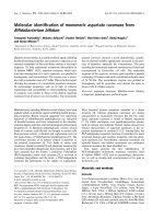
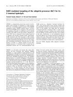
2). The patient was taken to angiography which revealed
near complete full length occlusion of the SMA and sev-
eral branches including mild renal artery stenosis (Fig-
ure 3). The angiography findings were consistent with
atypical fibromuscular dysplasia with a medial pattern.
The suspicion was high gi ven the lack of atherosclerotic
lesions, absence of hyperlipidemia, negative vasculitis
workup, and pattern of o cclusions seen on angiography.
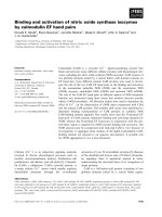
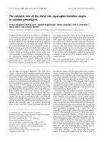
The patient’s severe SMA stenosis subsequently under-
went suboptimal angioplasty, which warranted a 6 × 15
Palmaz Blue Angioplus stent (Figures 4, 5, 6). Her
symptoms improved tremendously, in that her abdom-
inal pain subsided and she had an improved tolerance
to oral feeds.
Discussion
Due to the findings of severe proximal or distal SMA
stenosis, the diagnosis of subacute mesenteric ischemia
versus chronic mesenteric ischemia was considered.
Non -occlusive mesenteric ischemia was ruled out based
on the lack of hypotension and the patient’s overall clin-
ical history. Colonic ischemia, which is the most com-
mon form of ischemic vascular compromise, was ruled
out based on the colonic biopsy and clinical history [2].
This prompted the differential diagnoses of fibromuscu-
lar dysplasia versus a hypercoagulable state causing
mesenteric vascular thrombosis, or a chronic athero-
sclerotic process with an acute t hrombosis. The patient
did not have any risk factors for arterial embolic disease,
such as atrial fibrillation, valvular or heart surgery or
cardiac mural thrombosis.
The patient’s age and risk factors made the diagnosis
of chronic mesenteric ischemia from atherosclerosis
very unlikely. She had no known history of peripheral
vascular disease or an equivalent, which are high risk
fact ors for atheroscler osis [1]. Her body mass index was
Senadhi Journal of Medical Case Reports 2010, 4:373
/>Page 2 of 9
19.2 and she was considered in great health prior to this
hospitalization. Additionally, the patient did not have a
history of hyperlipidemia or smoking, known risk factors
for atherosclerotic mesenteric ischemia. Also, most
patients with chronic mesenteric ischemia from athero-
sclerosis are over the age of 60 [1]. Thus, atherosclerotic
chronicmesentericischemiawasthoughttobehighly
unlikely.
The consideration for venous thrombosis was then
evaluated. The patient was anticoagulated with IV
heparin, whic h could not be continu ed secondary to
bleeding. The hypercoagulable workup done in the
absence of anticoagulation, including antithrombin-3
antigen level, lupus anticoagulant, beta II glycoprotein
antibody, cardiolipin antibody, protein C/S and homo-
cystinuria, showed negative results. We ruled out venous
thrombosis from antithrombin-3 deficiency and all other
hypercoagulable states. Additionally, isolated abdominal
vein thrombosis in the absence of deep venous thro m-
bosis, pulmonary embolus and stro ke made a hypercoa-
gulable venous state very unlikely. It was agreed by
vascular surgery from t he MRA and CT angiography
findings that the patient did indeed have fibromuscular
dysplasia causing superior mesenteric artery stenosis as
well as renal artery stenosis, especially considering the
absence o f hyperlipidemia and inflammation (Figures 1
and 2). The patient was taken to angiography where she
underwe nt a suboptimal angioplasty of her SMA, which
Figure 1 Axial image showing superior mesenteric artery (SMA) stenosis.
Senadhi Journal of Medical Case Reports 2010, 4:373
/>Page 3 of 9
was followed up with an immediate 6 × 15 Palmaz Blue
Angioplus stent (Figure 3, figure 4, figure 5 and figure
6). Heparin was restarted after completing the hypercoa-
gulable work up and the patient’ s clinical status
improved with anticoagulation and clopidog rel bisulfate
(Plavix). After more than a month of hospitalization, she
was discharged with clopidogrel bisulfate alone for her
abdominal stent. She was tolerating oral feeds and is
back to her baseline lifestyle at a one-year follow-up.
Several times during her care, she was advised to have
significant abdominal surgery. However, due to the
recognition of her diagnosis, our team felt confident
with her management and advised a conservative
approach. Overall, she did very well and returned to a
normal lifestyle, while avoiding significant a bdominal
surgery.
Chronic mesenteric ischemia from atherosclerosis is
ideally managed with surgical correction via transaortic
endarterectomy, external iliac retrograde bypass or ante-
rograde bypass [3-9]. However, there are studies that
state stenting has been equivalent with respect to short-
term and long-term outcomes [8,9]. In our case, distal
small vessel disease of the SMA made surgery less favor-
able [10,11]. Warfarin and nitrates are typically used in
thetreatmentofchronicmesentericischemiafrom
atherosclerosis [4]. Our patient was managed with an
Figure 2 Sagittal image showing superior mesenteric artery (SMA) stenosis.
Senadhi Journal of Medical Case Reports 2010, 4:373
/>Page 4 of 9
Angioplus stent with clop idogrel bisulfate alone to pre-
vent stent thrombosis.
Fibromuscular dysplasia is a nonatherosclerotic, non-
inflammatory condition that leads to the narrowing of
medium sized arteries. Fibromuscular dysplasia classically
affects the renal arteries (63% to 89%) and prese nts most
commonly with secondary hypertension [12]. The least
common sites of involvement are the mesenteric (9%)
and iliac (5%) arteries. Fibromuscular dysplasia in atypical
sites such as the mesenteric arteries may go undiagnose d
Figure 3 Angiogram showing superior mesenteric artery (SMA) stenosis.
Senadhi Journal of Medical Case Reports 2010, 4:373
/>Page 5 of 9
unless the stenosis is severe, as in our patient [1]. The
classic diagnosis of fibromuscular dysplasia is made on
angiography as a ‘string of beads appearance’. However,
this only represents 60% to 70% of fibromuscul ar dyspla-
sia cases [12]. Atypical fibromuscular dysplasia, defined
as not representing the classic appearance, is a much
more difficult diagnosis [13,14]. Atypical fibromuscular
dysplasia is usually diagnosed on the basis of strong
clinical suspicion without signs of atherosclerotic dis-
ease or vasculitis induced inflammation, such as in our
Figure 4 Angiogram showing superior mesenteric artery (SMA) stenosis post angioplasty.
Senadhi Journal of Medical Case Reports 2010, 4:373
/>Page 6 of 9
patient [13,14]. However, acute infarction of the
involved arteries can mislead physicians, as it will raise
inflammatory markers. In atypical fibromuscular dyspla-
sia, the angiographic appearance can appear very similar
to atherosclerotic lesions as there are smooth concentric
lesions [12]. However, subtle distinctions can be made
as atherosclerosis is a diffuse process and does not
occur in the absence of other systemic arterial involve-
ment. Atypical fibromuscular dysplasia is much more
likely in the app ropriate cl inic al context, such as in our
patient.
Conclusion
Fibromuscular dysplasia most commonly presents with
renal artery stenosis, which rarely causes abdominal
pain. This case illustrates how fibromuscular dysplasia
can present with postprandial abdominal pain and as a
rare cause of chronic mesenteric ischemia, similar to
chronic mesenteric ischemia from atherosclerosis. Addi-
tionally, this case a lso illustrates the clinical and angio-
grap hic presentation of atypical fibromuscular dysplasia,
which does not present with the classic ‘string of beads ’
appearance on angiography. The management of
Figure 5 Sagittal image showing superior mesenteric artery (SMA) stent.
Senadhi Journal of Medical Case Reports 2010, 4:373
/>Page 7 of 9
fibromuscular dysplasia induced chronic me senteric
ischemia is similar to the management of atheroscleroti c
chronic mesenteric ischemia based on this case. How-
ever, surgical revascularization is not a bsolutely neces-
sary in fibromuscular dysplasia induced chronic
mesenteric ischemia and abdominal artery stents with
angioplasty can be placed with successful patient out-
comes. Lastly, this case illustrates that angioplasty and
Angioplusstentscanbeusedwith clopidogrel bisulfate
without warfarin or vasodilators, such as nitrates, in
chronic mesenteric ischemia secondary to fibromuscular
dysplasia.
Consent
Written consent was obtained from the patient for pub-
lication of the case report and accompanying images. A
copy of the written consent is available for review by
the Editor-in-Chief of the journal.
Abbreviations
ANA: antinuclear antibody test; ASCA: anti-saccharomyces cerevisiae
antibody; CA-125: cancer antigen 125; C-ANCA: cytoplasmic-anti-neutrophil
cytoplasmic antibody; CRP: C-reactive protein; CT: computed tomography;
ESR: erythrocyte sedimentation rate; IBD: inflammatory bowel disease; IV:
intravenous; MRA: magnetic resonance angiography; OCP: oral contraceptive
pills; P-ANCA: perinuclear-anti-neutrophil cytoplasmic antibody; SMA: superior
Figure 6 Volume rendering technique (VRT) image showing superior mesenteric artery (SMA) stent.
Senadhi Journal of Medical Case Reports 2010, 4:373
/>Page 8 of 9
mesenteric artery; TSH: thyroid stimulating hormone; UA: urinalysis; WBC:
white blood cell count.
Acknowledgements
I would like to give special thanks to Dr Peter Mackrell in the Department of
Vascular Surgery and Dr Harry Kaplan in the Department of Internal
Medicine for their contributions in the patient’s care. Their dedication to
improving the standard of patient care and advocating for our patient
resulted in a wonderful outcome for her
Authors’ contributions
VS was integral in the management of the patient, carried out the patient’s
medical care to the point of diagnosis/treatment, performed the literature
review and wrote the manuscript.
Competing interests
The author declares that they have no competing interests.
Received: 29 January 2010 Accepted: 19 November 2010
Published: 19 November 2010
References
1. Moawad J, Gewertz BL: Chronic mesenteric ischemia. Clinical
presentation and diagnosis. Surg Clin North Amr 1997, 77:357-369.
2. Cappell MS: Intestinal (mesenteric) vasculopathy. I. Acute superior
mesenteric arteriopathy and venopathy. Gastroenterol Clinics N America
1998, 27:783-825, vi.
3. Chahid T, Alfidja AT, Biard M, Ravel A, Garcer JM, Boyer L: Endovascular
treatment of chronic mesenteric ischemia: results in 14 patients.
Cardiovascular Interventional Radiol 2004, 27:637-642.
4. Chang JB, Stein TA: Mesenteric ischemia: acute and chronic. Ann Vasc
Surg 2003, 17:323-328.
5. English WP, Pearce JD, Craven TE, Edwards MS, Geary RL, Plonk GW,
Hansen J: Chronic visceral ischemia: symptom-free survival after open
surgical repair. Vasc Endovascular Surg 2004, 38:493-503.
6. Geroulakos G, Tober JC, Anderson L, Smead WL: Antegrade visceral
revascularisation via a thoracoabdominal approach for chronic
mesenteric ischemia. Eur J Vasc Endovasc Surg 1999, 17:56-59.
7. Hung KH, Lee CT, Lam KK, et al: Ischemic bowel disease in chronic
dialysis patients. Chang Keng I Hsueh Tsa Chih 1999, 22:82-87.
8. Kazmers A: Operative management of chronic mesenteric ischemia. Ann
Vasc Surg 1998, 12:299-308.
9. Kihara TK, Blebea J, Anderson KM, Freidman D, Atnp RG: Risk factors and
outcomes following revascularization for chronic mesenteric ischemia.
Ann Vasc Surg 1999, 13:37-44.
10. Lauenstein TC, Ajaj W, Narin B, Göhde SC, Kröger D, Debatn JF, Rühm SG:
MR imaging of apparent small-bowel perfusion for diagnosing
mesenteric ischemia: feasibility study. Radiology 2005, 234:569-575.
11. Mateo RB, O’Hara PJ, Hertzer NR, Mascha EJ, Beven EG, Krajewski LP:
Elective surgical treatment of symptomatic chronic mesenteric occlusive
disease: early results and late outcomes. J Vasc Surg 1999, 29:821-831,
discussion 832.
12. Luscher TF, Lie JT, Stanson AW, Houser OW, Hollier LH, Sheps SG: Arterial
fibromuscular dysplasia. Mayo Clin Proc 1987, 62:931-952.
13. den Butter G, van Bockel JH, Aarts JC: Arterial fibrodysplasia:rapid
progression complicated by rupture of a visceral aneurysm into the
gastrointestinal tract. J Vasc Surg 1988, 7:449-453.
14. Begelman SM, Olin JW: Fibromuscular dysplasia. Curr Opinions Rheumatol
2000, 12:41-47.
doi:10.1186/1752-1947-4-373
Cite this article as: Senadhi: A rare cause of chronic mesenteric
ischemia from fibromuscular dysplasia: a case report. Journal of Medical
Case Reports 2010 4:373.
Submit your next manuscript to BioMed Central
and take full advantage of:
• Convenient online submission
• Thorough peer review
• No space constraints or color figure charges
• Immediate publication on acceptance
• Inclusion in PubMed, CAS, Scopus and Google Scholar
• Research which is freely available for redistribution
Submit your manuscript at
www.biomedcentral.com/submit
Senadhi Journal of Medical Case Reports 2010, 4:373
/>Page 9 of 9