báo cáo khoa học: "Inguinal lymph node metastases from a testicular seminoma: a case report and a review of the literature" pptx
Bạn đang xem bản rút gọn của tài liệu. Xem và tải ngay bản đầy đủ của tài liệu tại đây (457.27 KB, 4 trang )
CAS E REP O R T Open Access
Inguinal lymph node metastases from a testicular
seminoma: a case report and a review of the
literature
Mohamed Ismail
1*
, Faruquz Zaman
1
, Sohail Baithun
2
, Venod Nargund
1,3
, Jhumur Pati
1,3
, Junaid Masood
1,3
Abstract
Introduction: We report the case of a true hermaphrodite with testicular seminoma with resulting metastases to
the inguinal lymph nodes eight month s after radical orchidectomy. This is an unusual presentation of testicular
cancer and, to the best of our knowledge, the first report of this kind in the literature.
Case presentation: A 45-year-old Caucasian true hermaphrodite, raised as a male, developed a testicular
seminoma. He had undergone a left orchidopexy at the age of 10 for undescended testes. Metastases from
testicular tumors to inguinal lymph nodes are a rare occurrence. It has been suggested that previous inguinal or
scrotal surgery may alter the pattern of nodal metastasis of testicular cancer. We review the literature to evaluate
the incidence of inguinal lymph node involvement in early stage testicular cancer and discuss possible routes of
metastases to this unusual site. We also discuss the management of the inguinal lymph nodes in patients with
testicular tumors and a previous history of inguinal or scrotal surgery, as this remains controversial.
Conclusion: Inguinal lymph node metastases from testicular cancer are rare. A history of inguinal or scrotal surgery
may predispose involvement of the inguinal nodes. During radical inguinal orchidectomy, the surgeon should be
careful to minimize the handling of the testis and ensure high ligati on of the spermatic cord up to the internal
inguinal ring to reduce the risk of inguinal lymph node metastasis.
Introduction
Testicular cancer is a relatively rare cancer and is
responsible for one to two percent of all male cancer. In
the UK, around 2000 new cases are d iagnosed every
year [1]. Seminoma is the most common of the germ
cell tumors (GCTs) that affect the testis. It constitutes
around 40 to 45 percent of all GCTs. Histologically it
can be s ubdivided into classic, anaplastic and spermato-
cytic subtypes [2]. Testicular seminoma has rarely been
reported in patients with true hermaphroditism [3].
Usually, the testicular lymphatics drain along the
gonadal vessels to the retroperitoneal nodes, which are
located between the lower thoracic and lumbar verteb-
rae, including the renal hili and around the inferior vena
cava and the aorta [4]. The lymphatics that accompany
the testicular vessels exit the testis through the inguinal
ring to the retroperitoneal para-aortic lymph nodes fol-
lowing typical pa tterns of spread according to the side
of the primary tumor [5]. Involvement of the iliac and
inguinal nodes can occasionally occur in a secondary
retrograde fashion, usually when there are bulky retro-
peritoneal metastases [4].
Primary involvement of the iliac and inguinal nodes is
rare and associate d with tumor extension into the epidi-
dymis, breaching of the tuni ca vaginalis through to the
scrotal wall or extension to the v as deferens. Direct
inguinal metastases are also reported as a result of pre-
vious surgical manipulation of the inguinoscrota l region
[6], as in our case.
Usually the superficial inguinal nodes drain the skin
from the lower abdomen, part of the buttocks and scro-
tum, the perineum and the penis. The deep inguinal
nodes, which can be found under the f ascia lata, are
drained from t he superficial nodes, legs and deep penile
structures. However, following surgery where the testi-
cular lymphatics are damaged and disrupted as a result
* Correspondence:
1
Department of Urology, Homerton University Hospital NHS Foundation
Trust, London, E9 6SR, UK
Full list of author information is available at the end of the article
Ismail et al . Journal of Medical Case Reports 2010, 4:378
/>JOURNAL OF MEDICAL
CASE REPORTS
© 2010 Ismail et al; licensee BioMed Central Ltd. This is an Open Access article distributed under the terms of the Creative Commons
Attribution License ( .0), which permits unrestricted use, distribution, and reproduction in
any medium, provided the original work is properly cited.
of dissection of the spermatic cord during orchidopexy,
orchidectomy, hydro cele repair, varicocel ectomy or her-
nia repair, these lymphatics seek new collateral vessels
for drainage. Injured lymphatics from scrotal incisions
re-anastomose with the testicular lymphatics and can
therefore provide a direct route of spread to the inguinal
nodes [7].
Ohtani and Gannon studied the microvasculature of
the rat vas deferens and have described the arterial and
venous drainage in great detail [8]. They found a sube-
pithelial capillary network and it has been postulated
that this capillary network exists in humans. Lockett
et al. p ostulated in his report that seminoma may have
spread along a similar subepithelial capillary network
along the vas [9]. For these reasons, radical inguinal
orchidectomy is the procedureofchoicefortesticular
tumors to avoid the sequelae associated with scrotal
contamination.
Case presentation
A 45-year-old Caucasian true hermaphrodite, who has
been raised as a male, presented with a hard left testicu-
lar mass which had significantly increased in size over
the preceding few months. His past medical history
included a left orchidopexy at the age of 10 years. He
hadalsopreviouslyundergoneahysterectomyanda
right oophorectomy and no testicular tissue had ever
been identified on the right side. On examination, he
had a left-sided inguinoscrotal scar. His left testis was
enlarged and hard. We could detect no other abnormal-
ity. His human chorionic gonadotrophin (HCG) level
was elevated (11 mIU/ml) and the other tumor markers,
including lactate dehydrogenase (LDH - 240 U/L) and
alpha-fetoprotein (AFP - 3 ng/ml), were normal. A sta-
ging computed tomography (CT) scan showed no evi-
dence of metastatic disease. A left radical orchidectomy
was performed. I ntra-operatively, his whole testis was
found to be hard and n o distinct mass was identified. A
histopathology examination revealed a homogenous fri-
able testis with no epididymis identified. The tumor
breached the tunica albuginea and tunica vaginalis.
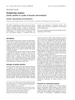
Microscopic examination showed a classical seminoma
with vascular and perineural invasion (Figure 1). The
spermatic cord margin appeared free of the tumor and
the tumor reached the excision margin. Therefore, his-
tological staging demonstrated a T2 lesion. His HCG
level was normal after the orchidectomy. He was com-
menced on a testosterone replacement therapy post-
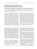
operatively. At a routine eight month follow up, he was
found to have an enlarged lymph node in the left ingu-
inal region. A CT sc an confirmed the presence of a 2.4
cm left inguinal lymph node (Figure 2). There was also
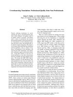
pelvic lymph node and chest involvement. An excision
biopsy of his inguinal node revealed a cl assical
metastatic seminoma with extra-capsular spread to the
surrounding adipose tissue (Figure 3). Treatment was
started with two cycles of carboplatin AUC10. He made
a good recovery after the chemotherapy and repea t CT
scans have shown no evidence of recurrence after two
years of follow up.
Discussion
In patients with a prior history of orchidopexy or scrotal
surgery who have a testicular tumor, the incidence of
inguinal metastases is unclear but has bee n reported in
series varying from two percent [10] up to 10 percent
Figure 1 A histology specimen shows classical seminoma
arising in the testis. Vascular and perineural invasion can be seen
(arrow). The spermatic cord margin was free of tumor.
Figure 2 A CT scan of the pelvis revealing a 2.4 cm left
inguinal lymph node (arrow).
Ismail et al . Journal of Medical Case Reports 2010, 4:378
/>Page 2 of 4
[11]. Daugaard et al. evaluated the incidence of inguinal
lymph node metastases in 695 patients with stage I testi-
cular cancer [10]. Two percent of patients developed
inguinal node metastasis. Non-seminomatous GCTs
more frequentl y invaded inguinal lymph node s than
seminoma.
The routine manage ment of the inguinal lymphatics
(palpable or not) in patients with testicular tumors and
a previous history of inguinal or scrotal surgery remains
controversial, as a result of insufficient data [6]. Prophy-
lactic inguinal lymphadenectomy is rarely mentioned in
the literature. In some series, patients have been found
to have positive inguinal nodes with no retroperitoneal
lymphadenopathy, supporting the need to perform rou-
tine ipsilateral inguinal lymphadenectomy even when
the retroperitoneal nodes are clear [6,12]. Wheeler et al.
advocated ipsilateral inguinalandbilateralretroperito-
neal node dissection as the primary therapy for non-
seminomatous testicular tumor with a previous history
of scrotal and inguinal procedures [6].
Another series in which 20 cases of testicular tumor
and previous scrotal surgery were presented, failed to
document the incidence of inguinal lymphadenopathy
[13]. They c oncluded that additional treatment to the
inguinal nodes was not required but most of their
patients underwent immediate radiation therap y or che-
motherapy with n one undergoing groin dissection. The
true incidence of inguinal metastases in their study is
therefore unknown. It was suggested that failure to per-
form prophylactic inguinal node dissection does not
adversely affect patient survival and regular groin palpa-
tion and dissection of any suspicious lymph nodes was
recommended. If positive, cisplatinum, vinblastine and
bleomycin chemotherapy is given [14]. Mianne et al.
also suggested that prophylactic ipsilateral inguinal dis-
section is not necessary i n patients with non-seminoma-
tous testicular tumors with a history of inguinal or
scrotal surgery, owing to the efficacy of primary and sec-
ondary chemotherapy [15]. However, for testicular semi-
noma they advocated additional inguinoscrotal
radiotherapy. The low incidence of inguinal lymph node
metastasis, morbidity rate following radical ilioinguinal
dissection, the accessibility of the inguinal nodes to fol-
low-up examination and the availability of highly success-
ful multimodal therapy make expectant management of
the clinically negative groin an a ttractive alternative. A
diagnosis of inguinal node metastases is usually made by
an excision biopsy of the nodes, but fine needle aspiration
(FNA) has also been used.
Conclusion
Inguinal lymph node metastases from testicular cancer
are rare. A history of inguinal or scrotal surgery may
predispose involvement of the inguinal nodes as a result
of altered patterns of lymphatic drainage. The routine
management of inguinal lymphatics (palpable or not) in
patients with testicular tumors and a previous history of
inguinal or scrotal surgery remains controversial, with
no consensus amongst those treating these patients.
During radical inguinal orchidectomy, the surgeon
should be careful to minimize the handli ng of the testis
and ensure high ligation of the spermatic cord up to the
internal inguinal ring to reduce the risk of inguinal
lymph node metastasis.
Consent
Written informed consent was obtained from the patient
for publication of this case report and any accompany-
ing images. A copy of the written c onsent is available
for review by the Editor-in-Chief of this journal.
Abbreviations
(AFP): Alpha-fetoprotein; (CT): Computed tomography ; (FNA): Fine needle
aspiration; (GCTs): Germ cell tumors; (HCG): Human chorionic gonadotrophin;
(LDH): Lactate dehydrogenase
Author details
1
Department of Urology, Homerton University Hospital NHS Foundation
Trust, London, E9 6SR, UK.
2
Department of Pathology, Bart’s and the London
NHS Trust, London, EC1A 7BE, UK.
3
Department of Urology, Bart’s and The
London NHS Trust, London EC1A 7BE, UK.
Figure 3 Metastases to the inguinal node consistent with the
original seminoma (large arrow) and invasion through the
capsule into the surrounding adipose tissue (small arrow).
Ismail et al . Journal of Medical Case Reports 2010, 4:378
/>Page 3 of 4
Authors’ contributions
MI wrote the original manuscript. FZ and JP analyzed and interpreted the
patient data with regard to the hematological and radiological diagnosis. SB
performed the histological examination of the testis. VN and JM were major
contributors in writing the manuscript. All authors read and approved the
final manuscript.
Competing interests
The authors declare that they have no competing interests.
Received: 7 December 2009 Accepted: 25 November 2010
Published: 25 November 2010
References
1. Cancer Research UK: Testicular cancer incidence and statistics. Cancer
Research UK; 2009 [ />incidence/].
2. Looijenga LH, Oosterhuis JW: Pathogenesis of testicular germ cell
tumours. Rev Reprod 1999, 4:90-100.
3. Verp MS, Simpson JL: Abnormal sexual differentiation and neoplasia.
Cancer Genet Cytogenet 1987, 25:191-218.
4. Jamieson JK, Dobson JF: The lymphatics of the testicle. Lancet 1910,
1:493-495.
5. Höltl L, Peschel R, Knapp R, Janetschek G, Steiner H, Hittmair A, Rogatsch H,
Bartsch G, Hobisch A: Primary lymphatic metastatic spread in testicular
cancer occurs ventral to the lumbar vessels. Urology 2002, 59:114-118.
6. Wheeler JS Jr, Babayan RK, Hong WK, Krane RJ: Inguinal node metastases
from testicular tumors in patients with prior orchiopexy. J Urol 1983,
129:1245-1247.
7. Corby HM, Lynch TH, Fitzpatrick JM, Smith JM: Inguinal lymph node
metastases from a testicular tumor. Br J Urol 1996, 77:923-924.
8. Ohtani O, Gannon BJ: The microvasculature of the rat vas deferens: a
scanning electron and light microscopic study. J Anat 1982, 135:521-529.
9. Lockett CJ, Nandwani GM, Stubington SR: Testicular seminoma - unusual
histology and staging with sub epithelial spread of seminoma along the
vas deferans. BMC Urol 2006, 6:5.
10. Daugaard G, Karas V, Sommer P: Inguinal metastases from testicular
cancer. BJU Int 2006, 97:724-726.
11. Batata MA, Whitmore WF Jr, Chu FC, Hilaris BS, Loh J, Grabstald H,
Golbey R: Cryptorchidism and testicular cancer. J Urol 1980, 124:382-387.
12. Johnson DE, Babaian RJ: The case for conservative surgical management
of the ilioinguinal region after inadequate orchiectomy. J Urol 1980,
123:44-46.
13. Lanteri VJ, Choudhury M, Pontes JE, Wajsman Z, Beckley S, Murphy GP:
Treatment of testicular tumors arising in patients with previous inguinal
and/or scrotal surgery. J Urol 1982, 127:58-59.
14. Ozen H, Altug U, Bakkaloglu MA, Remzi D: Significance of Scrotal Violation
in the Prognosis of Patients with Testicular Tumours. BJU 1988,
62(3):267-270.
15. Mianné DM, Barnaud P, Altobelli A, Masson J, Valeri A: Inguinal lymphatic
metastasis of cancer of the testis: staging and therapeutic approach.
Ann Urol (Paris) 1991, 25:199-202.
doi:10.1186/1752-1947-4-378
Cite this article as: Ismail et al.: Inguinal lymph node metastases from a
testicular seminoma: a case report and a review of the literature.
Journal of Medical Case Reports 2010 4:378.
Submit your next manuscript to BioMed Central
and take full advantage of:
• Convenient online submission
• Thorough peer review
• No space constraints or color figure charges
• Immediate publication on acceptance
• Inclusion in PubMed, CAS, Scopus and Google Scholar
• Research which is freely available for redistribution
Submit your manuscript at
www.biomedcentral.com/submit
Ismail et al . Journal of Medical Case Reports 2010, 4:378
/>Page 4 of 4