báo cáo khoa học: "Post-bronchoscopy fatal endobronchial hemorrhage in a woman with bronchopulmonary mucormycosis: a case report" docx
Bạn đang xem bản rút gọn của tài liệu. Xem và tải ngay bản đầy đủ của tài liệu tại đây (448.87 KB, 4 trang )
CAS E REP O R T Open Access
Post-bronchoscopy fatal endobronchial
hemorrhage in a woman with bronchopulmonary
mucormycosis: a case report
Paola Di Carlo
1*
, Daniela Cabibi
2
, Anton Maria La Rocca
3
, Dario De Luca
4
, Francesco La Licata
1
, Ennio Sacco
3
Abstract
Introduction: During infection, Mucorales fungi invade major blood vessels, leading to extensive necrosis, and in
cases of extensive pulmonary disease, bleeding into the lungs may occur.
Case presentation: We report an unexpected event of post-bronchoscopy fatal endobronchial hemorrhage in a
62-year-old HIV-negative Italian woman with well controlled diabetes mellitus who presented with diffuse cavitated
pulmonary lesions. Fiberoptic bronchoscopy revealed bilateral obstruction of the segmental bronchi. Fatal massive
bleeding occurred after standard biopsy procedures. Histologic examination showed that the hyphae were more
deeply colored by hematoxylin-e osin (H&E) than by other stains for fungi. Culture and autopsy confirmed
bronchopulmonary mucormycosis.
Conclusion: Infection by Mucorales fungi should be considered in the diabetes population regardless of the
degree of metabolic control. In these patients, particular caution should be taken during bronchoscopic procedures
because of the greater friability of the fungal lesions.
Introduction
“Zygomycosis” refers to infections caused by a class of
fungi called Zygomycetes, which includes the gene ra
Rhizopus, Absidia, and Rhizomucor. They had previously
been assigned to the genus Mucor and were considered
responsible for the disease known as “ mu cormycosis”
[1,2]. These fungi are ubiquitous in nature and are com-
mon inhabitants of decomposing matter. They can
cause serious and rapidly fatal infections, particularly in
individuals with compromised immune systems, such as
those with poorly controlled diabetes with ketoacidosis
[1-4].
The fungi invade major blood vessels, leading to
extensive necrosis, and in extensive pulmonary disease,
bleeding into the lungs may occur. In pati ents with dia-
betes mellitus, pulmonary mu cormycosis may develop,
withalessfulminantdiseasecoursebutwithatypical
presentation of a solitary nodule [5].
Biopsy (sur gical or transbronchial) of abnormal tissue
retrieved by bronchoscopic aspiration or bronchoalveo-
lar lavage (BAL) via a bro nchoscope and microbiologic
evaluation are the most efficient methods for detecting
endobronchial Mucor [3-5]. We report a rare case of
diff use pulmonary mucormycosis in a patient with well-
controlled type 2 diabetes who had a fatal pulmonary
hemorrhage during a fiberoptic bronchoscopy
procedure.
Case presentation
A 62-year-old woman of Italianoriginandnationality
with a history of fever and a persistent cough for three
weeks was admitted to our hospital for a scheduled
fiberoptic bronchoscopy (FB) to assess the nature of a
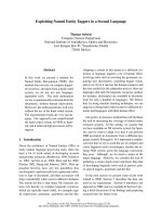
diffuse pulmonary lesion revealed by computed tomo-
graphy (CT) chest scan (Figure 1).
Multiple cavitated lesions in the lungs were diagnosed.
Some contained air and had a hyperdense capsule in the
bronchi, consistent with bronchiectasis, whereas others
resembled heteroplastic cavitary lesions (Figure 1). The
patient had no significant clin ical history except for type
2 diabetes mellitus (DM), contro lled with oral anti-
* Correspondence:
1
Infectious Diseases Section, Department of Health Promotion Sciences,
University of Palermo, Palermo, Italy
Full list of author information is available at the end of the article
Di Carlo et al. Journal of Medical Case Reports 2010, 4:398
/>JOURNAL OF MEDICAL
CASE REPORTS
© 2010 Di Carlo et al; licensee B ioMed Central Ltd. This is an Open Access article distributed under the terms of the Creative Comm ons
Attribution License ( g/licenses/by/2.0), which permits unrestricted use, distribution, and reproduction in
any medium, provided the original work is properly cited.
diabetic treatment and a suitable diet. In the three years
of follow-up, her hemoglobin A
1c
remained below 7%.
At the time of admission to hospital, the patient’ s
white blood cell count was 10 × 10
9
/L with 50% neutro-
phils; hemoglobin and blood glucose were 13.0 g and
260 mg/100 ml, respectively. No coagulation alterations
were observed, and the thrombocyte count was 200.000
cells/ml. A human immunodeficie ncy virus (HIV) test
performed on admission was negative.
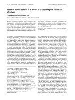
The bronchoscopy examination revealed a mucous and
necrotic plug completely occluding the lingular bronchus
and the apical segment of both lower-lobe bronchi (B6)
(Figure 2a). Grasping forceps were used to remove
mucous plugs for cytohistologic analysis, and other sam-
ples were obtained by bronchial aspiration. At the end of
the sampling procedures, performed by a team of
bronchologists with more than 20 years of experience in
these techniques, the patient had a massive hemorrhage
followed by cardiorespiratory arrest and was transferred
to the Intensive Care Unit (ICU) in a state of coma.
Histologic examination showed large areas of necrosis
in the bronchial mucosa, with peripheral granuloma tous
rea ction and several mul tinuclea ted cells (Figure 2b). In
the necrotic areas, numerous broad, very rarely septate,
haphazardly branched hyphae were evident. Signifi-
cantly, the hyphae were more deeply stained by hema-
toxylin-eosin (H&E) than by other special stains for
fungi, such as periodic acid Schiff (PAS) (Figure 2c, d)
and Gomori methenamine silver stain (GMS).
Morphologic aspects and stain affinity suggested
mucormycosis. This hypothesis was confirmed by the
results of the bronchial aspirate culture (histologic sam-
ple), which showed filamentous mycetes belonging to
the class Zygomycetes. Consequently, the patient was
treated with 5 mg of liposomal amphotericin B per
pound of body weight.
No evidence of dissem inated infection was found, and
analysis of cerebrospinal fluid did not reveal cerebral
mycoses. The patient died 20 days after being admitted
to the ICU . Autopsy confirmed bronchopulmonary
mucormycosis.
Discussion
Mucormycosis can be radiologically misdiagnosed as
active tuberculosis, chronic necrotizing aspergillosis,
coccidioidomycosis, or bronchiectasis. These have all
been reported in the diabetes population and may be
associated with massive or recurrent hemoptysis [6-8].
Therefore, fiber optic bronchoscopy wit h biopsy or
bronchial aspirate or both are needed to confirm a sus-
pected diagnosis and to start an appropriate therapy.
Previous reports of rhinocerebral or pulmonary
mucormycosis in HIV-negative diabetes patients
involved subjects with poorly controlled diabetes: acido-
sis and hyperglycemia provide an excellent environment
for the fungus to grow [9].
In our patient’ s case, no evide nce of severe or persis-
tent hyperglycemia was noted. Her general condition
and absence of coagulation alterations indicated that a
bronchoscopy examination could be carried out. How-
ever, the fatal hemorrhagic event occurred during stan-
dard bronchoscopic procedures performed t o obtain
specimens f or histologic and microbiologic assessment.
Autopsy findings confirmed that endobronchial mucous
and necrotic plugs seen during the bronchoscopic pro-
cedure were related to pulmonary vascular invasion by
mucoraceous hyphae.
Figure 1 Transverse computed tomography scan shows
circular masses in pulmonary fields.
Figure 2 Bronchoscopic image shows muconecrotic tissue. (a)
On histologic examination, necrotic tissue reveals large areas of
necrosis with peripheral granulomatous reaction and several
multinucleated cells; in the necrotic areas, numerous broad, very
rarely septate, haphazardly branched hyphae were evident; the
hyphae were deeply stained by H&E stain, more so than by PAS
staining (b-d). H&E (b) ×100; (c) ×400; PAS (d) ×400.
Di Carlo et al. Journal of Medical Case Reports 2010, 4:398
/>Page 2 of 4
Fiberoptic bronchoscopic examination is a useful proce-
dure for identifying bronchial obstructions and endolum-
inal le sions, as well as for assessing the tracheobronchial
tree beyond stenoses. Moreover, the procedure makes it
possible to restore normal airflow in airless areas around
the blockage (possible atelectasis or subatelectasis). Al
Majed [10] reported the removal of a mucormycosis lesion
through a rigid bronchoscope.
The risks associated with bronchoscopy procedures
are w ell known. However, our case study suggests that,
in the absence of a clear or well-defined diagnosis, parti-
cular caution should be exercised when conducting an
endoscopic examination in diabetes patients with sus-
pected pulmonary mucormycosis. Similar to other cases
of this group of fungi, angioinvasion, thrombosis, and
necrotic lesions are the hallmark features. Moreover,
diabetes patients have endothelial dysfunction, increased
arterial stiffness, or decreased arterial distensibility [11].
Therefore, any sampling procedure such as aspiration
may trigger vessel rupture, with massive bronchial
hemorrhage.
In these cases, BAL could be advocated as a less inva-
sive technique. Moreover, non-invasive techniques, such
as virtual bronchoscopy, have been found to be useful
for assessing lesion friability in cavitated disease [12].
It has been suggested that an air-crescent sign on a
chest radiograph is an important sign of potentially fatal
hemoptysis [13]. Moreo ver, lesion changes and progres-
sion should indicate the need to start early antifungal
and surgical therapy [4]. However, in our case, none of
these signs was observed.
Starting treatment early seems to be the significant
factor in reducing mortality associated with this dissemi-
nat ed pulmonary disease. Most patients with mucormy-
cosis have been treated with lipid preparations of
amphotericin (predominantly liposomal) with few reac-
tions or adverse events [3]. For years, amphotericin B
has b een the drug of choice for these highly aggres sive
infections. Recently , patients who were unresponsive to
monotherapy with liposomal amphotericin B responded
favorably to the addition of echinocandin caspofungin
acetate [14]. Newly introduced, second-generation tria-
zoles include voriconazole, which is not active against
the Zygomycetes, and posaconazole, which has been
demonstrated to be active in vitro,inanimalmodels,
and in case reports [15].
Other mechanisms to prevent or limit this fatal com-
plication are unclear.
Conclusion
Two interesting findings emerge from this case study.
First, that mucormycosis should be considered in all dia-
betes patients regardless of degree of metabolic control.
Second,thatfungallesionsmaybemorefriablein
these subjects, who might be at greater risk of complica-
tions associated with broncoscopy procedures.
Consent
Written informed consent was obt ained from the
patient’s family for publication of this case report and
accompanying images. A copy of the written consent is
available for review by the Editor-in-Chief of this journal.
Abbreviations
GMS: Gomori methenamine silver stain; H&E: hematoxylin-eosin; PAS:
periodic acid Schiff.
Author details
1
Infectious Diseases Section, Department of Health Promotion Sciences,
University of Palermo, Palermo, Italy.
2
Department of Human Pathology,
University of Palermo, Palermo, Italy.
3
Rehabilitation Unit, G.F. Ingrassia
Hospital, Palermo, Italy.
4
Bronchology Unit, V. Cervello Hospital, Palermo, Italy.
Authors’ contributions
PD, DC, and DD, participated in the conception of the idea, review of the
literature, writing of the manuscript, and interpretation of histologic assays.
FL collected and interpreted data. AML and ES wrote the pathologic section
and reviewed the manuscript. All authors have read and approved the final
manuscript.
Competing interests
The authors declare that they have no competing interests.
Received: 11 January 2010 Accepted: 9 December 2010
Published: 9 December 2010
References
1. Ellis DH: Systemic zygomycosis. In Microbiology and Microbial Infections,
Medical Mycology. 10 edition. Edited by: Merz WG, Hay RJ. London: Hodder
Arnold; 2005:659-686.
2. Ribes JA, Vanover-Sams CL, Baker DJ: Zygomycetes in human disease. Clin
Microbiol Rev 2000, 13:236-301.
3. Prabhu RM, Patel R: Mucormycosis and entomophthoramycosis: a review
of the clinical manifestations, diagnosis and treatment. Clin Microbiol
Infect 2004, 10:31-47.
4. Pagano L, Offidani M, Fianchi L, Nosari A, Candoni A, Piccardi M, Corvatta L,
D’Antonio D, Girmenia C, Martino P, Del Favero A: GIMEMA (Gruppo
Italiano Malattie Ematologiche dell’Adulto) infection program:
mucormycosis in hematologic patients. Haematologica 2004, 89:207-214.
5. Gale AM, Kleitsch WP: Solitary pulmonary nodule due to phycomycosis
(mucormycosis). Chest 1972, 62:752-755.
6. Winn RE, Johnson R, Galgiani JN, Butler C, Pluss J: Cavitary coccidioidomycosis
with fungus ball formation: diagnosis by fiberoptic bronchoscopy with
coexistence of hyphae and spherules. Chest 1994, 105:412-416.
7. Sugino K, Hasegawa C, Sano G, Shibuya K, Homma S: Pathophysiological
study of chronic necrotizing pulmonary aspergillosis. Jpn J Infect Dis
2008, 61:450-453.
8. Lee JH, Kwon SY, Yoon HI, Yoon CJ, Lee KW, Kang SG, Lee CT: Haemoptysis
due to chronic tuberculosis vs. bronchiectasis: comparison of long-term
outcome of arterial embolisation. Int J Tuberc Lung Dis 2007, 11:781-787.
9. Chakrabarti A, Das A, Mandal J, Shivaprakash MR, George VK, Tarai B, Rao P,
Panda N, Verma SC, Sakhuja V: The rising trend of invasive zygomycosis
in patients with uncontrolled diabetes mellitus. Med Mycol 2006,
44:335-342.
10. al-Majed S, al-Kassimi F, Ashour M, Mekki MO, Sadiq S: Removal of
endobronchial mucormycosis lesion through a rigid bronchoscope.
Thorax 1992, 47:203-204.
11. Cooper ME, Bonnet F, Oldfield M, Jandeleit-Dahm K: Mechanisms of
diabetic vasculopathy: an overview. Am J Hypertens 2001, 14:475-486.
Di Carlo et al. Journal of Medical Case Reports 2010, 4:398
/>Page 3 of 4
12. Finkelstein SE, Summers RM, Nguyen DM, Stewart JH, Tretler JA,
Schrump DS: Virtual bronchoscopy for evaluation of malignant tumors of
the thorax. J Thorac Cardiovasc Surg 2002, 123:967-972.
13. Dykhuizen RS, Kerr KN, Soutar RL: Crescent sign and fatal haemoptysis in
pulmonary mucormycosis. Scand J Infect Dis 1994, 26:498-501.
14. Vazquez L, Mateos JJ, Sanz-Rodriguez C, Perez E, Caballero D, San Miguel JF:
Successful treatment of rhinocerebral zygomycosis with a combination
of caspofungin and liposomal amphotericin B. Haematologica 2005,
90:39-42.
15. Page RL, Schwiesow J, Hilts A: Posaconazole as salvage therapy in a
patient with disseminated zygomycosis: case report and review of the
literature. Pharmacotherapy 2007, 27:290-298.
doi:10.1186/1752-1947-4-398
Cite this article as: Di Carlo et al.: Post-bronchoscopy fatal
endobronchial hemorrhage in a woman with bronchopulmonary
mucormycosis: a case report. Journal of Medical Case Reports 2010 4:398.
Submit your next manuscript to BioMed Central
and take full advantage of:
• Convenient online submission
• Thorough peer review
• No space constraints or color figure charges
• Immediate publication on acceptance
• Inclusion in PubMed, CAS, Scopus and Google Scholar
• Research which is freely available for redistribution
Submit your manuscript at
www.biomedcentral.com/submit
Di Carlo et al. Journal of Medical Case Reports 2010, 4:398
/>Page 4 of 4