báo cáo khoa học: "Refractory topiramate-induced angle-closure glaucoma in a man: a case report" pot
Bạn đang xem bản rút gọn của tài liệu. Xem và tải ngay bản đầy đủ của tài liệu tại đây (304.58 KB, 3 trang )
CAS E REP O R T Open Access
Refractory topiramate-induced angle-closure
glaucoma in a man: a case report
Matthew C Willett, Deepak P Edward
*
Abstract
Introduction: Topiramate is a sulphonamide derivative indicated in the treatment of epilepsy and migraine.
A known adverse affect is an idiosyncratic reaction that results in angle-closure glaucoma. We describe a patient
with bilateral glaucoma related to topiramate that showed some unusual clinical features.
Case presentation: A 39-year-old Caucasian man presented with acute angle-closure glaucoma; he initially
presented with intractable headaches after being treated with an escalating dose of topiramate. Clinical signs
included elevated intraocular pressure that was initially refractory to treatment, shallow anterior chambers, and
extensive bilateral choroidal effusions. After treatment with intravenous methylprednisolone, in conjunction with
conventional glaucoma treatment, there was rapid reduction of intraocular pressure, gradual delaye d resolution of
the choroidal effusion and induced myopic shift; and eventually a good outcome without optic nerve damage.
Conclusion: This case illustrates the importance of recognizing this entity in a non-ophthalmic setting and that
intravenous methylprednisolone may be useful in the treatment of the condition when it is not responsive to
conventional treatment. In addition, it is important to recognize that complete resolution of visual symptoms from
the myopic shift may be delayed, despite normalization of intraocular pressure.
Introduction
We report a case of t opiramate-induced angle-closure
glaucoma (TiACG) that was initially refractory to con-
ventional treatment. This case illustrates the importance
and method of timely and proper diagnosis, as well as
management of such cases that might initially respond
poorly to treatment.
Case presentation
A 39-year-old Caucasian man, with a past medical his-
tory of hypertension and migraine headaches, presented
to the emergency department with a one-day history o f
left-sided headache with blurred vision and haloes
around lights, which rapidly progressed to involve the
right side. After initial computed tomography of the
head, which was unremarkable, he was admitted to the
hospital with the diagnosis of intractable headaches and
treated with intravenous morphine and topiramate. The
fol lowing morning, the patient’ s ocular complaints wor-
sened, and the ophthalmology service was consulted.
Further questioning revealed that the patient had been
started on topiramate, 25 mg, one week before presenta-
tion for migraine headaches, and had been instructed to
double the do se one day before the onset of symptoms.
On initial examination, the best-corrected visual acuity
(BCVA) was 20/100 in both eyes. The intraocular pres-
sures (IOPs) by tonopen were 70 mm Hg in both eyes.
Slit-lamp examination revealed bilateral moderate con-
junctival injection, moderate to severe corneal edema,
and fixed mid-dilated pupils. The anterior chambers in
both eyes were shallow but without iridocorneal touch
or discernable inflammation. Gonioscopy with and with-
out compression revealed appositional angles. A limited
view to the posterior pole was available. B-scan ultra-
sound showed moderate serous choroidal effusions in
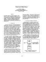
both eyes that extended to the posterior pole (Figure 1).
The patient was diagnosed with angle-closure glaucoma,
likely secondary to topiramate treatment.
After discontinuation of the topiramate, the patient
was treated with topical timolol maleate, 0.5%, dorzola-
mide hydrochloride, 2%, brimonidine tartrate, 0.15%,
homatropine, 2%, prednisolone acetate, 1%, and 500 mg
acetazolamide orally. After one hour, the topical
* Correspondence:
Summa Health Systems, 75 Arch St. Suite 512, Akron, OH 44304, USA
Willett and Edward Journal of Medical Case Reports 2011, 5:33
/>JOURNAL OF MEDICAL
CASE REPORTS
© 2011 Willett and Edward; licensee BioMed Central Ltd. This is an Open Access article distributed under the terms of the Creative
Commons Attri bution License ( which permits unrestricted use , distribution, and
reproduction in any m edium , provided the original work is properly cited.
treatment was repeated with the same combination, and
no change in IOP was noted. Intravenous 20% mannitol,
100 g in 500 ml, was admini stered, and three hours
later, IOP was 51 mmHg in the right eye and 83 mmHg
in the left eye. Given the continued IOP elevation and
the severity of the choroidal effusions, the patient was
given methylprednisolone, 250 mg IVPB, and the pre-
viously outlined regimen was continued. By the next
morning the patient’ s pain sco re was 0/10 in the right
eye and 4/10 in the left eye. The BCVA at that visit was
20/60, right eye, and counting fingers, left eye. Retino-
scopy of th e right eye was -5.00 D, consistent with ante-
rior displacement of the crystalline lens, and was
unsuccessful in the left eye secondary to corneal edema.
The patient was previously emmetropic. At that visit,
the IOP values were 38 mm Hg, right eye, and 37 mm
Hg in the left eye by pneumotonometer. Anterior-seg-
ment examination showed persistent bilateral shallowing
of the anterior chamber with micro cystic epithelial
edema and bullae bilaterally with descemet folds in the
left eye only. The patient was given a second dose of
methylprednisolone, 250 mg IVPB, and discharged
home with a regimen of atropine 1%, Cosopt (2% dorzo-
lamide; 0.5% timolol; Merck Inc, NJ), bimatoprost,
0.03%, and prednisolone acetate, 1% drops. The patient
was followed up daily with gradual reduction of intrao-
cular pressures. At one week after the initial presenta-
tion, the patient’s best corrected visual acuity was 20/15
in the right eye and 20/20 in the left eye with -4.00 D
sphere both eyes. The intraocular pressures were
12 mm Hg, right eye, and 16 mm Hg, left eye, and
gonioscopy showed open angles bilaterally. The ante-
rior-chamber depth improved, and choroidal effusions
were decreased on fundoscopic examination, eventually
resolving by the two-week visit. The myopic shift con-
tinued to decrease, and by one month, the patient’ s
uncorrected visual acuity had returned to 20/15 in both
eyes, and intraocular pressures without hypotensive
medications were 10 mm Hg right eye and 11 mm Hg
left eye. Anterior examination was significant for glau-
comflecken bilaterally and localized posterior synechiae
in the left eye. Dilated fundus examination showed that
the cup-to-disk ratio was 0.2 with intact rim and a full
visual field in both eyes.
Discussion
We report on a case of severe bilateral TiACG with
extensive choroidal effusions. Fraunfelder et al. [1]
reviewed 115 reports of TiACG and found that 85% of
cases occurred within the first two weeks of treatment.
This is consistent with the presentation in this patient,
who had been on treatment for on e week and experi-
enced the onset of symptoms within one day of dou-
bling his dose. Previous reports have not identified a
minimal dose of topiramate associated with ACG, and
the Fraunfelder et al. study showed that almost 50% of
cases occur with doses of 50 mg or less. It is possible
that the angle-closure glaucoma in our patients may
have been precipitated when the dose was doubled [1].
The principal step in appropriate management of
TiACG is making a c orrect diagnosis. We suggest that
an earlier diagnosis and treatment at the time of the
patient’ s presentation to the emergency department
might have allowed faster resolution of the episode of
angle closure. It is recommended that a patient with
angle-closure glaucoma, especially if it is bilateral,
should have a detailed history and review of the medica-
tion list for topiramate or other sulfonamide derivatives
that cause such idiosyncratic uveal effusions. Refraction
and B-scan ultrasonography should then be performed,
looking for myopic shift and ciliochoriodal effusion,
which help differentiate TiACG from typical pupillary
block ACG [2].
An interesting feature in our patient was the large and
extensive posterior choroidal effusion that was demon-
strated in both eyes. In TiACG, the uveal effusions
reported are typically anterior, causing shallowing of the
anterior chamber [1]. Our patient presumably also had
Figure 1 B-scan ultrasound of the left eye showing moderately elevated choroidal effusions that extend into the posterior pole.
Willett and Edward Journal of Medical Case Reports 2011, 5:33
/>Page 2 of 3
bilateral anterior uveal effusions, accounting for the
anterior chamber shallowing seen clinically. However,
ultrasound biomicroscopy was not performed in our
patient. Posterior uveal effusions have been reported in
TiACG but are often shall ow in nature. Large choroidal
effusions from other causes are often associated with
lowerIOP,whichmaybeassociated with aqueous
hypersecretion or c iliary body detachment [3]. It is
unclear whether the choroidal effusions in TiACG are
associated with normal aqueous humor production or
hyposecretion. It is, however, likely that mechanical
angle closure by ante rior displacement of the iris lens
diaphragm may override any other factors that affect
aqueous dynamics, resulting in IOP elevation.
The anterior displacement of the iris lens diaphragm
caused a myopic shift in the patient’s refraction and, as
illustrated by the temporal course in this patient, the
myopic shift may take several weeks to resolve, despite
normalization of IOP. This might be a useful observa-
tion to discuss with the patient during the recovery
phase.
The treatment of TiACG is tailored to the severity of
IOP elevation. The first step is immediate discontinua-
tion of topiramate. This step combined with topical
hypotensive medications and cycloplegics is most often
sufficient to reduce the IOP. In a large retrospective
case series, the typical course of IOP normalization in
TiACG was reported to be within hours to days after
cessation of topiramate and initiating of conventional
IOP-reducing therapy [1]. In contrast, our case of bilat-
eral TiACG necessitated further treatment to correct
the marked elevation of IOP that appeared to be refrac-
tory to conventional therapy over many hours. Rhee and
colleagues [4] suggested that in cases that do not
respond to conventional therapy, systemic corticoster-
oids and hyperosmolar agents should be used to speed
recovery and to avoid the need for surgical intervention.
This approach was used in our patient, and recovery fol-
lowed after administration of two doses of methylpred-
nisolone. Although one could argue that the cessation of
topiramate resulted in the resolution of the symptoms
and signs, given the severity of the presentation in this
case and the low side-effect profile associated with the
pulse doses of methylprednisolo ne, it is reasonable to
consider this as a treatment option.
Conclusion
The use of topiramate may lead to a spectrum of ocular
symptoms ranging from myopic shift to secondary
angle-closure glaucoma, secondary to ciliochoroidal effu-
sions. In cases of severe disease refractory to conven-
tional treatment, a lternative options co uld include t he
use of hyperosmotics and possibly intravenous methyl-
prednisolone to speed recovery.
Consent
Written informed consent was obtained from the
patients for publication of this case and any accompany-
ing images. A copy of the written consent is available
for review by the Editor-in-Chief of this journal.
Authors’ contributions
Both authors contributed equally to the manuscript. Both authors read and
approved the final manuscript.
Competing interests
Neither author has any conflicting interests.
Received: 27 February 2010 Accepted: 26 January 2011
Published: 26 January 2011
References
1. Fraunfelder FW, Fraunfelder FT, Keates EU: Topiramate-associated acute,
bilateral, secondary angle-closure glaucoma. Ophthalmology 2004,
111:109-111.
2. Lee GC, Tam CP, Danesh-Meyer HV, Myers JS, Katz LJ: Bilateral angle
closure glaucoma induced by sulphonamide-derived medications. Clin
Exp Ophthal 2007, 35:55-58.
3. Quinn CC, Greenfield DS: Choroidal effusion and hemorrhage. In
Glaucoma. Edited by: Shaarawy T. Philadelphia: Saunders/Elsevier;
2009:195-203.
4. Rhee DJ, Ramos-Esteban JC, Nipper KS: Rapid resolution of topiramate-
induced angle-closure glaucoma with methylprednisolone and mannitol.
Am J Ophthalmol 2006, 141:1133-1134.
doi:10.1186/1752-1947-5-33
Cite this article as: Willett and Edward: Refractory topiramate-induced
angle-closure glaucoma in a man: a case report. Journal of Medical Case
Reports 2011 5:33.
Submit your next manuscript to BioMed Central
and take full advantage of:
• Convenient online submission
• Thorough peer review
• No space constraints or color figure charges
• Immediate publication on acceptance
• Inclusion in PubMed, CAS, Scopus and Google Scholar
• Research which is freely available for redistribution
Submit your manuscript at
www.biomedcentral.com/submit
Willett and Edward Journal of Medical Case Reports 2011, 5:33
/>Page 3 of 3