Báo cáo y học: "Synchronous perforation of a duodenal and gastric ulcer: a case report" ppt
Bạn đang xem bản rút gọn của tài liệu. Xem và tải ngay bản đầy đủ của tài liệu tại đây (582.38 KB, 3 trang )
CAS E REP O R T Open Access
Synchronous perforation of a duodenal and
gastric ulcer: a case report
Dimos Karangelis
1,2*
, Georgios I Tagarakis
2
, Christos Karathanos
1
, Konstantinos Bouliaris
1
, Andony J Baddour
2
,
Anargyros Giaglaras
1
Abstract
Introduction: Peritonitis due to peptic ulcer perforation is a surgical emergency with a high risk of mortality and
morbidity.
Case presentation: We present a rare case of a 54-year-old Caucasian man who underwent an emergency
laparotomy for peritonitis caused by perforation of two peptic ulcers. The first was located on the anterior wall of
the duodenum and the second was posterior, pre-pyloric, close to the lesser curvature.
Conclusion: To the best of our knowledge, this is only the second report in the medical literature of a
simultaneous perforation of two peptic ulcers; though rare, every surgeon performing open or laparoscopic repair
of a perforated peptic ulcer should be aware of the possibility of simultaneous perforation.
Introduction
Peptic ulcer disease (PUD; gastric and duodenal ulcers)
remains one o f the most prevalent and costly gastroin-
testinal diseases [1] . The annual incidence of peptic ulcer
ranges from 0.1% to 0.3% [2]. Internationally, the fre-
quency varies among countries but there are two major
precipitating factors: Helicobacter pylori infection and the
consumption of non-steroidal anti-inflammatory drugs
(NSAIDs). Ulcer incidence increases with age for both
duodenal u lcers (DUs) and gastric ulcers (GUs) and DUs
[3] emerge two decades earlier than GUs, particularly in
men. Several factors pre dict increased risk with NSAIDs,
such as H. pylori infection, advanced age, comorbidities
and adjunct therapy with drugs such as co rticosteroids,
anticoagulants and bisphoshonates. Complications
(bleeding, perforation, obstruction) can occur in patients
with peptic ulcers of any etiology. Perforation occurs in
about 5% to 10% of patients with active ulcer disease.
Duodenal, antral and gastric body ulcers account for
60%, 20% and 20% of perforati ons, respectively, of peptic
ulcers [4,5]. Surgical abdominal exploration (both laparo-
scopic and laparotomic) is always indicated in gastroduo-
denal perforation. Hemodynamic instability, signs of
peritonitis and free extravasation of contrast material on
upper gastrointestinal tract contrast studies make the
decision for operation more urgent and imperative. Suc-
cessful treatment of perforated peptic ulcers with a
laparoscopic approach was first reported in 1990 [6,7].
Since then, various institutions have used this technique
to treat patients with perforated peptic ulcers. Contrain-
dications for laparoscopic repair for perforated peptic
ulcers include large perforations, prior abdominal
surgery, a posterior location of the perforation, and a
poor general state of health.
Case presentation
A 54-year-old Caucasian Greek man presented to the
Accident and Emergency department of our hospital with
a 20-day history of abdominal pain, vomiting and loss of
appetite. H e mentioned an eight kg weight loss ove r t he
last 20 days, as he had been drinking almost exclusively
water due to his symptoms. He had not pr esented to any
hospital facility earlier because he lived in a remote area in
the mountains. On admission, he had the septic image of
paleness, tachypnea, tachycardia (110 beats/minute) and a
fever of 38.5°C, as well as a rigid abdomen. Abdominal
and plain chest X-rays demo nstrated free gas under both
the hemidiaphragms. After initial resuscitation (placement
of intravenous lines and nasogastric tube followed by ade-
quate administration of fluids), our patient underwent an
emergency exploratory laparotomy. Our patient’s
* Correspondence:
1
Department of General Surgery, General Hospital of Larissa, Greece
Full list of author information is available at the end of the article
Karangelis et al. Journal of Medical Case Reports 2010, 4:272
/>JOURNAL OF MEDICAL
CASE REPORTS
© 2010 Karangelis et al; licensee BioMed Centr al Ltd. This is an Open Access article distributed under the terms of the Creative
Commons Attribution License ( which permits unrestricted u se, distribution, and
reproduction in any medium , provide d the original work is properly cited.
worsening clinical image and his deteriorating clinical
signs (tachypnea and tachycardia), along with the presence
of his acute abdomen led us to conclude that an emer-
gency laparotomy constituted the treatment of choice. In
the face of the emergency situation a computed tomogra-
phy (CT) scan was not performed. Laparotomy revealed
peritonitis due to a perforated ulcer on the anterior wall of
the duodenum, which was sutured, while the suture line
was reinforce d with an omental patch (Figure 1). After a
thorough lavage of the peritoneal cavity, further explora-
tion of the intra-abdominal organs revealed a second pos-
terior pre-pyloric ulcer on the lesser curvature of the
stomach, perforated into the lesser sac (Figure 2). A wedge
resection with staplers was carried out (Figure 3), while no
further acid reduction procedures were undertaken due to
sepsis. A Nissen fundoplication was performed as an anti-
reflux measure. Our patient recovered uneventfully a nd
was discharged home on the 13th post-operative day; at
this time we administered an appropriate eradication ther-
apy. More specifically, we followed the protocol of triple
therapy: a proton pump inhibitor, amoxici llin and clari-
thromycin were administered. After discharge our patient
was referred to gastrointestinal specialists. Our colleagues
planned a surveillance endoscopy according to their
protocol.
Discussion
Perforation of a peptic ulcer is a su rgical em ergency that
still carries a risk of mortality. We successfully managed a
rare and difficult case of simultaneous perforation of duo-
denal and gastric ulcers that could have been easily mis-
diagnosed and undertreated. Retrospectively studying our
case, we can state that there is a growing experience with
laparoscopic techniques for management of peptic ulcers.
A Graham patch, with or without a laparoscopic vagotomy
for perforated peptic ulcers is probably the most appropri-
ate minimally invasive approach when in experienced
hands [8,9]. Nevertheless, this case raises doubts as to the
extent laparoscopy would have been a safe procedure in
our case in terms of revealing both lesions.
Finally, every surgeon should strictly follow one of the
basic principles of abdominal surgery and perform a
thorough examination of the peritoneal cavity in every
case of diffuse peritonitis, even if the underlying pathol-
ogy appears to be obvious.
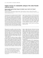
Figure 1 Omental patch-reinforced suture on the anterior wall
of the duodenum.
Figure 2 The second, posterior pre-pyloric ulcer on the lesser
curvature of the stomach.
Figure 3 Wedge resection with staplers.
Karangelis et al. Journal of Medical Case Reports 2010, 4:272
/>Page 2 of 3
Conclusions
In summary, emergency physicians and surgeons should
maintain a high level of clinical suspicion as a second
perforative peptic lesion, though a rare possibility, could
exist and could potentially be lethal.
Competing interests
The authors declare that they have no competing interests.
Authors’ contributions
KD was the primary surgeon for the case, conducted a thorough literature
research, and was the chief author in terms of writing the paper. TG
performed a consultation regarding the anti-reflux treatment, and co-
authored the paper. KC assisted with the linguistics and performed literature
research. BK assisted with the literature research. BA performed literature
research and checked the final version of the manuscript. GA was the
attending surgeon for the case and checked the paper. All authors read and
approved the final manuscript.
Consent statement
Written informed consent was obtained from the patient for publication of
this case report and accompanying images. A copy of the written consent is
available for review by the Editor-in-Chief of this journal.
Author details
1
Department of General Surgery, General Hospital of Larissa, Greece.
2
Department of Cardiovascular and Thoracic Surgery, University Hospital of
Thessaly, Larissa, Greece.
Received: 19 January 2010 Accepted: 18 August 2010
Published: 18 August 2010
References
1. Sonnenberg A, Everhart JE: Health impact of peptic ulcer in the United
States. Am J Gastroenterol 1997, 92:614.
2. Garcia Rodriguez LA, Hernandez-Diaz S: Risk of uncomplicated peptic
ulcer among users of aspirin and nonaspirin nonsteroidal
antiinflammatory drugs. Am J Epidemiol 2004, 159:23.
3. Sonnenberg A: Temporal trends and geographical variations of peptic
ulcer disease. Aliment Pharmacol Ther 1995, 9(Suppl 2):3.
4. Graham DY: Ulcer complications and their nonoperative treatment.
Gastrointestinal Disease Philadelphia, PA: WB SaundersSleisenge, M, Fordtran
J , 5 1993, 698.
5. Gunshefski L, Flancbaum L, Brolin RE, Frankel A: Changing patterns in
perforated peptic ulcer disease. Am Surg 1990, 56:270.
6. Nathanson LK, Easter DW, Cuschieri A: Laparoscopic repair/peritoneal
toilet of perforated duodenal ulcer. Surg Endosc 1990, 4:232-233.
7. Mouret P, Francois Y, Vignal J, Barth X, Lombard-Platet R: Laparoscopic
treatment of perforated peptic ulcer. Br J Surg 1990, 77:1006.
8. Lau WY, Leung KL, Kwong KH, Davey IC, Robertson C, Dawson JJ,
Chung SC, Li AK: A randomized study comparing laparoscopic versus
open repair of perforated peptic ulcers using suture or sutureless
technique. Ann Surg 1996, 224:131.
9. Zittel TT, Jehle EC, Becker HD: Surgical management of peptic ulcer
disease today - indication, technique and outcome. Langenbecks Arch
Surg 2000, 385:84.
doi:10.1186/1752-1947-4-272
Cite this article as: Karangelis et al.: Synchronous perforation of a
duodenal and gastric ulcer: a case report. Journal of Medical Case Reports
2010 4:272.
Submit your next manuscript to BioMed Central
and take full advantage of:
• Convenient online submission
• Thorough peer review
• No space constraints or color figure charges
• Immediate publication on acceptance
• Inclusion in PubMed, CAS, Scopus and Google Scholar
• Research which is freely available for redistribution
Submit your manuscript at
www.biomedcentral.com/submit
Karangelis et al. Journal of Medical Case Reports 2010, 4:272
/>Page 3 of 3