Báo cáo y học: "Atypical presentation of acute pancreatitis in a man with pancreatic insufficiency and cystic fibrosis: a case report" pptx
Bạn đang xem bản rút gọn của tài liệu. Xem và tải ngay bản đầy đủ của tài liệu tại đây (445.72 KB, 3 trang )
CAS E REP O R T Open Access
Atypical presentation of acute pancreatitis in a
man with pancreatic insufficiency and cystic
fibrosis: a case report
Malcolm Turner
1
, Hugh Jackson
2
, Robin Harle
3
, Rob Bohmer
4
, David W Reid
1*
Abstract
Introduction: Whether acute pancreatitis can occur in pancreatically insufficient individuals with cystic fibrosis
remains a matter of debate.
Case presentation: We describe a case of acute pancreatitis occurring in a 52-year-old Caucasian Australian man
with moderately severe cystic fibrosis lung disease and pancreatic insufficiency. An inflammatory mass within the
head of his pancreas was confirmed using computed tomography, magnetic resonance imaging and pancreatic
biopsy, but serum amylase and lipase remained normal throughout the acute phase of his illness. His symptoms
and the pancreatic mass resolved following the insertion of a biliary stent and the introduction of ursodeoxycholic
acid.
Conclusion: Our case report highlights the potential for acute pancreatitis to occur in patients with pancreatic
insufficiency and cystic fibrosi s. We further demonstrate that conventional biochemical markers that are normally
assessed to confirm the diagnosis may not be of particular use. As patients with cystic fibrosis survive into their
fourth and fifth decades of life, atypical presentations of acute pancreatitis may become more common.
Introduction
Acute pancreatitis in cystic fibrosis occurs almost exclu-
sively in young p atients with pancreatic sufficiency [1].
We describe the case of a 53-year-old man with cystic
fibrosis and pancreatic insufficiency who presented with
abdominal pain and a diagnosis of acute pancreatitis
despite normal amylase and lipase levels in his periph-
eral blood.
Case presentation
A 52-year-old Australian Caucasian male with cystic
fibrosis was admitted to our hospital with an exacerba-
tion of pulmonary sepsis accompanied by a vague
abdominal pain. Abdominal X-ray revealed faecal load-
ing in his caecum and ascending colon with proximal
small bowel dilata tion consistent with meconium ileus
equivalent. His relevant medical history consisted of
pancreatic insufficiency and bronchiectasis with
moderately severe lung function impairment (FEV
1
2.18
L/s; 53% predicted). He had multiple hospital admis-
sions over the preceding two years with exacerbations of
chronic airway sepsis.
The diagnosis of cystic fibrosis had been made during
his early childhood when he presented with failure to
thrive, and this had been confirmed with a n elevated
sweat test and genotyping that revealed him to be a het-
erozygote for G542X, with the other allele unidentified.
G542X is a Class I mutation that results in the complete
failure to synthesize fu nctional cystic fibrosis transmem-
brane conductance regulator (CFTR) and is usually asso-
ciated with pancreatic insufficiency. On this particular
admission, his chest and abdominal symptoms resolved
after a course of intravenous antibiotics, fluid replace-
ment, a nd oral administration of N-acetyl cysteine. His
24-hour fecal fat levels were elevated at 41 grams and
his pancreatic enzymes were thus increased.
He presented again after six weeks due to a recurrence
of severe abdominal pain, anorexia and weight loss, but
without any alteration to his bowel habit. Examination
revealed epigastric tenderness and active bowe l sounds,
* Correspondence:
1
Department of Respiratory Medicine, Royal Hobart Hospital, Tasmania,
Australia
Full list of author information is available at the end of the article
Turner et al. Journal of Medical Case Reports 2010, 4:275
/>JOURNAL OF MEDICAL
CASE REPORTS
© 2010 Turner et al; licensee BioMe d Central Ltd. This is an Open Access article distributed under the terms of the Creative Commons
Attribution License ( which permits unrestricted use, distribution, and reproduct ion in
any medium, pro vided the original work is properly cited.
but no guarding. Results of respiratory examination
were unchanged. On this occasion, abdominal X-ray was
normal, as were his full blood count, urea and electro-
lytes, serum amylase (46 IU/L; NR < 100 IU/L), and
liv er function tests. However, he continued to complai n
of severe abdominal pain radiating through his back and
lower chest. An abdominal ultrasound demonstrated an
increased echogenicity consistent with inspissated secre-
tions in the pancreatic duct and an 8 mm common bile
duct with no other abnormalities.
Two days after admission, our patient became jaun-
diced. Repeat blood tests demonstrated a cholestatic pic-
ture: total protein 66 g/L, albumin 36 g/L, alkaline
phosphates (ALP) 809IU/L, alanine transaminase (ALT)
543IU/L, glutamyl transaminase (GGT) 334IU/L, and
bilirubin 23 mmol/L. His serum amylase (42IU/L) and
lipase (4IU/L; NR: < 10IU/L), as well as urinary lipase
(96IU/L, NR: < 500IU/L) levels remained normal. An
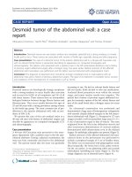
abdominal computed tomography (CT) scan demon-
strated that he had a heterogeneous mass measuring
5×5×5 cm and located at the head of the pancreas with
biliary and pancreatic duct dilation (Figure 1). Magnetic
resonance cholangiopancre atography confirmed the
enlargement of the head of his pancreas with dilated
intrahepatic and extrahepatic biliary ducts. These find-
ings were thought to be c onsistent with either acute
pancreatitis or a pancreatic malignancy.
Results of his liver function tests remained abnormal
(ALP 1085IU/L, ALT 249IU/L, GGT 383IU/L, and bilir-
ubin 37 μmol/L) and the patient proceeded to endo-
scopic retrograde cholangiopancreatography, where a
narrowing of the common bile duct at the head of the
pancreas was identified with more distal anatomical dis-
tortion of the entire pancreatic ductal system. A biliary
stent was consequently inserted. CT g uided biopsy of
the pancreatic mass demonstrated reactive pancreatic
ductal epithelium with an infiltrate of macrop hages and
lymphocytes, but no evidence of malignancy was found.
The patient contin ued to e xperience abdominal pain
and ursodeoxycholic acid was introduced to treat any
potential contribution of biliary sludging to cholestasis
and also to minimize the risk of stent occlusion. Follow-
ing stent insertion and commencement of ursodeoxy-
cholic acid, his symptoms and liver function tests slowly
improved, and then returned to normal over the next
6 weeks. A repeat CT scan showed a resolution of bili-
ary dilation, but no change in the pancreatic mass was
noted. After 18 months he remains well with normal
liver function tests and no abdominal pain. A repeat CT
scan at this time demonstrated a complete resolution of
the pancreatic mass (Figure 2).
Discussion
Although chronic pancreatitis is common in cystic fibro-
sis, acute pancreatitis is rare and usually occurs in young
patients who are pancreatically sufficient [1,2]. We are
unaware of any previous reports of acute pancreatitis
occurring in an older adult with pancreatic insufficiency
and cystic fibrosis but in the setting of normal amylase
and l ipase. The few reports available concerning acute
pancreatitis in patients with pancreatic insufficiency
concern children or young adults and always with raised
amylase and lipase blood levels [1,3,4].
In our patient, acute pancreatitis was possibly related
to his abnormal pancreatic duct anatomy and the che-
mical insult of bile constituents causing direct damage
Figure 1 Computed tom ography scan demonstrating mass at
the head of the pancreas.
Figure 2 Computed tomography scan at 18 months showing
resolution of the pancreatic inflammatory mass.
Turner et al. Journal of Medical Case Reports 2010, 4:275
/>Page 2 of 3
to his pancreatic tissue, which was then followed by a
local inflammatory response. Despite these proposed
mechanisms there was no classical bioc hemical evidence
of pancreatic injury, although imaging confirmed a typi-
cal inflammatory mass and gross edema of the pancreas.
Clinical improvement was relatively rapid following bili-
ary stenting and the introduction of ursodeoxycholic
acid. A repeat CT scan demonstrated a complete resolu-
tion of the previous inflammatory mass.
When all other diagnoses have been excluded, the
poor diagnostic sensiti vity of amylase and lipase in both
blood and urine samples have to be considered in older
patients with cystic fibrosis who present with abdominal
pain.
Conclusion
As pa tients with cysti c fibrosis survive into their fourth
and fifth de cades of life, atypical presentations of acute
pancreatitis may become more common. Caution needs
to be exercised when diagnosing acute pancreatitis in
patients with pancreatic insufficiency, as the biochemical
parameters normally used may not accurately reflect the
disease process.
Consent
Written informed consent was obtained from the patient for publication of
this case report and accompanying images. A copy of the written consent is
available for review by the Editor-in-Chief of this journal.
Competing interests
The authors declare that they have no competing interests.
Authors’ contributions
DR was the consultant physician who cared for the patient at the time of
presentation and diagnosis. MT was the junior doctor attached to the
respiratory unit at the time. MT identified the unusual nature of the case
and wrote the first draft of this case report. DR contributed to the writing of
the case report as did his colleagues HJ, RB and RH, all of whom were
involved in diagnosing and managing the patient. RH interpreted the
radiology results. All authors read and approved the final manuscript.
Author details
1
Department of Respiratory Medicine, Royal Hobart Hospital, Tasmania,
Australia.
2
Department of Gastroenterology, Royal Hobart Hospital, Tasmania,
Australia.
3
Department of Radiology, Royal Hobart Hospital, Tasmania,
Australia.
4
Department of Surgery, Royal Hobart Hospital, Collins Street,
Hobart, 7001, Tasmania, Australia.
Received: 29 October 2009 Accepted: 18 August 2010
Published: 18 August 2010
References
1. De Boeck K, Weren M, Proesmans M, Kerem E: Pancreatitis among patients
with cystic fibrosis: correlation with pancreatic status and genotype.
Pediatrics 2005, 115:e463-e469.
2. Walkowiak J, Lisowska A, Blaszczynski M: The changing faces of the
exocrine pancreas in cystic fibrosis: pancreatic sufficiency, pancreatitis
and genotype. Eur J Gastroenterol Hepatol 2008, 20:157-160.
3. Maiz L, Kirchschläger E, Suárez L, Escobar H: Acute pancreatitis in a patient
with cystic fibrosis and pancreatic insufficiency. Rev Esp Enferm Dig 1996,
88:581-582.
4. Moreno Gonzalez E, Ibaoez Aguirre J, Rico Selas P, Vorwald P,
Santoyo Santoyo J, Gomez Sanz R, Seoane Gonzalez J, Figueroa Andollo J,
Sciadini M: Recurrent acute pancreatitis as a complication of cystic
fibrosis: report of one case treated surgically. Ann Ital Chir 1991,
62:345-347.
doi:10.1186/1752-1947-4-275
Cite this article as: Turner et al.: Atypical presentation of acute
pancreatitis in a man with pancreatic insufficiency and cystic fibrosis: a
case report. Journal of Medical Case Reports 2010 4:275.
Submit your next manuscript to BioMed Central
and take full advantage of:
• Convenient online submission
• Thorough peer review
• No space constraints or color figure charges
• Immediate publication on acceptance
• Inclusion in PubMed, CAS, Scopus and Google Scholar
• Research which is freely available for redistribution
Submit your manuscript at
www.biomedcentral.com/submit
Turner et al. Journal of Medical Case Reports 2010, 4:275
/>Page 3 of 3