Applied Surgical Physiology Vivas - part 4 pptx
Bạn đang xem bản rút gọn của tài liệu. Xem và tải ngay bản đầy đủ của tài liệu tại đây (293.26 KB, 19 trang )
6. What is the rate of cerebral blood flow?
50 ml per 100 g of brain tissue. It accounts for 15% of
the CO, or about 750 mlmin
Ϫ1
.
7. How does this rate of flow vary with the arterial
pressure?
The rate of flow remains essentially stable owing to
local autoregulation of flow. This is a characteristic
feature of some specialised vascular beds, such as the
renal system.
8. What is the basic mechanism of autoregulation?
There are two principle reasons:
᭹
Myogenic response: an increase in the arteriolar wall
tension brought on by an increase in the arterial
pressure stimulates contraction of the mural smooth
muscle cells. The resulting vasoconstriction
stabilises the flow in the face of these pressure
changes
᭹
Vasodilator ‘washout’: if flow is suddenly and
momentarily increased by a sudden rise in the
driving pressure, locally-produced vasodilating
mediators are washed out of the vessel, leading to
vasoconstriction and a return of the flow to the
steady state
9. What are the main factors that govern the cerebral
blood flow?
᭹
PaCO
2
: hypercarbia increases the cerebral flow
through an increase of the [H
ϩ
]. The reverse occurs
with hypocarbia
᭹
PaO
2
: hypoxia produces cerebral vasodilatation,
increasing the flow. This influence is less important
than the above
᭹
Sympathetic stimulation: causes some vasoconstriction,
but this is the least important influence
APPLIED SURGICAL PHYSIOLOGY VIVAS
C
CEREBROSPINAL FLUID AND CEREBRAL
BLOOD FLOW
᭢
45
C
CEREBROSPINAL FLUID AND CEREBRAL
BLOOD FLOW
10. What is meant by the cerebral perfusion pressure?
This is defined as the difference between the mean
arterial pressure and the intracranial pressure. It must
remain above around 70 mmHg for adequate cerebral
perfusion.
APPLIED SURGICAL PHYSIOLOGY VIVAS
46
COLON
1. What are the major functions of the colon?
᭹
Absorption of water: the most important
᭹
Absorption of minerals: predominantly sodium. There
is, however, net secretion of potassium and
bicarbonate
᭹
Expulsion of faeces
᭹
Indirect role: bacterial flora in the colon are able to
synthesise vitamin K and some of the B vitamins.
They also produce some important fatty acids
2. What types of contraction does the colon have in
common with the small bowel?
᭹
Segmentation: this mixes the contents of the colon,
facilitating absorption
᭹
Peristalsis: propelling the contents distally
3. What type of contraction is peculiar to the colon?
Mass action contraction. There is simultaneous contrac-
tion of the smooth muscle over a very long length. This
moves material from one portion of the colon to
another in one movement. It occurs between 1
–3 times
per day.
4. Identify one way in which the basic electric rhythm
of the colon differs from that of the small bowel.
Unlike in the small bowel, the frequency of the wave of
contraction increases along the colon. At the ileocaecal
valve it is 2 per minute, and in
the sigmoid colon, up to
6 per minute.
5. What is the gastro-colic reflex?
This occurs after a meal enters the stomach, leading to
an increase in the motility of the proximal and distal
APPLIED SURGICAL PHYSIOLOGY VIVAS
C
COLON
᭢
47
C
COLON
colon, together with an increase in the frequency of
mass movements.
6. Outline the events that occur during defecation.
᭹
The defecation reflex is triggered by the distension
of the rectal walls by faeces entering from a mass
contraction proximally
᭹
The intra-rectal pressure has to reach 18 mmHg
before the reflex is triggered
᭹
Afferent impulses pass to sacral segments 2, 3 and 4.
This leads to stimulation of the efferent reflex
pathway, together with stimulation of the thalamus
and cortical sensory areas producing the conscious
desire to defecate
᭹
Efferent impulses pass back to the myenteric plexus
of the rectum, activating postganglionic PNS
neurones
᭹
This leads to contraction, propelling the faeces
forward
᭹
PNS stimulation also leads to relaxation of the
internal anal sphincter
᭹
The external sphincter relaxes, reducing the
pressure in the anal canal. Further peristalsis in the
rectum pushes the faeces out
᭹
This is augmented by voluntary contractions of the
pelvic floor muscles when performing the Valsalva
manoeuvre
7. What happens to the reflex pathway when there is
conscious desire not to defecate?
When faecal material enters the upper anal canal, there
is stimulation of S1, 2 and 3, as mentioned. If the desire
to defecate is resisted, then this leads to activation of
the pudendal nerve, which sends signals to the external
anal sphincter, increasing its tone. There is also acti-
vation of ascending pathways to the sensory cortex,
enabling the subject to distinguish between solid and
APPLIED SURGICAL PHYSIOLOGY VIVAS
᭢
48
gaseous material in the rectum. If there is solid,
descending pathways reinforce the external sphincter.
If the content is gas, the descending pathways lead to
relaxation of the sphincter and expulsion of the gas.
8. When does involuntary defecation occur?
This occurs when the rectal pressure is greater than
55 mmHg. This may occur either because of a volumin-
ous content, or in the presence of colo
nic spasm and
diarrhoea.
The reflex defecation triggered by this pressure rise
also occurs in the spinal patient.
9. Summarise the involvement of ANS in the
maintenance of continence and defecation.
᭹
PNS: relaxes the internal sphincter
᭹
SNS: stimulates tonic contraction of the internal
sphincter
10. Which physiologic mechanisms are involved in the
maintenance of faecal continence?
᭹
Sympathetically-mediated tonic contraction of the
internal anal sphincter
᭹
The pudendal nerve also maintains tonic
contraction of the external sphincter
᭹
Thus, contraction of the sphincters maintains an
anal pressure of 40–90 mmHg
᭹
The pubo-rectalis sling of the pelvic floor maintains
an anorectal angle of 120Њ
᭹
Resting intra-abdominal pressure provides a lateral
force on the slit-like anal canal, closing it off
APPLIED SURGICAL PHYSIOLOGY VIVAS
C
COLON
49
C
CONTROL OF VENTILATION
CONTROL OF VENTILATION
1. What are the main functions of the lung?
᭹
Oxygenation
᭹
Ventilation: elimination of carbon dioxide
᭹
Acid-base balance: forms the respiratory component
to acid-base homeostasis
᭹
Endocrine: production of angiotensin converting
enzyme
2. Broadly speaking, which parts of the brain are
responsible for controlling the rate and depth of
ventilation?
᭹
The brainstem: pons and medulla involved mainly.
These give ventilation its automacity and rhythmical
nature
᭹
Cerebral cortex: this gives some voluntary control
3. Which par ts of the brainstem have been identified
as being particularly important? Outline the role that
each plays in control.
Note that these areas of the brainstem have collectively
been termed the respiratory centre. They consist of:
᭹
Medullary respiratory centre: found in the reticular
formation. Composed of a dorsal group (involved in
inspiration) and a ventral group (involved in
expiration). The expiratory area in the ventral
group is not normally active during quiet
respiration, since expiration is predominantly a
passive process
᭹
Apneustic area: located in the pons. This area is
thought to prolong the inspiratory phase of the
respiratory cycle
᭹
Pneumotaxic area: also located in the pons. This
inhibits the activity of the inspiratory area of the
medulla. It may be involved in ‘fine tuning’ of
respiratory rate, depth and rhythm
APPLIED SURGICAL PHYSIOLOGY VIVAS
᭢
50
4. Which physiologic variables form the basis for
control of ventilation? Place them in order of
impor tance.
᭹
PaCO
2
: the most important regulatory factor
᭹
PaO
2
᭹
pH of the blood and CSF: has some influence above
and beyond the PaCO
2
5. How are changes in these parameters detected?
Through central and peripheral chemoreceptors
that stimulate the activity of the brainstem respiratory
centre.
6. Where are these receptors located?
᭹
Central chemoreceptors: located at the ventral surface
of the medulla. These are sensitive to changes in
PaCO
2
᭹
Peripheral chemoreceptors: found in the carotid and
aortic bodies. These are sensitive mainly to a fall of
PaO
2
and pH, and sensitive to a rise in PaCO
2
7. By what mechanism are central chemoceptors
sensitive to changes in the PaCO
2
?
These chemoreceptors are influenced indirectly.
Arterial CO
2
diffuses into the CSF and dissolves. This
produces protons (H
ϩ
), which then stimulate the cen-
tral chemoceptor. Therefore, the increased ventilation
blows off CO
2
.
8. Do you know of any other factors influencing the
pattern of ventilation?
Yes! The pattern of ventilation is also influenced by the
signals from a number of receptors located in and
around the respiratory apparatus.
᭹
Mechanical receptors: such as pulmonary stretch
receptors and J receptors. The former are involved
in the Hering-Breuer inflation reflex, where distension
APPLIED SURGICAL PHYSIOLOGY VIVAS
C
CONTROL OF VENTILATION
᭢
51
C
CONTROL OF VENTILATION
of the lung leads to slowing of inspiration and
increased expiratory time. The J receptors are
located in the airways close to capillaries, and are
thought to stimulate inspiration following and
increase in pulmonary blood flow
᭹
Others: such irritant receptors in the lungs and
nasopharynx, as well as chest wall receptors
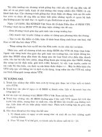
9. Below is a graph of the variation in the minute
ventilation with the PaO
2
. What do the lines A, B and
C represent?
APPLIED SURGICAL PHYSIOLOGY VIVAS
᭢
52
A
B
C
100908070
PaO
2
(mmHg)
50 604030
From Berne RM, Levy MN. Principles of Physiology, 3rd edition, 2000,
London, with permission from Elsevier
2010
0
20
40
Ventilation (L/min)
60
The three lines represent the ventilatory response to
changes in the PaO
2
at different PaCO
2
s. From line A to
C there is a progressive increase in the PaCO
2
.
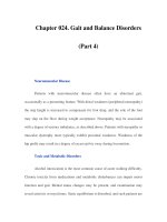
10. Draw a similar graph of how the ventilatory
response varies with the PaCO
2
at different PaO
2
s.
APPLIED SURGICAL PHYSIOLOGY VIVAS
C
CONTROL OF VENTILATION
᭢
53
PaCO
2
(mmHg)
PaO
2
Ventilation (L/min)
5
40
Effect of O
2
and CO
2
ventilatory response. The normal
ventilatory response to CO
2
is enhanced by hypoxia;
both the threshold (extrapolated X-intercept) and the
sensitivity (slope of response) are affected.
From Berne RM, Levy MN. Principles of Physiology,
3rd edition, 2000, London, with permission from Elsevier
100
70
11. What happens to the PaO
2
, PaCO
2
and ar terial pH
during exercise?
᭹
PaO
2
: there is usually a slight increase, but during
strenuous and persistent exercise, it may fall slightly
᭹
PaCO
2
: this changes little and in strenuous exercise
may fall
᭹
pH: this remains constant. Even during heavy
exercise, buffer systems ensure that lactic acidosis
has minimal impact on the overall pH of the blood
Therefore, during moderate exercise, there is surpris-
ingly little variation in all of the above parameters,
despite vast increases in the minute ventilation.
C
CONTROL OF VENTILATION
12. If these physiologic parameters are so consistent
during exercise, then what is the stimulus for a rise in
the minute ventilation during exercise?
This is not known, but a number of suggestions have
been put forward, such as increased limb movement, or
oscillations in the partial pressures of the respiratory
gases.
APPLIED SURGICAL PHYSIOLOGY VIVAS
54
CORONARY CIRCULATION
1. Where do the coronary arteries originate?
Both the right and left coronary arteries arise directly
from the ascending aorta at the aortic sinuses located
just above the leaflets of the aortic valve (also known as
the sinuses of Valsalva).
2. What is the rate of coronary flow at rest?
70–80 ml/min per 100 g of cardiac tissue. During exer-
cise, this can increase to 300–400 ml/min per 100 g.
3. What perc
entage of the CO does the heart receive?
4–5%.
4. Given that there is a high myocardial oxygen
demand at rest, what functional adaptations ensure
that supply meets demand?
Note that the myocardial oxygen consum
ption is in the
order of 8ml per 100 g of tissue. This is around 20 times
that of skeletal muscle. Functional adaptations to
ensure adequate oxygen delivery include:
᭹
High capillary density: producing a very high surface
area for oxygen delivery, and there is high blood
flow per unit weight of myocardium
᭹
High oxygen extraction ratio: the myocardium extracts
around 70% of the oxygen that is delivered to it
from the coronary flow. In contrast, the body
average is only 25%
᭹
Efficient metabolic hyperaemia: myocardial metabolites
generated during situations of increased exercise
and oxygen demand have a strong influence on
control of blood flow
᭹
During exercise, the increased oxygen demand is
met predominantly through an increase in the rate
of flow rather than an increase in the oxygen
extraction ratio
APPLIED SURGICAL PHYSIOLOGY VIVAS
C
CORONARY CIRCULATION
᭢
55
C
CORONARY CIRCULATION
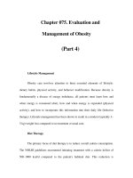
5. Look at the graph below, showing the pattern of
coronary flow during different phases of the cardiac
cycle. What is your interpretation of what is
happening? What causes this phenomenon?
APPLIED SURGICAL PHYSIOLOGY VIVAS
᭢
56
Systole Diastole
Arterial blood
pressure
120
100
80
Left coronary
blood flow
Zero flow
Right coronary
blood flow
Zero flow
From Smith JJ, Kampire JP. Circulatory Physiology–the
Essentials, 3rd edition, Lippincott, Williams & Wilkins
᭹
This shows that coronary flow is greatest during
diastole (accounting for 80% of the flow), unlike
other vascular beds. The lowest flow is during
isovolumetric contraction
᭹
This occurs due to mechanical compression of the
coronary vessels during systole, such that there is
reversal of the transmural pressure gradient across
the wall of the vessel, leading to momentary
occlusion
6. What factors are important in the control of
coronary blood flow (CBF)?
There are two main influential factors:
᭹
Metabolic factors: the dominant controlling process.
Some of the products of myocardial metabolism,
such as CO
2
, prostaglandins and adenosine produce
coronary vasodilatation
᭹
Neural control: 
2
-adrenoceptor stimulation
by vasomotor sympathetic nerves leads to coronary
vasodilatation. Any neurally-induced coronary
vasodilatation is overcome by metabolic factors
7. How does the coronary flow alter with changes of
perfusion pressure?
Between perfusion pressures of 60–180 mmHg, the
coronary flow is relatively constant. T
his is known as
autoregulation.
8. How does this come about?
There are a number of theories. Theses include:
᭹
Myogenic theory: increased transmural pressure
caused by a rise in the perfusion pressure stretches
arteriole myocytes. This stimulates their reflex
contraction, producing vasoconstriction. This
phenomenon maintains a steady flow despite the
rising pressure
᭹
Vasdilator washout: transient arteriolar dilatation
following a rise in the perfusion pressure also
washes out some vasdilators, such as adenosine.
Therefore, they can no longer promote further
dilatation in the face of rising pressures
9. Why does a sudden occlusion of CBF lead to MI?
Coronary vessels can be considered to be end vessels
with little anastom
oses between them. At the arteriolar
level, branches of the coronaries do communicate, but
APPLIED SURGICAL PHYSIOLOGY VIVAS
C
CORONARY CIRCULATION
᭢
57
C
CORONARY CIRCULATION
not enough to sustain the blood supply during acute
occlusion. Chronic obstruction, however, leads to the
progressive development of collateral vessels that
relieve some of the occlusive effects.
APPLIED SURGICAL PHYSIOLOGY VIVAS
58
FETAL CIRCULATION
1. Describe the stages in the passage of blood through
the fetal circulation.
᭹
Oxygenated blood enters the fetus from the
placenta through the umbilical vein
᭹
About 50% of the blood in the umbilical vein passes
into the liver, and goes through the hepatic
sinusoids. This eventually enters the inferior vena
cava (IVC)
᭹
The other 50% of the umbilical venous blood
bypasses the liver via the ductus venosus to enter the
IVC directly
᭹
From the IVC, the blood enters the right atrium
᭹
It is directed by the septum secundum through the
foramen ovale and into the left atrium. It undergoes
mixing with the small amount of deoxygenated
blood returning from the lungs through the
pulmonary veins
᭹
From the left atrium, blood is ejected into the left
ventricle, and eventually into the systemic
circulation through the aorta
᭹
A small amount of right atrial blood does not pass
through the foramen ovale, but is ejected into the
right ventricle, and into the pulmonary trunk
᭹
The vast majority of this pulmonary arterial blood
enters the aorta through the ductus arteriosus. The
rest enters the lungs
᭹
Of the blood that eventually enters the descending
aorta, about half supplies the lower body, and the
other half enters the umbilical arteries for return
back to the placenta
APPLIED SURGICAL PHYSIOLOGY VIVAS
F
FETAL CIRCULATION
᭢
59
F
FETAL CIRCULATION
᭹
The diagram below summarises these events:
APPLIED SURGICAL PHYSIOLOGY VIVAS
60
The number refers to oxygen saturation of the blood in %.
From Berne RM, Levy MN. Principles of Physiology, 3rd edition, 2000,
London, with permission from Elsevier
Unbilical arteries
58
Descending aorta
58
Inferior vena cava
27
Ductus venosus
Left ventricle
Right ventricle
Pulmonary veins
42
Pulmonary artery
52
Ductus arteriosus
52
Aorta
62
Superior vena cava
25
Foramen ovale
67
Right atrium
Inferior vena cava
67
Superior vena cava 25
Inferior vena cava 67
Portal vein
Umbilical vein
80
2. What changes occur to the fetal circulation
following birth?
᭹
Closure of the three main shunts: foramen ovale, ductus
venosus and ductus arteriosus
᭹
Closure and degeneration of the umbilical vessels
᭹
Fall in pulmonary vascular resistance (PVR): following
aeration of the lungs. This leads to a rise in the
pulmonary blood flow. In the first few postnatal
months, there is a progressive fall in the PVR
᭹
Reversal of ventricular wall thickness: the reduction in
the PVR to sub-systemic levels leads to thinning of
right ventricular wall compared to the left
GLOMERULAR FILTRATION AND
RENAL CLEARANCE
1. What factors determine whether a molecule is
filtered at the glomerulus?
᭹
Molecular size: small molecules such as urea
and glucose are filtered freely. The cut off size is
about 40 Å
᭹
Molecular charge: due to the negative charge of the
glomerular basement membrane, negatively
charged molecules, like proteins, are not filtered
2. What is meant by the renal clearance of a substance?
The renal clearance represents the volume of the
plasma from which all of a substance has been removed
and excreted in the urine, per un
it time.
3. How can the renal clearance be calculated?
From the equation:
where
C ϭ clearance of substance in mlmin
Ϫ1
U ϭ urinary concentration of the substance
V ϭ urine flow rate per minute
P ϭ plasma concentration of the substance
4. From which physical law is this equation derived?
The concept of renal clearance is based on the law of
mass balance. With respect to the kidney, this states that:
Amount of substance X excreted ϭ
Amoun
t filtered Ϫ Amount reabsorbed
ϩ Amount secreted
C ϭ
иUV
P
APPLIED SURGICAL PHYSIOLOGY VIVAS
G
GLOMERULAR FILTRATION AND
RENAL CLEARANCE
᭢
61
G
GLOMERULAR FILTRATION AND
RENAL CLEARANCE
5. Under what circumstances does the renal clearance
of a substance equal the glomerular filtration rate
(GFR)?
The clearance of a substance is the same as the GFR
when that substance is excreted purely by the process of
filtration.
In that situation, the amount filtered ϭ amount
excreted
6. Do you k
now of a substance that is excreted purely
through glomerular filtration, enabling it to be used
for measurements of the GFR?
Yes, the polyfructose molecule inulin. It is used for
measurement of the GFR because the volume of plasma
completely cleared of inulin per unit time is equal to
the volume of plasma filtered per unit time.
7. What physiologic properties make a substance
suitable for measurements of the GFR?
᭹
It must be filtered freely at the glomerulus
᭹
It must not undergo secretion or reabsorption by
the tubules
᭹
It must not be metabolised by the kidney
᭹
It must not inherently alter the GFR
8. What is the practical disadvantage to using inulin
for GFR measurements?
It must undergo continuous intravenous infusion.
GFR PUV
Thus, GFR
иϭ и
ϭ
иUV
P
APPLIED SURGICAL PHYSIOLOGY VIVAS
᭢
62
9. What is the most common way of measuring the
GFR?
By measuring 24-hour urinary creatinine excretion.
This molecule is endogenously produced at a generally
constant rate. It is not perfect for GFR measurements
since it is secreted by the tubules to a slight degree.
10. What is the difference between creatine and
creatinine?
᭹
Creatine: this is an amino acid stored in muscle and
brain tissue. It acts as a ready and rapid source of
phosphate groups when muscle exercises. During
periods of minimal activity, it is phosphorylated into
creatine phosphate. This phosphate is released
during activity
᭹
Creatinine: this is the anhydride of creatine and is
formed as a waste product of creatine metabolism
11. What is the normal range of creatinine clearance?
The normal range is 80–110 mlmin
Ϫ1
and declines with
increasing age.
12. What normally happens to glucose following
filtration at the glomerulus?
The filtered load of glucose normally undergoes com-
plete reabsorption by cells of the proximal tubule.
Therefore none is found in the urine.
APPLIED SURGICAL PHYSIOLOGY VIVAS
G
GLOMERULAR FILTRATION AND
RENAL CLEARANCE
᭢
63