Applied Surgical Physiology Vivas - part 6 doc
Bạn đang xem bản rút gọn của tài liệu. Xem và tải ngay bản đầy đủ của tài liệu tại đây (304.1 KB, 19 trang )
The slope B during expiration is ‘effort independent’ in
any one individual, and reaches a ceiling irrespective of
the expiratory force generated. This is due to the effects
of dynamic airways compression limiting the rate of
expiration. The greater the expiratory force generated,
the greater the airway compression limiting flow.
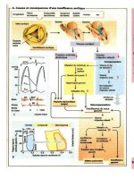
9. Draw a graph showing how the flow-volume loop
alt
ers in COPD and restrictive lung disease compared
to normality. What happens to the FEV and FEV
1
under these circumstances?
APPLIED SURGICAL PHYSIOLOGY VIVAS
M
MECHANICS OF BREATHING IV –
AIRWAY RESISTANCE
83
᭹
Note that in COPD, the total lung capacity (TLC),
FRC and RV are greater due to gas trapping
following loss of radial traction. Peak flow is
reduced due to airways obstruction and reduced
lung elastance. FEV
1
/FVC is reduced
᭹
In restrictive lung disease, all the lung volumes are
reduced, but the FEV
1
/FVC is normal or increased
RLDϭRestrictive lung disease
From NMS: Physiology, 4th edition, Bullock, Boyle & Wang,
2001, Lippincott, Williams & Wilkins
0
0
Lung volume (L)
VE max
VE max
VE max
COPD
RLD
Normal
(Inspiratory)
Flow (L/sec)
(Expiratory)
M
MICROCIRCULATION I
MICROCIRCULATION I
1. What is this equation, and in simple terms, what is
it describing?
J
v
ϭ L
p
S {(P
c
Ϫ P
i
) Ϫ(
p
Ϫ
i
)}
This is the Starling equation and describes the factors
that determine the flow of water across capillary walls.
2. So, basically, what is it saying?
It states that the net filtration of water across a capillary
wall is proportional to the difference between the
hydraulic and osmotic forces across the vessel wall
where:
᭹
P
c
: capillary filtration pressure
᭹
P
i
: interstitial pressure
᭹
p
p
: colloid oncotic (osmotic) pressure
᭹
p
i
: interstitial oncotic pressure
3. What are the other symbols in the equation, and
what do they mean?
᭹
L
p
: hydraulic conductance. This is the filtration rate
per unit change of pressure across the membrane
᭹
S: surface area of the vessel wall
᭹
s: the reflection coefficient. This is simply a measure
of how leaky the membrane is. This measures about
0.8, meaning that only 80% of the potential oncotic
pressure is exerted across the vessel wall
4. Can you name some factors that determine the
P
c
across a capillary wall?
᭹
Distance along the capillary: going from the arterial to
the venous side of the capillary, there is a fall in the
pressure. Typically, at the arterial end it is 35 mmHg,
and at the venous end, 20 mmHg
᭹
The resistances of the arterioles and venules at
either end of the capillary
APPLIED SURGICAL PHYSIOLOGY VIVAS
᭢
84
᭹
Gravity: both arterial and venous pressures increase
below the heart
5. Can you elaborate on how the resistances of
surrounding arterioles and venules affect the P
c
of
the capillary?
In basic terms the greater the resistance of the sur-
rounding vessel, the lower the P
c
. What is important
though, is the ratio of the resistance of the arteriole to
the venule (R
a
/R
v
).
᭹
The greater the R
a
/R
v
the lower the P
c
. When the
arteriole is constricted, the P
c
is closer to the
(lower) pressure in the venule
᭹
The lower the R
a
/R
v
the higher the P
c
, because the
arteriole is less constricted, its pressure has a greater
influence on the P
c
And so it follows that, from the Starling equation, the
greater the P
c
, the greater rate of filtration of water
across the vessel wall into the interstitium.
6. Can you name another filtration process that is
influenced heavily by the resistance ratios?
The net filtration of water across the glomerulus is also
influenced by the pre-to-post capillary resistance ratios.
This leads to alterations in not only the GFR, but also
the filtration fraction (the proportion of water passing
through the glomerulus that is filtered through).
Although other Starling forces are important in deter-
mining filtration across the glomerulus, the main point
of control of the GFR is through alterations in the
vascular resistances.
7. Give a normal value for the colloid osmotic
pressure.
25 mmHg.
APPLIED SURGICAL PHYSIOLOGY VIVAS
M
MICROCIRCULATION I
᭢
85
M
MICROCIRCULATION I
8. Which proteins are most important in exerting the
plasma colloid osmotic pressure?
᭹
Albumin: with a molecular weight of 69,000
᭹
g-globulins: with a combined molecular weight of
150,000
9. What about the interstitium?
The major proteins in the interstitium are:
᭹
Collagen
᭹
Proteoglycans
᭹
Hyaluronate
These have a positive influence on both the osmotic
pressure and the interstitial fluid pressure. (As the
interstitial proteins take up water, they swell, increasing
the interstitial pressure.)
APPLIED SURGICAL PHYSIOLOGY VIVAS
86
MICROCIRCULATION II
1. What is oedema (edema)?
This is defined as the abnormal accumulation of fluid
in the extravascular space.
2. What are two broad types, and how may they be
distinguished?
᭹
Transudate: due to imbalances in the hydrostatic
forces of the Starling equation
᭹
Exudate: occurs following an increase in the capillary
permeability
The main difference (that can be used to aid diagnosis
of the aetiology) is that an exudates is rich in protein
and fibrinogen.
3. What are the main causes?
The main causes are categorised according to the vari-
ables in the Starling equation:
᭹
Reduced colloid osmotic pressure (p
p
): that occurs with
hypoproteinaemic states, such as malnutrition,
protein-losing enteropathy and the nephrotic
syndrome
᭹
Incr eased capillary filtration pressure (P
c
): as in cardiac
failure where there is peripheral dependant oedema,
ascites and pulmonary oedema. Most commonly, the
main culprit is an elevation of the venous pressure,
as in deep venous thrombosis. Increased filtration
pressure also arises from abnormal retention of salt
and water, e.g. renal failure an other causes of
hypervolaemia
᭹
Increased capillary permeability: leading to the
formation of an exudates – which follows an
inflammatory process where there is an immune
mediated increase in the capillary permeability
APPLIED SURGICAL PHYSIOLOGY VIVAS
M
MICROCIRCULATION II
᭢
87
M
MICROCIRCULATION II
᭹
Lymphatic occlusion: leading to an accumulation of
fluid in the interstitial compartment, e.g. malignant
occlusion following lymphatic compression or
lymphadenopathy
4. Apart from the increase in the capillary permeability,
why else does inflammation promote oedema
?
The vasodilatation associated with inflammation
increases the capillary filtration pressure (i.e. there is a
decrease in the pre-to-post capillary resistance ratio). As
seen in Microcirculation I, the P
c
is closely determined by
the pre-to-post capillary resistance ratio.
5. During the inflammatory process, which mediators
are responsible for the increase in the capillary
permeability?
᭹
Histamine: released from mast cells and basophils
᭹
5-HT: from platelets
᭹
Platelet-activating factor: from neutrophils, basophils
and macrophages
᭹
Others: C5
a
, PGE
2
, and bradykinin
APPLIED SURGICAL PHYSIOLOGY VIVAS
88
MICTURITION
1. What are the functions of the bladder?
᭹
Collection and low pressure storage of urine
᭹
Expulsion of urine at an appropriate time and place
᭹
Aids in preventing organisms from ascending to the
upper urinary tract
2. Outline the innervation of the bladder.
᭹
PNS: the bladder’s detrusor muscle has a rich
parasympathetic supply that causes contraction.
These nerves run from spinal segments S2, 3 and 4.
It also causes sphincter relaxation
᭹
SNS: these travel with the hypogastric nerves from
L1, 2 and 3. Leads to ␣
1
mediated contraction of
the sphincter and 
2
mediated relaxation of the
detrusor
᭹
These nerves combine to form a plexus at the base
of the bladder
3. How is the bladder’s sphincteric mechanism
arranged in the male?
In males, there are two distinctive systems:
᭹
Bladder neck mechanism: this is proximally placed.
This not only provides urinary continence, but also
prevents retrograde ejaculation
᭹
Distal sphincter mechanism: this is a urethra-based
system that lies at the apex of the prostate gland.
This is able to maintain continence even in the face
of injury to the bladder neck mechanism
4. How does this arrangement differ from that of the
female?
᭹
Bladder neck mechanism: in females, this system is
poorly defined and may even be incompetent in the
nulliparous
APPLIED SURGICAL PHYSIOLOGY VIVAS
M
MICTURITION
᭢
89
M
MICTURITION
᭹
Distal sphincter mechanism: this is relatively more
important in females. It is longer than the male
counterpart, extending along two-thirds of the
urethra
5. At what bladder volume is the first urge to
micturate felt?
About 150 ml. At 400 ml, there is a marked sense of
fullness.
6. What is the
capacity of the bladder?
Around 500 ml.
7. What are the two phases of bladder function?
᭹
Storage phase
᭹
Initiation and controlled voiding
8. What is the important feature of the first phase?
During the storage phase, the bladder shows receptive
relaxation. This means that the bladder progressively
fills and expands without much increase in the intra-
vesical pressure.
9. Outline the events during the voiding
phase.
᭹
As the bladder fills, afferent activity from stretch
receptors increase and passes via the posterior roots
of the sacral cord to the brain, thereby mediating
the desire to void
᭹
The higher centres are able to intervene at any time
during the voiding reflex to stop or re-initiate the
process
᭹
During voiding, urethral relaxation precedes
detrusor contraction
APPLIED SURGICAL PHYSIOLOGY VIVAS
᭢
90
᭹
There is simultaneous relaxation of the pelvic floor
muscles
᭹
The neuronal control of this coordinated activity is
not fully understood. It is thought that central
inhibitory influences acting on sacral centres are
removed and voiding is initiated under the
influence of pontine medullary centres. This is
associated with increased PNS flow to the detrusor
muscle, leading to sphincter relaxation and detrusor
contraction
10. What happens to the voiding
cycle in the spinal
patient?
If the spinal cord is transacted above the 5th lumbar
segment, the state of cord bladder develops. This leads to
a state of detrusor-sphincter dyssynergia, where there is
simultaneous contraction of the detrusor and urethral
sphincter. Voiding still occurs since the sphincter con-
tractions are not prolonged, but there is still a consid-
erable urinary retention.
APPLIED SURGICAL PHYSIOLOGY VIVAS
M
MICTURITION
91
M
MOTOR CONTROL
MOTOR CONTROL
1. What kinds of coordinated movements does
skeletal muscle contraction lead to?
᭹
Voluntary movement
᭹
Reflexes
᭹
Maintenance of posture
᭹
Repetitive and rhythmical movements, e.g. breathing
All of these types of movement are under the control of
an integrated motor system.
2. What are the components of the motor system that
initiate, coordinate and execute these movements?
The components can b
e thought of as forming an inter-
active hierarchy. They consist of:
᭹
Cerebral cortex: consisting of the motor cortex and
associated areas
᭹
Subcortical areas: the cerebellum, basal ganglia and
brainstem
᭹
Spinal cord: this carries fibres from the cerebral
cortex to motoneurones, but is also capable of its
own intrinsic reflex activity
᭹
Motoneurones: these form the final common pathway
᭹
Motor units: the functional contractile unit
᭹
Receptors and afferent pathways: these sensory
pathways relay information back to the other
components, which can in turn adjust movement,
e.g. proprioceptive information
3. Where is the motor cortex located?
This is found at the precentral gyrus (Brodmann
’s area 4).
This controls contralateral muscular activity. There is
also an associated motor cortex, found in Brodmann’s
areas 6. This helps control movement on both sides of
the body.
APPLIED SURGICAL PHYSIOLOGY VIVAS
᭢
92
4. Where in the spinal cord are cell bodies of the
motoneurones located?
These are located in the ventral horns of the spinal
cord. They congregate together as motor nuclei in spe-
cific parts of this ventral horn depending on whether
they supply muscles of the axial or appendicular skel-
eton, and whether they supply proximal or distal limb
muscles.
Note that they may also be found in the brainstem, as
the motor nuclei of cranial nerves III, IV, VI and XII.
5. What types of motoneurone are there, and what
types of skeletal muscle fibre do they innervate?
᭹
a-motoneurons: these are large diameter fibres that
innervate the majority of worker fibre. Such fibres
are also known as extrafusal fibre since they are not
encased within connective tissue sheaths. Such
␣ fibres have multiple dendritic processes
᭹
g-motoneurons: these have smaller axons than the
above and innervate the intrafusal fibres of the
muscle spindle
6. Apar t from skeletal muscle, what other connections
do motoneurones make?
Motoneurones synapse with a number of other type of
cell through connections on their cell bodies:
᭹
Afferent sensory fibres: such as the afferents from
cutaneous receptors that mediate cutaneous
reflexes, and muscle spindle afferent fibres that
mediate muscle reflexes
᭹
Descending pathways: these make synaptic
connections directly from higher centres. Such
connections may run down in pyramidal or
extrapyramidal pathways
᭹
Interneurones: these are the most common kind of
synaptic connection onto motoneurones. They are
usually found between afferent neurones and
APPLIED SURGICAL PHYSIOLOGY VIVAS
M
MOTOR CONTROL
᭢
93
M
MOTOR CONTROL
motoneurones. They may form excitatory, or
inhibitory connections, and so influence
motoneurone activity. One important inhibitory
interneurone is the Renshaw cell, which is vital for
controlling motoneurone firing
7. Define the motor unit.
This consists of a motoneurone a
nd all of the muscle
fibres that it innervates. The sizes of the unit vary
greatly depending on the type of muscle. Large muscles
and those involved in maintaining posture consist of
very large units, with many fibres being innervated by
one axon. Muscles involved in delicate and precise
movements have small units, where only a few fibres are
inn
ervated by a single motoneurone.
Note that all of the fibres in any individual unit are of
the same type, i.e. fast-twitch, slow-twitch, or fast fatigue-
resistant fibres. Thus, whenever a motoneurone fires,
all of the muscle fibres in that unit contract.
8. What is a reflex?
This is defined as an automatic response to a stimulus.
9. What are the two main types of spinal cord reflex
that involve skeletal muscle activity?
᭹
Withdrawal reflex: this is mediated by cutaneous
nociceptors that connect to afferent pathways that
stimulate ␣-motoneurones. Thus there is automatic
contraction of a muscle in response to a painful
stimulus. This is a complex polysynaptic pathway
that also leads to inhibition of antagonistic muscles
to the flexors
᭹
Stretch reflex: there is reflex muscle contraction
following stretch of the fibres. This is seen most
clearly in the knee jerk reflex. It is mediated by the
action of muscle spindle receptors interspersed
among the regular muscle fibres
APPLIED SURGICAL PHYSIOLOGY VIVAS
᭢
94
APPLIED SURGICAL PHYSIOLOGY VIVAS
M
MOTOR CONTROL
᭢
95
10. What types of muscle fibre form muscle spindles?
These are formed from intrafusal muscle fibres. Unlike
regular muscle fibres, these special fibres that form spin-
dles are located within connective tissue capsules. The
ratio of regular fibres to spindle fibres varies according
to the function of each muscle.
Note that such spindle fibres lie in parallel with the
regular, extrafusal fib
res.
11. What types of muscle spindle are there?
There are two types, depending on the morphology of
the fibre within the spindle capsule:
᭹
Nuclear bag fibres: so-called because of the central
clustering of their nuclei. They are generally longer
and thicker than the nuclear chain fibres
᭹
Nuclear chain fibres: the nuclei are arranged as a
chain along the fibre
12. How does the afferent innervation arising from
each of these differ?
᭹
Nuclear bag fibres are connected mainly to Group Ia
afferents
᭹
Nuclear chain fibres are connected mainly to Group II
sensory afferents, which are smaller and slower
conducting than the above
13. Describe the steps involved in the muscle stretch
(knee jerk) reflex.
᭹
The patellar tendon is stretched following contact
with the tendon hammer. This also results in stretch
of the quadriceps muscle
᭹
The muscle spindle fibres, which lie in parallel to
the regular muscle fibres, are also stretched
᭹
The afferents arising from the spindles discharge,
relaying back directly to the ␣-motoneurone in the
ventral horn of the spinal cord
M
MOTOR CONTROL
᭹
Thus, there is a monosynaptic pathway of connection
᭹
This excitatory connection leads to firing of the
␣-motoneurone, which leads to reflex contraction of
the quadriceps
᭹
The spindle afferent fibres also synapse with
inhibitory interneurones that inhibit the
contraction of the hamstrings
14. What is the role of the ␥-motoneurones that
innervate muscle spindles?
Stimulation of these fibres causes stretch of the fibres
within the spindle without affecting the length of the
surroundin
g extrafusal fibres. Therefore, by altering
the initial length of the fibre, there is an alteration in
the sensitivity of the spindle to the stretching of the rest
of the muscle.
APPLIED SURGICAL PHYSIOLOGY VIVAS
96
MUSCLE I – SKELETAL AND
SMOOTH MUSCLE
1. What types of muscle are there in the body?
᭹
Skeletal: Striated and voluntary
᭹
Cardiac: Striated and involuntary
᭹
Smooth: Involuntary
2. What is mechanical summation?
This is when the force of contraction increases through
the stimulation of multiple twitch contractions whose
individual forces accumulate. This only occurs when the
muscle is stimulated to contract before it has fully
relaxed from a contraction preceding it.
3. What happens to the fibre if the
re is continuous
stimulation?
If the muscle is stimulated at increasing frequency, a
twitch contraction becomes a long and continuous
tetanic contraction. The force generated by tetanus is
much greater than that of a twitch. The frequency
required to generate a tetanic contraction is called the
tetanic frequency.
4. What are the basic types
of skeletal muscle fibre
and mention briefly some of their differences.
᭹
Type I: slow twitch fibre that is also slow to fatigue.
Contains a high concentration of myoglobin, e.g.
soleus muscle
᭹
Type II: fast twitch that also fatigues quickly. They
have large reserves of glycogen as an energy source,
e.g. extraocular muscles. There are two types of fast-
twitch fibre depending on their degree of activity
APPLIED SURGICAL PHYSIOLOGY VIVAS
M
MUSCLE I – SKELETAL AND
SMOOTH MUSCLE
᭢
97
M
MUSCLE I – SKELETAL AND
SMOOTH MUSCLE
6. What is the function of the T tubule system, and
where is it located?
This system is an invagination of the sarcolemma (mus-
cle cell membrane). In skeletal muscle, it is located at
the junction of the A and I bands. It also lies adjacent
to the sarcoplasmic reticulum (SR), so that there is
rapid release of Ca
2ϩ
.
It is important for the transmission of the action poten-
tial across the myofibril.
7. What is the source of intracellular calcium?
Calcium ions are stored in the SR. This is a network of
tubules (akin to the endoplasmic reticulum of other
cells) that separates the myofibrils. The SR lies against
the T tubule network. This point of con
tact is called the
lateral cistern of the SR.
Therefore, the action potentials running within the
T tubule system can stimulate a rapid release of Ca
2ϩ
from the SR.
APPLIED SURGICAL PHYSIOLOGY VIVAS
᭢
98
5. Draw a sarcomere, and label it.
H
Z
Line
A Band
I Band
Z Line
8. List the architectural hierarchy of the skeletal
muscle cell.
᭹
The muscle is divided into bundles of fascicles that
are separated by a connective tissue sheath
᭹
Each fascicle is composed of bundles of individual
muscle fibres separated by an endomysium
᭹
Each muscle fibre is composed of bundles of
myofibrils separated by the SR network
᭹
The functional unit of the myofibril is called the
sarcomere. There are many of these in each
myofibril, being separated at the Z line
᭹
The sarcomeres are formed from an arrangement of
thick and thin filaments
᭹
The thick and thin filaments are contractile
proteins
᭹
Thick filaments are formed from myosin
᭹
Thin filaments are formed from actin, troponin and
tropomysin
9. How does the action potential reaching the fibre
finally give rise to a contraction?
The action potential brings about contraction through
the process of excitation-contraction c
oupling:
᭹
The action potential spreads out from the motor
endplate through the T tubule system
᭹
This causes the mobilisation of Ca
2ϩ
from the SR
into the cytoplasm
᭹
Ca
2ϩ
binds to Troponin C on the light chains
᭹
This leads to the displacement of Tropomysin, so that
myosin-binding sites are exposed on the actin chain
᭹
Actin and myosin chains then cross-link onto one
another
᭹
Thick filaments of myosin slide on the actin thin
filaments
᭹
This final stage is made possible by the energy
generated from the hydrolysis of adenosine
APPLIED SURGICAL PHYSIOLOGY VIVAS
M
MUSCLE I – SKELETAL AND
SMOOTH MUSCLE
᭢
99
M
MUSCLE I – SKELETAL AND
SMOOTH MUSCLE
triphosphate (ATP) to adenosine diphosphate
(ADP) by ATPase activity of the myosin head
10. Give some examples of where you might find
smooth muscle in the body.
Examples include, the inner circular and outer longi-
tudinal muscles in the wall of the gastrointestinal tract,
muscle in the walls of blood vessels especially arterioles,
detrusor muscle of the bladder, the myo
metrium of the
uterus, and sphincter pupillae of the iris. There are
numerous and varied examples.
11. How does the structure of smooth muscle differ
from skeletal muscle?
There are several fundamental differences:
᭹
Smooth muscle, as can be expected from such a
wide distribution in the body, show great variability
in the size and morphology of the fibres – reflecting
the wide variation in tasks required in different
systems
᭹
Smooth muscle cells are often bunched into
interweaving bundles of fibres bound together with
collagen
᭹
Gap junctions separate individual fusiform muscle
cells. This type of connection causes a rapid
transmission of excitation throughout the smooth cell
population in an organ, e.g. during a coordinated
contraction wave along a segment of bowel
᭹
Actin and myosin filaments are not arranged as
sarcomeres in smooth muscle. Instead, these
filaments are irregularly arranged throughout the cell
᭹
The SR is more poorly developed in smooth
muscle
᭹
T tubules are absent from smooth muscle
᭹
Thin (actin) filaments are bound to ‘dense’ bodies
which anchor them to the cell membrane
APPLIED SURGICAL PHYSIOLOGY VIVAS
᭢
100
12. How is contraction generated in smooth muscle?
᭹
Contraction can be generated spontaneously by
some types of smooth muscle cells, e.g. in the bowel
wall. Like cardiac cells, such cells have unstable
membrane potentials that decay spontaneously,
producing contraction. Such spontaneous
depolarisations that can be large enough to
generate action potential are called slow waves and
can be readily demon
strated in the bowel wall
᭹
Contraction may be generated by mechanical stretch
of muscle fibres, e.g. in blood vessel walls. This is
partly the basis for autoregulation of blood flow in
the cerebral, coronary and renal vascular beds
᭹
Stimulation is by neurotransmitter activation, e.g.
acetlycholine-mediated activation of bronchial
smooth muscle cell and bowel contractions
Note that unlike skeletal muscle, in smooth muscle, actin-
myosin interaction and subsequent contraction occur fol-
lowing calcium-induced phosphorylation of myosin,
mediated by the enzy
me myosin light-chain kinase.
13. Which calcium-binding protein distinguishes
smooth from skeletal muscle?
᭹
In smooth muscle, the important Ca
2ϩ
-binding
protein is calmodulin. This essentially permits
phosphorylation of myosin filaments
᭹
With skeletal muscle, the Ca
2ϩ
-binding protein is
troponin, which is associated with thin (actin)
filaments
APPLIED SURGICAL PHYSIOLOGY VIVAS
M
MUSCLE I – SKELETAL AND
SMOOTH MUSCLE
101