MEDICAL PHYSIOLOGY - PART 9 pot
Bạn đang xem bản rút gọn của tài liệu. Xem và tải ngay bản đầy đủ của tài liệu tại đây (1.61 MB, 71 trang )
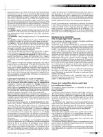
cells, which face the lumen, are covered with microvilli.
Pseudopods formed from the apical membrane extend into
the lumen. The lateral membranes of the follicular cells are
connected by tight junctions, which provide a seal for the
contents of the lumen. The basal membranes of the follicu-
lar cells are close to the rich capillary network that pene-
trates the stroma between the follicles.
The lumen of the follicle contains a thick, gel-like sub-
stance called colloid (see Fig. 33.1). The colloid is a solu-
tion composed primarily of thyroglobulin, a large protein
that is a storage form of the thyroid hormones. The high
viscosity of the colloid is due to the high concentration (10
to 25%) of thyroglobulin.
The thyroid follicle produces and secretes two thyroid
hormones, thyroxine (T
4
) and triiodothyronine (T
3
).
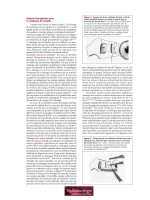
Their molecular structures are shown in Figure 33.2. Thy-
roxine and triiodothyronine are iodinated derivatives of
the amino acid tyrosine. They are formed by the coupling
of the phenyl rings of two iodinated tyrosine molecules in
an ether linkage. The resulting structure is called an
iodothyronine. The mechanism of this process is dis-
cussed in detail later.
Thyroxine contains four iodine atoms on the 3, 5, 3Ј,
and 5Ј positions of the thyronine ring structure, whereas
triiodothyronine has only three iodine atoms, at ring posi-
tions 3, 5, and 3Ј (see Fig. 33.2). Consequently, thyroxine
is usually abbreviated as T
4
and triiodothyronine as T
3
. Be-
cause T
4
and T
3
contain the element iodine, their synthesis
by the thyroid follicle depends on an adequate supply of
iodine in the diet.
Parafollicular Cells Are the Sites of
Calcitonin Synthesis
In addition to the epithelial cells that secrete T
4
and T
3
, the
wall of the thyroid follicle contains small numbers of
parafollicular cells (see Fig. 33.1). The parafollicular cell is
usually embedded in the wall of the follicle, inside the basal
lamina surrounding the follicle. However, its plasma mem-
brane does not form part of the wall of the lumen. Parafol-
licular cells produce and secrete the hormone calcitonin.
Calcitonin and its effects on calcium metabolism are dis-
cussed in Chapter 36.
SYNTHESIS, SECRETION, AND METABOLISM
OF THE THYROID HORMONES
T
4
and T
3
are not directly synthesized by the thyroid folli-
cle in their final form. Instead, they are formed by the
chemical modification of tyrosine residues in the peptide
structure of thyroglobulin as it is secreted by the follicular
cells into the lumen of the follicle. Therefore, the T
4
and T
3
formed by this chemical modification are actually part of
the amino acid sequence of thyroglobulin.
The high concentration of thyroglobulin in the colloid
provides a large reservoir of stored thyroid hormones for
later processing and secretion by the follicle. The synthesis
of T
4
and T
3
is completed when thyroglobulin is retrieved
through pinocytosis of the colloid by the follicular cells.
Thyroglobulin is then hydrolyzed by lysosomal enzymes
carry out their physiological functions. The thyroid hor-
mones exert their regulatory functions by influencing gene
expression, affecting the developmental program and the
amount of cellular constituents needed for the normal rate
of metabolism.
FUNCTIONAL ANATOMY OF THE
THYROID GLAND
The human thyroid gland consists of two lobes attached to
either side of the trachea by connective tissue. The two
lobes are connected by a band of thyroid tissue or isthmus,
which lies just below the cricoid cartilage. A normal thy-
roid gland in a healthy adult weighs about 20 g.
Each lobe of the thyroid receives its arterial blood sup-
ply from a superior and an inferior thyroid artery, which
arise from the external carotid and subclavian artery, re-
spectively. Blood leaves the lobes of the thyroid by a series
of thyroid veins that drain into the external jugular and in-
nominate veins. This circulation provides a rich blood sup-
ply to the thyroid gland, giving it a higher rate of blood
flow per gram than even that of the kidneys.
The thyroid gland receives adrenergic innervation from
the cervical ganglia and cholinergic innervation from the
vagus nerves. This innervation regulates vasomotor func-
tion to increase the delivery of TSH, iodide, and metabolic
substrates to the thyroid gland. The adrenergic system can
also affect thyroid function by direct effects on the cells.
Thyroxine and Triiodothyronine Are Synthesized
and Secreted by the Thyroid Follicle
The lobes of the thyroid gland consist of aggregates of
many spherical follicles, lined by a single layer of epithelial
cells (Fig. 33.1). The apical membranes of the follicular
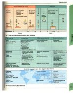
CHAPTER 33 The Thyroid Gland 597
Colloid
Follicular
cell
Capillary
Parafollicular
cell
A cross-sectional view through a portion of
the human thyroid gland.
FIGURE 33.1
598 PART IX ENDOCRINE PHYSIOLOGY
to its constituent amino acids, releasing T
4
and T
3
mole-
cules from their peptide linkage. T
4
and T
3
are then se-
creted into the blood.
Follicular Cells Synthesize
Iodinated Thyroglobulin
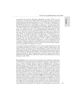
The steps involved in the synthesis of iodinated thyroglob-
ulin are shown in Figure 33.3. This process involves the
synthesis of a thyroglobulin precursor, the uptake of io-
dide, and the formation of iodothyronine residues.
Synthesis and Secretion of the Thyroglobulin Precursor.
The synthesis of the protein precursor for thyroglobulin is
the first step in the formation of T
4
and T
3
. This substance
is a 660-kDa glycoprotein composed of two similar 330-
kDa subunits held together by disulfide bridges. The sub-
units are synthesized by ribosomes on the rough ER and
then undergo dimerization and glycosylation in the
smooth ER. The completed glycoprotein is packaged into
vesicles by the Golgi apparatus. These vesicles migrate to
the apical membrane of the follicular cell and fuse with it.
The thyroglobulin precursor protein is then extruded onto
the apical surface of the cell, where iodination takes place.
Iodide Uptake. The iodide used for iodination of the thy-
roglobulin precursor protein comes from the blood perfus-
ing the thyroid gland. The basal plasma membranes of fol-
licular cells, which are near the capillaries that supply the
follicle, contain iodide transporters. These transporters
move iodide across the basal membrane and into the cy-
tosol of the follicular cell. The iodide transporter is an ac-
tive transport mechanism that requires ATP, is saturable,
and can also transport certain other anions, such as bro-
mide, thiocyanate, and perchlorate. It enables the follicular
cell to concentrate iodide many times over the concentra-
tion of iodide present in the blood; therefore, follicular
cells are efficient extractors of the small amount of iodide
circulating in the blood. Once inside follicular cells, the io-
dide ions diffuse rapidly to the apical membrane, where
they are used for iodination of the thyroglobulin precursor.
Formation of the Iodothyronine Residues. The next step
in the formation of thyroglobulin is the addition of one or
two iodine atoms to certain tyrosine residues in the precur-
sor protein. The precursor of thyroglobulin contains 134
tyrosine residues, but only a small fraction of these become
iodinated. A typical thyroglobulin molecule contains only
20 to 30 atoms of iodine.
The iodination of thyroglobulin is catalyzed by the en-
zyme thyroid peroxidase, which is bound to the apical
membranes of follicular cells. Thyroid peroxidase binds
an iodide ion and a tyrosine residue in the thyroglobulin
precursor, bringing them in close proximity. The enzyme
oxidizes the iodide ion and the tyrosine residue to short-
lived free radicals, using hydrogen peroxide that has been
generated within the mitochondria of follicular cells. The
free radicals then undergo addition. The product formed
is a monoiodotyrosine (MIT) residue, which remains in
peptide linkage in the thyroglobulin structure. A second
iodine atom may be added to a MIT residue by this same
enzymatic process, forming a diiodotyrosine (DIT)
residue (see Fig. 33.3).
Iodinated tyrosine residues that are close together in
the thyroglobulin precursor molecule undergo a coupling
reaction, which forms the iodothyronine structure. Thy-
roid peroxidase, the same enzyme that initially oxidizes
iodine, is believed to catalyze the coupling reaction
through the oxidation of neighboring iodinated tyrosine
residues to short-lived free radicals. These free radicals
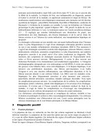
undergo addition, as shown in Figure 33.4. The addition
reaction produces an iodothyronine residue and a dehy-
droalanine residue, both of which remain in peptide link-
age in the thyroglobulin structure. For example, when two
neighboring DIT residues couple by this mechanism, T
4
is
formed (see Fig. 33.4). After being iodinated, the thy-
roglobulin molecule is stored as part of the colloid in the
lumen of the follicle.
Only about 20 to 25% of the DIT and MIT residues in
the thyroglobulin molecule become coupled to form
iodothyronines. For example, a typical thyroglobulin mol-
ecule contains five to six uncoupled residues of DIT and
two to three residues of T
4
. However, T
3
is formed in only
about one of three thyroglobulin molecules. As a result, the
thyroid secretes substantially more T
4
than T
3
.
Thyroid Hormones Are Formed From the
Hydrolysis of Thyroglobulin
When the thyroid gland is stimulated to secrete thyroid
hormones, vigorous pinocytosis occurs at the apical mem-
branes of follicular cells. Pseudopods from the apical mem-
brane reach into the lumen of the follicle, engulfing bits of
the colloid (see Fig. 33.3). Endocytotic vesicles or colloid
droplets formed by this pinocytotic activity migrate to-
ward the basal region of the follicular cell. Lysosomes,
which are mainly located in the basal region of resting fol-
3' 3
HO
HO O
O
5'
5
HH
CC
COOH
HNH
2
Thyroxine (T
4
)
3'
3
5
HH
H
C C COOH
NH
2
Triiodothyronine (T
3
)
The molecular structure of the thyroid hor-
mones. The numbering of the iodine atoms on
the iodothyronine ring structure is shown in red.
FIGURE 33.2
licular cells, migrate toward the apical region of the stimu-
lated cells. The lysosomes fuse with the colloid droplets
and hydrolyze the thyroglobulin to its constituent amino
acids. As a result, T
4
and T
3
and the other iodinated amino
acids are released into the cytosol.
Secretion of Free T
4
and T
3
. T
4
and T
3
formed from the
hydrolysis of thyroglobulin are released from the follicular
cell and enter the nearby capillary circulation, however, the
mechanism of transport of T
4
and T
3
across the basal
plasma membrane has not been defined. The DIT and MIT
generated by the hydrolysis of thyroglobulin are deiodi-
nated in the follicular cell. The released iodide is then re-
utilized by the follicular cell for the iodination of thy-
roglobulin (see Fig. 33.3).
Binding of T
4
and T
3
to Plasma Proteins. Most of the T
4
and T
3
molecules that enter the bloodstream become
bound to plasma proteins. About 70% of the T
4
and 80% of
the T
3
are noncovalently bound to thyroxine-binding
globulin (TBG), a 54-kDa glycoprotein that is synthesized
and secreted by the liver. Each molecule of TBG has a sin-
gle binding site for a thyroid hormone molecule. The re-
maining T
4
and T
3
in the blood are bound to transthyretin
or to albumin. Less than 1% of the T
4
and T
3
in blood is in
the free form, and it is in equilibrium with the large protein-
bound fraction. It is this small amount of free thyroid hor-
mone that interacts with target cells.
The protein-bound form of T
4
and T
3
represents a
large reservoir of preformed hormone that can replenish
the small amount of circulating free hormone as it is
cleared from the blood. This reservoir provides the body
with a buffer against drastic changes in circulating thyroid
hormone levels as a result of sudden changes in the rate of
T
4
and T
3
secretion. The protein-bound T
4
and T
3
mole-
cules are also protected from metabolic inactivation and
excretion in the urine. As a result of these factors, the thy-
roid hormones have long half-lives in the bloodstream.
The half-life of T
4
is about 7 days; the half-life of T
3
is
about 1 day.
Thyroid Hormones Are Metabolized by
Peripheral Tissues
Thyroid hormones are both activated and inactivated by
deiodination reactions in the peripheral tissues. The en-
zymes that catalyze the various deiodination reactions are
regulated, resulting in different thyroid hormone concen-
trations in various tissues in different physiological and
pathophysiological conditions.
Conversion of T
4
to T
3
. As noted earlier, T
4
is the major se-
cretory product of the thyroid gland and is the predominant
thyroid hormone in the blood. However, about 40% of the
T
4
secreted by the thyroid gland is converted to T
3
by enzy-
matic removal of the iodine atom at position 5Ј of the thyro-
nine ring structure (Fig. 33.5). This reaction is catalyzed by a
5Ј-deiodinase (type 1) located in the liver, kidneys, and thy-
roid gland. The T
3
formed by this deiodination and that se-
creted by the thyroid react with thyroid hormone receptors
in target cells; therefore, T
3
is the physiologically active form
of the thyroid hormones. A second 5Ј-deiodinase (type 2) is
CHAPTER 33 The Thyroid Gland 599
Follicular cell
Lumen
Blood
Iodide
transporter
Tight junction
I
-
I
-
I
-
ER
Golgi
Thyroglobulin (Tg)
precursor
Deiodination
DIT
MIT
Secretion
Proteolysis
T
4
T
3
Lysosomes
Colloid
droplet
Pseudopod
Endosomes
Micropinocytosis
Macropinocytosis
MIT
DIT
Iodination and
coupling
T
g
H
2
O
2
T
g
T
4
T
3
T
4
T
3
Thyroid hormone synthesis and secretion. (See text for details.) DIT, diiodotyrosine;
MIT, monoiodotyrosine.
FIGURE 33.3
600 PART IX ENDOCRINE PHYSIOLOGY
present in skeletal muscle, the CNS, the pituitary gland, and
the placenta. Type 2 deiodinase is believed to function pri-
marily to maintain intracellular T
3
in target tissues, but it may
also contribute to the generation of circulating T
3
. All of the
deiodinases contain selenocysteine in the active center. This
rare amino acid has properties that make it ideal to catalyze
deiodination reactions.
Deiodinations That Inactivate T
4
and T
3
. Whereas the
5Ј-deiodination of T
4
to produce T
3
can be viewed as a
metabolic activation process, both T
4
and T
3
undergo en-
zymatic deiodinations, particularly in the liver and kidneys,
which inactivate them. For example, about 40% of the T
4
secreted by the human thyroid gland is deiodinated at the
5 position on the thyronine ring structure by a 5-deiodi-
nase. This produces reverse T
3
(see Fig. 33.5). Since reverse
T
3
has little or no thyroid hormone activity, this deiodina-
tion reaction is a major pathway for the metabolic inactiva-
tion or disposal of T
4
. Triiodothyronine and reverse T
3
also
undergo deiodination to yield 3,3Ј-diiodothyronine. This
inactivate metabolite may be further deiodinated before be-
ing excreted.
Regulation of 5Ј-Deiodination. The 5Ј-deiodination reac-
tion is a regulated process influenced by certain physiolog-
ical and pathological factors. The result is a change in the
relative amounts of T
3
and reverse T
3
produced from T
4
.
For example, a human fetus produces less T
3
from T
4
than
a child or adult because the 5Ј-deiodination reaction is less
active in the fetus. Also, 5Ј-deiodination is inhibited during
fasting, particularly in response to carbohydrate restriction,
but it can be restored to normal when the individual is fed
again. Trauma, as well as most acute and chronic illnesses,
also suppresses the 5Ј-deiodination reaction. Under all of
these circumstances, the amount of T
3
produced from T
4
is
reduced and its blood concentration falls. However, the
amount of reverse T
3
rises in the circulation, mainly be-
cause its conversion to 3,3Ј-diiodothyronine by 5Ј-deiodi-
nation is reduced. A rise in reverse T
3
in the blood may sig-
nal that the 5Ј-deiodination reaction is suppressed.
Note that during fasting or in the disease states mentioned
above, the secretion of T
4
is usually not increased, despite the
decrease of T
3
in the circulation. This response indicates that,
under these circumstances, a T
3
decrease in the blood does
not stimulate the hypothalamic-pituitary-thyroid axis.
Minor Degradative Pathways. T
4
and, to a lesser extent,
T
3
are also metabolized by conjugation with glucuronic
acid in the liver. The conjugated hormones are secreted
into the bile and eliminated in the feces. Many tissues also
metabolize thyroid hormones by modifying the three-car-
bon side chain of the iodothyronine structure. These mod-
ifications include decarboxylation and deamination. The
derivatives formed from T
4
, such as tetraiodoacetic acid
(tetrac), may also undergo deiodinations before being ex-
creted (see Fig. 33.5).
TSH Regulates Thyroid Hormone Synthesis
and Secretion
When the concentrations of free T
4
and T
3
fall in the
blood, the anterior pituitary gland is stimulated to secrete
thyroid-stimulating hormone (TSH), raising the concen-
tration of TSH in the blood. This action results in increased
interactions between TSH and its receptors on thyroid fol-
licular cells.
TSH Receptors and Second Messengers. The receptor for
TSH is a transmembrane glycoprotein thought to be located
on the basal plasma membrane of the follicular cell. These re-
ceptors are coupled by G
s
proteins, mainly to the adenylyl cy-
clase-cAMP-protein kinase A pathway, however, there is also
evidence for effects via phospholipase C (PLC), inositol
trisphosphate, and diacylglycerol (see Chapter 1). The phys-
iological importance of TSH-stimulated phospholipid me-
tabolism in human follicular cells is unclear, since very high
concentrations of TSH are needed to activate PLC.
TSH and Thyroid Hormone Formation and Secretion.
TSH stimulates most of the processes involved in thyroid
hormone synthesis and secretion by follicular cells. The
rise in cAMP produced by TSH is believed to cause many
of these effects. TSH stimulates the uptake of iodide by fol-
licular cells, usually after a short interval during which io-
O
O
CHCH
2
CH
CO
CH
2
NH
NH
CO
2 DIT free
radicals
Radical addition
Quinoid
intermediate
O
O
CO
NH
NH
CO
CHCH
2
CHCH
2
Electronic rearrangement
Dehydroalanine
residue
NH
CO
CHCH
2
CHCH
2
NH
CO
Thyroxine
residue
HO O
+
Theoretical model for the coupling reaction
between two diiodotyrosine (DIT) residues
in iodinated thyroglobulin. This model is based on free radical
formation catalyzed by thyroid peroxidase. (Adapted from Tau-
rog AM. Hormone synthesis: Thyroid iodine metabolism. In:
Braverman LE, Utiger RD, eds. Werner & Ingbar’s The Thyroid: A
Fundamental and Clinical Text. 8th Ed. Philadelphia: Lippincott
Williams & Wilkins, 2000;61–85.)
FIGURE 33.4
dide transport is actually depressed. TSH also stimulates
the iodination of tyrosine residues in the thyroglobulin pre-
cursor and the coupling of iodinated tyrosines to form
iodothyronines. Moreover, it stimulates the pinocytosis of
colloid by the apical membranes, resulting in a great in-
crease in endocytosis of thyroglobulin and its hydrolysis.
The overall result of these effects of TSH is an increased re-
lease of T
4
and T
3
into the blood. In addition to its effects
on thyroid hormone synthesis and secretion, TSH rapidly
increases energy metabolism in the thyroid follicular cell.
TSH and Thyroid Size. Over the long term, TSH pro-
motes protein synthesis in thyroid follicular cells, main-
taining their size and structural integrity. Evidence of this
trophic effect of TSH is seen in a hypophysectomized pa-
tient, whose thyroid gland atrophies, largely as a result of a
reduction in the height of follicular cells. However, the
chronic exposure of an individual to excessive amounts of
TSH causes the thyroid gland to increase in size. This en-
largement is due to an increase in follicular cell height and
number. Such an enlarged thyroid gland is called a goiter.
These trophic and proliferative effects of TSH on the thy-
roid are primarily mediated by cAMP.
Dietary Iodide Is Essential for the
Synthesis of Thyroid Hormones
Because iodine atoms are constituent parts of the T
4
and T
3
molecules, a continual supply of iodide is required for the
synthesis of these hormones. If an individual’s diet is se-
verely deficient in iodide, as in some parts of the world, T
4
and T
3
synthesis is limited by the amount of iodide avail-
able to the thyroid gland. As a result, the concentrations of
T
4
and T
3
in the blood fall, causing a chronic stimulation of
TSH secretion, which, in turn, produces a goiter. Enlarge-
ment of the thyroid gland increases its capacity to accumu-
late iodide from the blood and to synthesize T
4
and T
3
.
However, the degree to which the enlarged gland can pro-
duce thyroid hormones to compensate for their deficiency
in the blood depends on the severity of the deficiency of io-
dide in the diet. To prevent iodide deficiency and the con-
sequent goiter formation in the human population, iodide
is added to the table salt (iodized salt) sold in most devel-
oped countries.
THE MECHANISM OF THYROID
HORMONE ACTION
Most cells of the body are targets for the action of thyroid
hormones. The sensitivity or responsiveness of a particular
cell to thyroid hormones correlates to some degree with
the number of receptors for these hormones. The cells of
the CNS appear to be an exception. As is discussed later,
the thyroid hormones play an important role in CNS de-
velopment during fetal and neonatal life, and developing
nerve cells in the brain are important targets for thyroid
hormones. In the adult, however, brain cells show little re-
sponsiveness to the metabolic regulatory action of thyroid
hormones, although they have numerous receptors for
these hormones. The reason for this discrepancy is unclear.
CHAPTER 33 The Thyroid Gland 601
O
HO
HO
H
H
H
C C COOH
NH
2
Triiodothyronine (T
3
)
Thyroxine (T
4
)
Reverse T
3
O
H
H
H
C C COOH
NH
2
3,3'-Diiodothyronine
H
H
H
C C COOH
NH
2
O
HO
H
H
H
C C COOH
NH
2
HO
O
H
H
C COOH
HO
O
Tetraiodoacetic acid (tetrac)
5'-Deiodinase
5-Deiodinase
The metabolism of thyroxine. Thyroxine is
deiodinated by 5Ј-deiodinase to form T
3
, the
physiologically active thyroid hormone. Some T
4
is also enzy-
matically deiodinated at the 5 position to form the inactive
metabolite, reverse T
3
. T
3
and reverse T
3
undergo additional
FIGURE 33.5
deiodinations (e.g., to 3,3Ј-diiodothyronine) before being ex-
creted. A small amount of T
4
is also decarboxylated and deami-
nated to form the metabolite, tetraiodoacetic acid (tetrac). Tetrac
may then be deiodinated before being excreted.
602 PART IX ENDOCRINE PHYSIOLOGY
Thyroid hormone receptors (TR) are located in the nu-
clei of target cells bound to thyroid hormone response el-
ements (TRE) in the DNA. TRs are protein molecules of
about 50 kDa that are structurally similar to the nuclear re-
ceptors for steroid hormones and vitamin D. Thyroid re-
ceptors bound to the TRE in the absence of T
3
generally act
to repress gene expression.
The free forms of T
3
and T
4
are taken up by target cells
from the blood through a carrier-mediated process that re-
quires ATP. Once inside the cell, T
4
is deiodinated to T
3
,
which enters the nucleus of the cell and binds to its recep-
tor in the chromatin. The TR with bound T
3
forms a com-
plex with other nuclear receptors (called a heterodimer) or
with another TR (homodimer) to activate transcription.
Other transcription factors may also complex with the TR
heterodimer or homodimer. As a result, the production of
mRNA for certain proteins is either increased or decreased,
changing the cell’s capacity to make these proteins
(Fig. 33.6). T
3
can influence differentiation by regulating
the kinds of proteins produced by its target cells and can in-
fluence growth and metabolism by changing the amounts
of structural and enzymatic proteins present in the cells.
The mechanisms by which T
3
alters gene expression con-
tinue to be investigated.
The gene expression response to T
3
is slow to appear.
When T
3
is given to an animal or human, several hours
elapse before its physiological effects can be detected. This
delayed action undoubtedly reflects the time required for
changes in gene expression and consequent changes in the
synthesis of key proteins to occur. When T
4
is adminis-
tered, its course of action is usually slower than that of T
3
because of the additional time required for the body to
convert T
4
to T
3
.
Thyroid hormones also have effects on cells that occur
much faster and do not appear to be mediated by nuclear
TR receptors, including effects on signal transduction path-
ways that alter cellular respiration, cell morphology, vascu-
lar tone, and ion homeostasis. The physiological relevance
of these effects is currently being investigated.
ROLE OF THE THYROID HORMONES
IN DEVELOPMENT, GROWTH, AND
METABOLISM
Thyroid hormones play a critical role in the development
of the central nervous system (CNS). They are also essen-
tial for normal body growth during childhood, and in basal
energy metabolism.
Thyroid Hormones Are Essential for
Development of the Central Nervous System
The human brain undergoes its most active phase of growth
during the last 6 months of fetal life and the first 6 months
of postnatal life. During the second trimester of pregnancy,
the multiplication of neuroblasts in the fetal brain reaches a
peak and then declines. As pregnancy progresses and the
rate of neuroblast division drops, neuroblasts differentiate
into neurons and begin the process of synapse formation
that extends into postnatal life.
Thyroid hormones first appear in the fetal blood during
the second trimester of pregnancy, and levels continue to
rise during the remaining months of fetal life. Thyroid hor-
mone receptors increase about 10-fold in the fetal brain at
about the time the concentrations of T
4
and T
3
begin to rise
in the blood. These events are critical for normal brain de-
velopment because thyroid hormones are essential for tim-
ing the decline in nerve cell division and the initiation of
differentiation and maturation of these cells.
If thyroid hormones are deficient during these prenatal
and postnatal periods of differentiation and maturation of
the brain, mental retardation occurs. The cause is thought
to be inadequate development of the neuronal circuitry of
the CNS. Thyroid hormone therapy must be given to a
thyroid hormone-deficient child during the first few
months of postnatal life to prevent mental retardation.
Starting thyroid hormone therapy after behavioral deficits
have occurred cannot reverse the mental retardation (i.e.,
thyroid hormone must be present when differentiation nor-
mally occurs). Thyroid hormone deficiency during infancy
causes both mental retardation and growth impairment, as
discussed below. Fortunately, this occurs rarely today be-
cause thyroid hormone deficiency is usually detected in
newborn infants and hormone therapy is given at the
proper time.
The exact mechanism by which thyroid hormones influ-
ence differentiation of the CNS is unknown. Animal stud-
ies have demonstrated that thyroid hormones inhibit nerve
cell replication in the brain and stimulate the growth of
nerve cell bodies, the branching of dendrites, and the rate
of myelinization of axons. These effects of thyroid hor-
mones are presumably due to their ability to regulate the
expression of genes involved in nerve cell replication and
differentiation. However, the details, particularly in the hu-
man, are unclear.
T
4
T
3
T
3
RXR
5'-Deiodinase
TR
Coactivator
RNA polymerase II
Transcription
Corepressor
TRE
DNA
The activation of transcription by thyroid
hormone. T
4
is taken up by the cell and deiod-
inated to T
3
, which then binds to the thyroid hormone receptor
(TR). The activated TR heterodimerizes with a second transcrip-
tion factor, 9-cis retinoic acid receptor (RXR), and binds to the
thyroid hormone response element (TRE). The binding of
TR/RXR to the TRE displaces repressors of transcription and re-
cruits additional coactivators. The final result is the activation of
RNA polymerase II and the transcription of the target gene.
FIGURE 33.6
Thyroid Hormones Are Essential for
Normal Body Growth
The thyroid hormones are important factors regulating the
growth of the entire body. For example, an individual who
is deficient in thyroid hormones, who does not receive thy-
roid hormone therapy during childhood, will not grow to a
normal adult height.
Thyroid Hormones and the Gene for GH. A major way
thyroid hormones promote normal body growth is by
stimulating the expression of the gene for growth hor-
mone (GH) in the somatotrophs of the anterior pituitary
gland. In a thyroid hormone-deficient individual, GH
synthesis by the somatotrophs is greatly reduced and con-
sequently GH secretion is impaired; therefore, a thyroid
hormone-deficient individual will also be GH-deficient. If
this condition occurs in a child, it will cause growth retar-
dation, largely a result of the lack of the growth-promot-
ing action of GH (see Chapter 32).
Other Effects of Thyroid Hormones on Growth. The
thyroid hormones have additional effects on growth. In tis-
sues such as skeletal muscle, the heart, and the liver, thyroid
hormones have direct effects on the synthesis of a variety
of structural and enzymatic proteins. For example, they
stimulate the synthesis of structural proteins of mitochon-
dria, as well as the formation of many enzymes involved in
intermediary metabolism and oxidative phosphorylation.
Thyroid hormones also promote the calcification and,
hence, the closure, of the cartilaginous growth plates of the
bones of the skeleton. This action limits further linear body
growth. How the thyroid hormones promote calcification
of the growth plates of bones is not understood.
Thyroid Hormones Regulate the Basal
Energy Economy of the Body
When the body is at rest, about half of the ATP produced
by its cells is used to drive energy-requiring membrane
transport processes. The remainder is used in involuntary
muscular activity, such as respiratory movements, peri-
stalsis, contraction of the heart, and in many metabolic
reactions requiring ATP, such as protein synthesis. The
energy required to do this work is eventually released as
body heat.
Basal Oxygen Consumption and Body Heat Production.
The major site of ATP production is the mitochondria,
where the oxidative phosphorylation of ADP to ATP takes
place. The rate of oxidative phosphorylation depends on
the supply of ADP for electron transport. The ADP supply
is, in turn, a function of the amount of ATP used to do work.
For example, when more work is done per unit time, more
ATP is used and more ADP is generated, increasing the rate
of oxidative phosphorylation. The rate at which oxidative
phosphorylation occurs is reflected in the amount of oxygen
consumed by the body because oxygen is the final electron
acceptor at the end of the electron transport chain.
Activities that occur when the body is not at rest, such
as voluntary movements, use additional ATP for the work
involved; the amounts of oxygen consumed and body heat
produced depend on total body activity.
Thermogenic Action of the Thyroid Hormones. Thyroid
hormones regulate the basal rate at which oxidative phos-
phorylation takes place in cells. As a result, they set the
basal rate of body heat production and of oxygen con-
sumed by the body. This is called the thermogenic action
of thyroid hormones.
Thyroid hormone levels in the blood must be within
normal limits for basal metabolism to proceed at the rate
needed for a balanced energy economy of the body. For ex-
ample, if thyroid hormones are present in excess, oxidative
phosphorylation is accelerated, and body heat production
and oxygen consumption are abnormally high. The con-
verse occurs when the blood concentrations of T
4
and T
3
are lower than normal. The fact that thyroid hormones af-
fect the amount of oxygen consumed by the body has been
used clinically to assess the status of thyroid function. Oxy-
gen consumption is measured under resting conditions and
compared with the rate expected of a similar individual
with normal thyroid function. This measurement is the
basal metabolic rate (BMR) test.
Tissues Affected by the Thermogenic Action of Thyroid
Hormones.
Not all tissues are sensitive to the thermo-
genic action of thyroid hormones. Tissues and organs that
give this response include skeletal muscle, the heart, the
liver, and the kidneys. These are also tissues in which thy-
roid hormone receptors are abundant. The adult brain,
skin, lymphoid organs, and gonads show little thermogenic
response to thyroid hormones. With the exception of the
adult brain, these tissues contain few thyroid hormone re-
ceptors, which may explain their poor response.
Molecular and Cellular Mechanisms. The thermo-
genic action of the thyroid hormones is poorly under-
stood at the molecular level. The thermogenic effect
takes many hours to appear after the administration of
thyroid hormones to a human or animal, probably be-
cause of the time required for changes in the expression
of genes involved. T
3
is known to stimulate the synthesis
of cytochromes, cytochrome oxidase, and Na
ϩ
/K
ϩ
-AT-
Pase in certain cells. This action suggests that T
3
may
regulate the number of respiratory units in these cells, af-
fecting their capacity to carry out oxidative phosphory-
lation. A greater rate of oxidative phosphorylation would
result in greater heat production.
Thyroid hormone also stimulates the synthesis of uncou-
pling protein-1 (UCP-1) in brown adipose tissue. ATP is
synthesized by ATP synthase in the mitochondria when pro-
tons flow down their electrochemical gradient. UCP-1 acts
as a channel in the mitochondrial membrane to dissipate the
ion gradient without making ATP. As the protons move
down their electrochemical gradient uncoupled from ATP syn-
thesis, energy is released as heat. Adult humans have little
brown adipose tissue, so it is not likely that UCP-1 makes a
significant contribution to nutrient oxidation or body heat
production. However, several uncoupling proteins (UCP-2
and UCP-3) have recently been discovered in many tissues,
and their expression is regulated by thyroid hormones.
CHAPTER 33 The Thyroid Gland 603
604 PART IX ENDOCRINE PHYSIOLOGY
These novel uncoupling proteins may be involved in the
thermogenic action of thyroid hormones.
Thyroid Hormones Stimulate Intermediary
Metabolism
In addition to their ability to regulate the rate of basal en-
ergy metabolism, thyroid hormones influence the rate at
which most of the pathways of intermediary metabolism
operate in their target cells. When thyroid hormones are
deficient, pathways of carbohydrate, lipid, and protein me-
tabolism are slowed, and their responsiveness to other reg-
ulatory factors, such as other hormones, is decreased. How-
ever, these same metabolic pathways run at an abnormally
high rate when thyroid hormones are present in excess.
Thyroid hormones, therefore, can be viewed as amplifiers
of cellular metabolic activity. The amplifying effect of thy-
roid hormones on intermediary metabolism is mediated
through the activation of genes encoding enzymes in-
volved in these metabolic pathways.
Thyroid Hormones Regulate Their Own Secretion
An important action of the thyroid hormones is the ability
to regulate their own secretion. As discussed in Chapter 32,
T
3
exerts an inhibitory effect on TSH secretion by thy-
rotrophs in the anterior pituitary gland by decreasing thy-
rotroph sensitivity to thyrotropin-releasing hormone
(TRH). Consequently, when the circulating concentration
of free thyroid hormones is high, thyrotrophs are relatively
insensitive to TRH, and the rate of TSH secretion de-
creases. The resulting fall of TSH levels in the blood re-
duces the rate of thyroid hormone release from the follicu-
lar cells in the thyroid. When the free thyroid hormone
level falls in the blood, however, the negative-feedback ef-
fect of T
3
on thyrotrophs is reduced, and the rate of TSH
secretion increases. The rise in TSH in the blood stimulates
the thyroid gland to secrete thyroid hormones at a greater
rate. This action of T
3
on thyrotrophs is thought to be due
to changes in gene expression in these cells.
The physiological actions of the thyroid hormones de-
scribed above are summarized in Table 33.1.
THYROID HORMONE DEFICIENCY AND
EXCESS IN ADULTS
A deficiency or an excess of thyroid hormones produces
characteristic changes in the body. These changes result
from dysregulation of nervous system function and altered
metabolism.
Thyroid Hormone Deficiency Causes Nervous
and Metabolic Disorders
Thyroid hormone deficiency in humans has a variety of
causes. For example, iodide deficiency may result in a re-
duction in thyroid hormone production. Autoimmune dis-
eases, such as Hashimoto’s disease, impair thyroid hor-
mone synthesis (see Clinical Focus Box 33.1). Other causes
of thyroid hormone deficiency include heritable diseases
that affect certain steps in the biosynthesis of thyroid hor-
mones and hypothalamic or pituitary diseases that interfere
with TRH or TSH secretion. Obviously, radioiodine abla-
tion or surgical removal of the thyroid gland also causes
thyroid hormone deficiency. Hypothyroidism is the dis-
ease state that results from thyroid hormone deficiency.
Thyroid hormone deficiency impairs the functioning
of most tissues in the body. As described earlier, a defi-
ciency of thyroid hormones at birth that is not treated
during the first few months of postnatal life causes irre-
versible mental retardation. Thyroid hormone deficiency
later in life also influences the function of the nervous sys-
tem. For example, all cognitive functions, including
speech and memory, are slowed and body movements
may be clumsy. These changes can usually be reversed
with thyroid hormone therapy.
Metabolism is also reduced in thyroid hormone-defi-
cient individuals. Basal metabolic rate is reduced, resulting
in impaired body heat production. Vasoconstriction occurs
in the skin as a compensatory mechanism to conserve body
heat. Heart rate and cardiac output are reduced. Food in-
take is reduced, and the synthetic and degradative
processes of intermediary metabolism are slowed. In severe
hypothyroidism, a substance consisting of hyaluronic acid
and chondroitin sulfate complexed with protein is de-
posited in the extracellular spaces of the skin, causing wa-
ter to accumulate osmotically. This effect gives a puffy ap-
pearance to the face, hands, and feet called myxedema. All
of the above disorders can be normalized with thyroid hor-
mone therapy.
An Excess of Thyroid Hormone Produces
Nervous and Other Disorders
The most common cause of excessive thyroid hormone
production in humans is Graves’ disease, an autoimmune
TABLE 33.1
The Physiological Actions of
Thyroid Hormones
Development of CNS Inhibit nerve cell replication
Stimulate growth of nerve cell bodies
Stimulate branching of dendrites
Stimulate rate of axon myelinization
Body growth Stimulate expression of gene for
GH in somatotrophs
Stimulate synthesis of many
structural and enzymatic proteins
Promote calcification of growth
plates of bones
Basal energy economy of Regulate basal rates of oxidative
the body phosphorylation, body heat
production, and oxygen
consumption (thermogenic effect)
Intermediary metabolism Stimulate synthetic and degradative
pathways of carbohydrate, lipid,
and protein metabolism
Thyroid-stimulating Inhibit TSH secretion by decreasing
hormone (TSH) secretion sensitivity of thyrotrophs to
thyrotropin-releasing hormone
(TRH)
disorder caused by antibodies directed against the TSH re-
ceptor in the plasma membranes of thyroid follicular cells.
These antibodies bind to the TSH receptor, resulting in an
increase in the activity of adenylyl cyclase. The consequent
rise in cAMP in follicular cells produces effects similar to
those caused by the action of TSH. The thyroid gland en-
larges to form a diffuse toxic goiter, which synthesizes and
secretes thyroid hormones at an accelerated rate, causing
thyroid hormones to be chronically elevated in the blood.
Feedback inhibition of thyroid hormone production by the
thyroid hormones is also lost.
Less common conditions that cause chronic elevations
in circulating thyroid hormones include adenomas of the
thyroid gland that secrete thyroid hormones and excessive
TSH secretion caused by malfunctions of the hypothala-
mic-pituitary-thyroid axis. The disease state that develops
in response to excessive thyroid hormone secretion, called
hyperthyroidism or thyrotoxicosis, is characterized by
many changes in the functioning of the body that are the
opposite of those caused by thyroid hormone deficiency.
Hyperthyroid individuals are nervous and emotionally
irritable, with a compulsion to be constantly moving
around. However, they also experience physical weakness
and fatigue. Basal metabolic rate is increased and, as a re-
sult, body heat production is increased. Vasodilation in
the skin and sweating occur as compensatory mechanisms
to dissipate excessive body heat. Heart rate and cardiac
output are increased. Energy metabolism increases, as
does appetite. However, despite the increase in food in-
take, a net degradation of protein and lipid stores occurs,
resulting in weight loss. All of these changes can be re-
versed by reducing the rate of thyroid hormone secretion
with drugs or by removal of the thyroid gland by radioac-
tive ablation or surgery.
CHAPTER 33 The Thyroid Gland 605
CLINICAL FOCUS BOX 33.1
Autoimmune Thyroid Disease—Postpartum Thyroiditis
Certain diseases affecting the function of the thyroid gland
occur when an individual’s immune system fails to recog-
nize particular thyroid proteins as “self” and reacts to the
proteins as if they were foreign. This usually triggers both
humoral and cellular immune responses. As a result, anti-
bodies to these proteins are generated, which then alter
thyroid function. Two common autoimmune diseases with
opposite effects on thyroid function are Hashimoto’s dis-
ease and Graves’ disease. In Hashimoto’s disease, the thy-
roid gland is infiltrated by lymphocytes, and elevated lev-
els of antibodies against several components of thyroid
tissue (e.g., antithyroid peroxidase and antithyroglobulin
antibodies) are found in the serum. The thyroid gland is de-
stroyed, resulting in hypothyroidism. In Graves’ disease,
stimulatory antibodies to the TSH receptor activate thyroid
hormone synthesis, resulting in hyperthyroidism (see text
for details).
A third, fairly common autoimmune disease is postpar-
tum thyroiditis, which usually occurs within 3 to 12 months
after delivery. The disease is characterized by a transient
thyrotoxicosis (hyperthyroidism) often followed by a pe-
riod of hypothyroidism lasting several months. Many pa-
tients eventually return to the euthyroid state. Often only
the hypothyroid phase of the disease may be observed, oc-
curring in more than 30% of women with antibodies to thy-
roid peroxidase detectable preconception. The disease is
also observed in patients known to have Graves’ disease.
The postpartum occurrence of the disorder is likely due to
increased immune system function following the suppres-
sion of its activity during pregnancy.
It has been estimated that 5 to 10% of women develop
postpartum thyroiditis. Of these women, about 50% have
transient thyrotoxicosis alone, 25% have transient hy-
pothyroidism alone, and the remaining 25% have both
phases of the disease. The prevalence of the disease has
prompted a clinical recommendation suggesting that thy-
roid function (serum T
4
, T
3
, and TSH levels) be surveyed
postpartum at 2, 4, 6, and 12 months in all women with thy-
roid peroxidase antibodies or symptoms suggestive of thy-
roid dysfunction. Patients who have experienced one
episode of postpartum thyroiditis should also be consid-
ered at risk for recurrence after pregnancy.
Treatment for thyrotoxicosis commonly involves in-
hibiting thyroid hormone synthesis and secretion. Thion-
amides are a class of drugs that inhibit the oxidation and
organic binding of thyroid iodide to reduce thyroid hor-
mone production. Some drugs in this class also inhibit the
conversion of T
4
to T
3
in the peripheral tissues. Thyroid
hormone replacement is required to treat hypothyroidism.
DIRECTIONS: Each of the numbered
items or incomplete statements in this
section is followed by answers or by
completions of the statement. Select the
ONE lettered answer or completion that is
the BEST in each case.
1. The effects of TSH on thyroid
follicular cells include
(A) Stimulation of endocytosis of
thyroglobulin stored in the colloid
(B) Release of a large pool of T
4
and
T
3
stored in secretory vesicles in the
cell
(C) Stimulation of the uptake of iodide
from the thyroglobulin stored in the
colloid
(D) Increase in perfusion by the blood
(E) Stimulation of the binding of T
4
and T
3
to thyroxine-binding globulin
(F) Increased cAMP hydrolysis
2. A child is born with a rare disorder in
which the thyroid gland does not
respond to TSH. What would be the
predicted effects on mental ability, body
growth rate, and thyroid gland size
when the child reaches 6 years of age?
REVIEW QUESTIONS
(continued)
606 PART IX ENDOCRINE PHYSIOLOGY
(A) Mental ability would be impaired,
body growth rate would be slowed,
and thyroid gland size would be larger
than normal
(B) Mental ability would be unaffected,
body growth rate would be slowed,
and thyroid gland size would be
smaller than normal
(C) Mental ability would be impaired,
body growth rate would be slowed,
and thyroid gland size would be
smaller than normal
(D) Mental ability would be
unaffected, body growth rate would be
unaffected, and thyroid gland size
would be smaller than normal
(E) Mental ability would be impaired,
body growth rate would be slowed,
and thyroid gland size would be
normal
(F) Mental ability would be unaffected,
body growth rate would be unaffected,
and thyroid gland size would be
unaffected
3. If the 6-year-old child described in the
previous question is now treated with
thyroid hormones, how would mental
ability, body growth rate, and thyroid
gland size be affected?
(A) Mental ability would remain
impaired, body growth rate would be
improved, and thyroid gland size
would be smaller than normal
(B) Mental ability would be improved,
body growth rate would be improved,
and thyroid gland size would be
normal
(C) Mental ability would remain
impaired, body growth rate would be
improved, and thyroid gland size
would be normal
(D) Mental ability would remain
impaired, body growth rate would be
improved, and thyroid gland size
would be larger than normal
(E) Mental ability would be improved,
body growth rate would remain
slowed, and thyroid gland size would
be normal
(F) Mental ability would be improved,
body growth rate would remain
slowed, and thyroid gland size would
larger than normal
4. Uncoupling proteins
(A) Utilize the proton gradient across
the mitochondrial membrane to
facilitate ATP synthesis
(B) Are decreased by thyroid hormones
(C) Dissipate the proton gradient
across the mitochondrial membrane to
generate heat
(D) Are present exclusively in brown fat
(E) Uncouple fatty acid oxidation from
glucose oxidation in mitochondria
(F) Are essential for maintaining body
temperature in mammals
5. Triiodothyronine (T
3
)
(A) Is produced in greater amounts by
the thyroid gland than T
4
(B) Is bound by the thyroid receptor
present in the cytosol of target cells
(C) Is formed from T
4
through the
action of a 5-deiodinase
(D) Has a half-life of a few minutes in
the bloodstream
(E) Is released from thyroglobulin
through the action of thyroid
peroxidase
(F) Can be produced by the
deiodination of T
4
in pituitary
thyrotrophs
6. A 40-year-old man complains of
chronic fatigue, aching muscles, and
occasional numbness in his fingers.
Physical examination reveals a modest
weight gain but no goiter is detected.
Laboratory findings include TSH Ͼ 10
U/L (normal range, 0.5 to 5 U/L);
free T
4
, low to low-normal. These
findings are most consistent with a
diagnosis of
(A) Hypothyroidism secondary to a
hypothalamic-pituitary defect
(B) Hyperthyroidism secondary to a
hypothalamic-pituitary defect
(C) Hyperthyroidism as a result of
iodine excess
(D) Hypothyroidism as a result of
autoimmune thyroid disease
(E) Hypothyroidism as a result of
iodine deficiency
(F) Hyperthyroidism as a result of
autoimmune thyroid disease
7. The reaction catalyzed by thyroid
peroxidase
(A) Produces hydrogen peroxide as an
end-product
(B) Couples two iodotyrosine residues
to form an iodothyronine residue
(C) Occurs on the basal membrane of
the follicular cell
(D) Catalyzes the release of thyroid
hormones into the circulation
(E) Couples MIT and DIT to
thyroglobulin
(F) Couples dehydroalanine with a
thyroxine residue
8. A 25-year-old woman complains of
weight loss, heat intolerance, excessive
sweating, and weakness. TSH and
thyroid hormones are elevated, goiter
is present, but no antithyroid
antibodies are detected. Which of the
following diagnoses is consistent with
these symptoms?
(A) Graves’ disease
(B) Resistance to thyroid hormone
action
(C) Plummer’s disease (thyroid gland
adenoma)
(D) A 5Ј-deiodinase deficiency
(E) Acute Hashimoto’s disease
(F) TSH-secreting pituitary tumor
SUGGESTED READING
Apriletti JW, Ribeiro RC, Wagner RL, et
al. Molecular and structural biology of
thyroid hormone receptors. Clin Exp
Pharmacol Physiol Suppl
1998;25:S2–S11.
Braverman LE, Utiger RD. Werner and
Ingbar’s The Thyroid: A Fundamental
and Clinical Text. 8th Ed.
Philadelphia: Lippincott Williams &
Wilkins, 2000.
Goglia F, Moreno M, Lanni A. Action of
thyroid hormones at the cellular level:
The mitochondrial target. FEBS Lett
1999;452:115–120.
Larsen PR, Davies TF, Hay ID. The thy-
roid gland. In: Wilson JD, Foster DW,
Kronenberg HM, Larsen PR, eds:
Williams Textbook of Endocrinology.
9th Ed. Philadelphia: WB Saunders,
1998.
Meier CA. Thyroid hormone and develop-
ment: Brain and peripheral tissue In:
Hauser P, Rovet J, eds. Thyroid Dis-
eases of Infancy and Childhood. Wash-
ington, DC: American Psychiatric
Press, 1999.
Motomura K, Brent GA. Mechanisms of
thyroid hormone action. Endocrinol
Metab Clin North Am 1998;27:1–23.
Munoz A, Bernal J. Biological activities of
thyroid hormone receptors. Eur J En-
docrinol 1997;137:433–445.
Reitman ML, He Y, Gong D-W. Thyroid
hormone and other regulators of un-
coupling proteins. Int J Obes Relat
Metab Disord 1999;23(Suppl
6):S56–S59.
The Adrenal Gland
Robert V. Considine, Ph.D.
34
CHAPTER
34
T
o remain alive, the organs and tissues of the human
body must have a finely regulated extracellular envi-
ronment. This environment must contain the correct con-
centrations of ions to maintain body fluid volume and to
enable excitable cells to function. The extracellular envi-
ronment must also have an adequate supply of metabolic
substrates for cells to generate ATP. Salts, water, and other
organic substances are continually lost from the body as a
result of perspiration, respiration, and excretion. Metabolic
substrates are constantly used by cells. Under normal con-
ditions, these critical constituents of the body’s extracellu-
lar environment are replenished by the intake of food and
liquids. However, a person can survive for weeks on little
else but water because the body has a remarkable capacity
for adjusting the functions of its organs and tissues to pre-
serve body fluid volume and composition.
The adrenal glands play a key role in making these ad-
justments. This is readily apparent from the fact that an
adrenalectomized animal, unlike its normal counterpart,
cannot survive prolonged fasting. Its blood glucose supply
diminishes, ATP generation by the cells becomes inade-
quate to support life, and the animal eventually dies. Even
■ FUNCTIONAL ANATOMY OF THE ADRENAL GLAND
■ HORMONES OF THE ADRENAL CORTEX
■ PRODUCTS OF THE ADRENAL MEDULLA
CHAPTER OUTLINE
1. The adrenal gland is comprised of an outer cortex sur-
rounding an inner medulla. The cortex contains three his-
tologically distinct zones (from outside to inside): the zona
glomerulosa, zona fasciculata, and zona reticularis.
2. Hormones secreted by the adrenal cortex include glucocor-
ticoids, aldosterone, and adrenal androgens.
3. The glucocorticoids cortisol and corticosterone are synthe-
sized in the zona fasciculata and zona reticularis of the ad-
renal cortex.
4. The mineralocorticoid aldosterone is synthesized in the
zona glomerulosa of the adrenal cortex.
5. Cholesterol, used in the synthesis of the adrenal cortical
hormones, comes from cholesterol esters stored in the
cells. Stored cholesterol is derived mainly from low-den-
sity lipoprotein particles circulating in the blood, but it can
also be synthesized de novo from acetate within the adre-
nal gland.
6. The conversion of cholesterol to pregnenolone in mito-
chondria is the common first step in the synthesis of all ad-
renal steroids and occurs in all three zones of the cortex.
7. The liver is the main site for the metabolism of adrenal
steroids, which are conjugated to glucuronic acid and ex-
creted in the urine.
8. ACTH increases glucocorticoid and androgen synthesis in
adrenal cortical cells in the zona fasciculata and zona retic-
ularis by increasing intracellular cAMP. ACTH also has a
trophic effect on these cells.
9. Angiotensin II and angiotensin III stimulate aldosterone
synthesis in the cells of the zona glomerulosa by increas-
ing cytosolic calcium and activating protein kinase C.
10. Glucocorticoids bind to glucocorticoid receptors in the cy-
tosol of target cells. The glucocorticoid-bound receptor
translocates to the nucleus and then binds to glucocorti-
coid response elements in the DNA to increase or decrease
the transcription of specific genes.
11. Glucocorticoids are essential to the adaptation of the body
to fasting, injury, and stress.
12. The catecholamines epinephrine and norepinephrine are
synthesized and secreted by the chromaffin cells of the ad-
renal medulla.
13. Catecholamines interact with four adrenergic receptors (␣
1
,
␣
2
, 
1
, and 
2
) that mediate the cellular effects of the hor-
mones.
14. Stimuli such as injury, anger, pain, cold, strenuous exer-
cise, and hypoglycemia generate impulses in the choliner-
gic preganglionic fibers innervating the chromaffin cells,
resulting in the secretion of catecholamines.
15. To counteract hypoglycemia, catecholamines stimulate
glucose production in the liver, lactate release from mus-
cle, and lipolysis in adipose tissue.
KEY CONCEPTS
607
608 PART IX ENDOCRINE PHYSIOLOGY
when fed a normal diet, an adrenalectomized animal typi-
cally loses body sodium and water over time, and eventu-
ally dies of circulatory collapse. Its death is caused by a lack
of certain steroid hormones that are produced and secreted
by the cortex of the adrenal gland.
The glucocorticoid hormones, cortisol and corticos-
terone, play essential roles in adjusting the metabolism of
carbohydrates, lipids, and proteins in liver, muscle, and adi-
pose tissues during fasting, which assures an adequate sup-
ply of glucose and fatty acids for energy metabolism de-
spite the absence of food. The mineralocorticoid hormone
aldosterone, another steroid hormone produced by the ad-
renal cortex, stimulates the kidneys to conserve sodium
and, hence, body fluid volume.
The glucocorticoids also enable the body to cope with
physical and emotional traumas or stresses. The physiological
importance of this action of the glucocorticoids is empha-
sized by the fact that adrenalectomized animals lose their
ability to cope with physical or emotional stresses. Even when
given an appropriate diet to prevent blood glucose and body
sodium depletion, an adrenalectomized animal may die when
exposed to traumas that are not fatal to normal animals.
Hormones produced by the other endocrine component
of the adrenal gland, the medulla, are also involved in com-
pensatory reactions of the body to trauma or life-threaten-
ing situations. These hormones are the catecholamines, ep-
inephrine and norepinephrine, which have widespread
effects on the cardiovascular system and muscular system
and on carbohydrate and lipid metabolism in liver, muscle,
and adipose tissues.
FUNCTIONAL ANATOMY OF THE
ADRENAL GLAND
The human adrenal glands are paired, pyramid-shaped or-
gans located on the upper poles of each kidney. The ad-
renal gland is actually a composite of two separate en-
docrine organs, one inside the other, each secreting
separate hormones and each regulated by different mech-
anisms. The outer portion or cortex of the adrenal gland
completely surrounds the inner portion or medulla and
makes up most of the gland. During embryonic develop-
ment, the cortex forms from mesoderm; the medulla arises
from neural ectoderm.
The Adrenal Cortex Consists of
Three Distinct Zones
In the adult human, the adrenal cortex consists of three his-
tologically distinct zones or layers (Fig. 34.1). The outer
zone, which lies immediately under the capsule of the
gland, is called the zona glomerulosa and consists of small
clumps of cells that produce the mineralocorticoid aldos-
terone. The zona fasciculata is the middle and thickest
layer of the cortex and consists of cords of cells oriented ra-
dial to the center of the gland. The inner layer is comprised
of interlaced strands of cells called the zona reticularis.
The zona fasciculata and zona reticularis both produce the
physiologically important glucocorticoids, cortisol and
corticosterone. These layers of the cortex also produce the
androgen dehydroepiandrosterone, which is related chem-
ically to the male sex hormone testosterone. The molecu-
lar structures of these hormones are shown in Figure 34.2.
Like all endocrine organs, the adrenal cortex is highly
vascularized. Many small arteries branch from the aorta and
renal arteries and enter the cortex. These vessels give rise to
capillaries that course radially through the cortex and ter-
minate in venous sinuses in the zona reticularis and adrenal
medulla; therefore, the hormones produced by the cells of
the cortex have ready access to the circulation.
The cells of the adrenal cortex contain abundant lipid
droplets. This stored lipid is functionally significant be-
cause cholesterol esters present in the droplets are an im-
portant source of the cholesterol used as a precursor for the
synthesis of steroid hormones.
The Adrenal Medulla Is a Modified
Sympathetic Ganglion
The adrenal medulla can be considered a modified sympa-
thetic ganglion. The medulla consists of clumps and strands
of chromaffin cells interspersed with venous sinuses. Chro-
maffin cells, like the modified postganglionic neurons that
receive sympathetic preganglionic cholinergic innervation
from the splanchnic nerves, produce catecholamine hor-
mones, principally epinephrine and norepinephrine. Epi-
nephrine and NE are stored in granules in chromaffin cells
and discharged into venous sinuses of the adrenal medulla
when the adrenal branches of splanchnic nerves are stimu-
lated (see Fig. 6.5).
HORMONES OF THE ADRENAL CORTEX
Only small amounts of the glucocorticoids, aldosterone,
and adrenal androgens are found in adrenal cortical cells at
Aldosterone
Cortisol
Androgens
Catecholamines
Medulla: 10–20%
Cortex: 80–90%
Zona reticularis
Zona fasciculata
Zona glomerulosa
The three zones of the adrenal cortex and
corresponding hormone secretion.
FIGURE 34.1
a given time because those cells produce and secrete these
hormones on demand, rather than storing them. Table 34.1
shows the daily production of adrenal cortex hormones in
a healthy adult under resting (unstimulated) conditions. Be-
cause the molecular weights of these substances do not vary
greatly, comparing the amounts secreted indicates the rel-
ative number of molecules of each hormone produced
daily. Humans secrete about 10 times more cortisol than
corticosterone during an average day, and corticosterone
has only one fifth of the glucocorticoid activity of cortisol
(Table 34.2). Cortisol is considered the physiologically im-
portant glucocorticoid in humans. Compared with the glu-
cocorticoids, a much smaller amount of aldosterone is se-
creted each day.
Because of similarities in their structures, the glucocorti-
coids and aldosterone have overlapping actions. For exam-
ple, cortisol and corticosterone have some mineralocorti-
coid activity; conversely, aldosterone has some glucocorti-
coid activity. However, given the amounts of these hor-
mones secreted under normal circumstances and their
relative activities, glucocorticoids are not physiologically
important mineralocorticoids, nor does aldosterone func-
tion physiologically as a glucocorticoid.
As discussed in detail later, the amounts of glucocorti-
coids and aldosterone secreted by an individual can vary
greatly from those given in Table 34.1. The amount se-
creted depends on the person’s physiological state. For ex-
ample, in an individual subjected to severe physical or emo-
tional trauma, the rate of cortisol secretion may be 10 times
greater than the resting rate shown in Table 34.1. Certain
diseases of the adrenal cortex that involve steroid hormone
biosynthesis can significantly increase or decrease the
amount of hormones produced.
The adrenal cortex also produces and secretes substan-
tial amounts of androgenic steroids. Dehydroepiandros-
terone (DHEA) in both the free form and the sulfated form
(DHEAS) is the main androgen secreted by the adrenal
cortex of both men and women (see Table 34.1). Lesser
amounts of other androgens are also produced. The adrenal
cortex is the main source of androgens in the blood in hu-
man females. In the human male, however, androgens pro-
duced by the testes and adrenal cortex contribute to the
male sex hormones circulating in the blood. Adrenal an-
drogens normally have little physiological effect other than
a role in development before the start of puberty in both
girls and boys. This is because the male sex hormone activ-
ity of the adrenal androgens is weak. Exceptions occur in
individuals who produce inappropriately large amounts of
certain adrenal androgens as a result of diseases affecting
the pathways of steroid biosynthesis in the adrenal cortex.
Adrenal Steroid Hormones Are Synthesized
From Cholesterol
Cholesterol is the starting material for the synthesis of
steroid hormones. A cholesterol molecule consists of four
interconnected rings of carbon atoms and a side chain of
eight carbon atoms extending from one ring (Fig. 34.3). In
all, there are 27 carbon atoms in cholesterol, numbered as
shown in the figure.
Sources of Cholesterol. The immediate source of choles-
terol used in the biosynthesis of steroid hormones is the
abundant lipid droplets in adrenal cortical cells. The cho-
CHAPTER 34 The Adrenal Gland 609
Aldosterone
Cortisol Corticosterone
Dehydroepiandrosterone
Zona glomerulosa
Zona fasciculata and zona reticularis
Molecular structures of the important hor-
mones secreted by the adrenal cortex.
FIGURE 34.2
TABLE 34.1
The Average Daily Production of Hor-
mones by the Adrenal Cortex
Hormone Amount Produced (mg/day)
Cortisol 20
Corticosterone 2
Aldosterone 0.1
Dehydroepiandrosterone 30
TABLE 34.2
Comparison of Shared Activities of
Adrenal Cortical Hormones
Glucocorticoid Mineralocorticoid
Hormone Activity
a
Activity
b
Cortisol 100 0.25
Corticosterone 20 0.5
Aldosterone 10 100
a
Percentage activity, with cortisol being 100%
b
Percentage activity, with aldosterone being 100%
610 PART IX ENDOCRINE PHYSIOLOGY
lesterol present in these lipid droplets is mainly in the form
of cholesterol esters, single molecules of cholesterol ester-
ified to single fatty acid molecules. The free cholesterol
used in steroid biosynthesis is generated from these choles-
terol esters by the action of cholesterol esterase (choles-
terol ester hydrolase [CEH]), which hydrolyzes the ester
bond. The free cholesterol generated by that cleavage en-
ters mitochondria located in close proximity to the lipid
droplet. The process of remodeling the cholesterol mole-
cule into steroid hormones is then initiated.
The cholesterol that has been removed from the lipid
droplets for steroid hormone biosynthesis is replenished in
two ways (Fig. 34.4). Most of the cholesterol converted to
steroid hormones by the human adrenal gland comes from
cholesterol esters contained in low-density lipoprotein
(LDL) particles circulating in the blood. The LDL particles
consist of a core of cholesterol esters surrounded by a coat
of cholesterol and phospholipids. A 400-kDa protein mol-
ecule called apoprotein B
100
is also present on the surface
of the LDL particle; it is recognized by LDL receptors lo-
calized to coated pits on the plasma membrane of adrenal
cortical cells (see Fig. 34.4). The apoprotein binds to the
LDL receptor, and both the LDL particle and the receptor
are taken up by the cell through endocytosis. The endo-
cytic vesicle containing the LDL particles fuses with a lyso-
some and the particle is degraded. The cholesterol esters in
the core of the particle are hydrolyzed to free cholesterol
and fatty acid by the action of CEH.
Any cholesterol not immediately used by the cell is con-
verted again to cholesterol esters by the action of the en-
zyme acyl-CoA:cholesterol acyltransferase (ACAT). The
esters are then stored in the lipid droplets of the cell to be
used later.
When steroid biosynthesis is proceeding at a high rate,
cholesterol delivered to the adrenal cell may be diverted di-
rectly to mitochondria for steroid production rather than
reesterified and stored. Accumulating evidence suggests
that high-density lipoprotein (HDL) cholesterol may also
be used as a substrate for adrenal steroidogenesis.
In humans, cholesterol that has been synthesized de novo
from acetate by the adrenal glands is a significant but minor
source of cholesterol for steroid hormone formation. The
rate-limiting step in this process is catalyzed by the enzyme
3-hydroxy-3-methylglutaryl CoA reductase (HMG CoA
reductase). The newly synthesized cholesterol is then in-
corporated into cellular structures, such as membranes, or
O
O
HO
OH
15
22
27
26
25
24
23
20
18
12
21
11
19
4
1
2
3
5
6
7
10
9
8
D
BA
C
14
13
17
16
OH
The formation of pregnenolone from cho-
lesterol by the action of cholesterol side-
chain cleavage enzyme (CYP11A1). Note the chemical struc-
ture of cholesterol, how the four rings are lettered (A to D), and
how the carbons are numbered. The hydrogen atoms on the car-
bons composing the rings are omitted from the figure.
FIGURE 34.3
ACAT CEH
CEH
Lipid
droplet
HMG CoA
reductase
Acetate
Cholesterol ester
Cholesterol ester
Endocytosis
Plasma membrane
Blood
Fatty acid ϩ cholesterol Steroids
Apoprotein
Coated pit
LDL
Adrenal cortical cell
Sources of cholesterol for steroid biosyn-
thesis by the adrenal cortex. Most choles-
terol comes from low-density lipoprotein (LDL) particles in the
blood, which bind to receptors in the plasma membrane and are
taken up by endocytosis. The cholesterol in the LDL particle is
used directly for steroidogenesis or stored in lipid droplets for
later use. Some cholesterol is synthesized directly from acetate.
CEH, cholesterol ester hydrolase; ACAT, acyl-CoA:cholesterol
acyltransferase; HMG, 3-hydroxy-3-methylglutaryl.
FIGURE 34.4
converted to cholesterol esters through the action of
ACAT and stored in lipid droplets (see Fig. 34.4).
Pathways for the Synthesis of Steroid Hormones.
Adrenal steroid hormones are synthesized by four CYP
enzymes. The CYPs are a large family of oxidative en-
zymes with a 450 nm absorbance maximum when com-
plexed with carbon monoxide; hence, these molecules
were once referred to as cytochrome P450 enzymes. The
adrenal CYPs are more commonly known by their trivial
names, which denote their function in steroid biosynthe-
sis (see Table 34.3).
The conversion of cholesterol into steroid hormones be-
gins with the formation of free cholesterol from the cho-
lesterol esters stored in intracellular lipid droplets. Free
cholesterol molecules enter the mitochondria, which are
located close to the lipid droplets, by a mechanism that is
not well understood. Evidence indicates that free choles-
terol associates with a small protein called sterol carrier
protein 2, which facilitates its entry into the mitochon-
drion in some manner. Several other proteins, as well as
cAMP, appear to be involved in cholesterol transport into
mitochondria, but the process is still unclear.
Once inside a mitochondrion, single cholesterol mole-
cules bind to the cholesterol side-chain cleavage enzyme
(CYP11A1), embedded in the inner mitochondrial mem-
brane. This enzyme catalyzes the first and rate-limiting re-
action in steroidogenesis, which remodels the cholesterol
molecule into a 21-carbon steroid intermediate called preg-
nenolone. The reaction occurs in three steps, as shown in
Figure 34.3. The first two steps consist of the hydroxylation
of carbons 20 and 22 by cholesterol side-chain cleavage en-
zyme. Then the enzyme cleaves the side chain of choles-
terol between carbons 20 and 22, yielding pregnenolone
and isocaproic acid.
Once formed, pregnenolone molecules dissociate from
cholesterol side-chain cleavage enzyme, leave the mito-
chondrion, and enter the smooth ER nearby. This mecha-
nism is not understood. At this point, the further remodel-
ing of pregnenolone into steroid hormones can vary,
depending on whether the process occurs in the zona fas-
ciculata and zona reticularis or the zona glomerulosa. We
first consider what occurs in the zona fasciculata and zona
reticularis. These biosynthetic events are summarized in
Figure 34.5.
In cells of the zona fasciculata and zona reticularis, most
of the pregnenolone is converted to cortisol and the main
adrenal androgen dehydroepiandrosterone (DHEA). Preg-
nenolone molecules bind to the enzyme 17␣-hydroxylase
(CYP17), embedded in the ER membrane, which hydroxy-
lates pregnenolone at carbon 17. The product formed by
this reaction is 17␣-hydroxypregnenolone (see Fig. 34.5).
The 17␣-hydroxylase has an additional enzymatic ac-
tion that becomes important at this step in the steroido-
genic process. Once the enzyme has hydroxylated carbon
17 of pregnenolone to form 17␣-hydroxypregnenolone,
it has the ability to lyse or cleave the carbon 20–21 side
chain from the steroid structure. Some molecules of 17␣-
hydroxypregnenolone undergo this reaction and are con-
verted to the 19-carbon steroid DHEA. This action of
17␣-hydroxylase is essential for the formation of andro-
gens (19 carbon steroids) and estrogens (18 carbon
steroids), which lack the carbon 20–21 side chain. There-
fore, this lyase activity of 17␣-hydroxylase is important in
the gonads, where androgens and estrogens are primarily
made. 17␣-hydroxylase does not exert significant lyase
activity in children before age 7 or 8. As a result, young
boys and girls do not secrete significant amounts of adre-
nal androgens. The appearance of significant adrenal an-
drogen secretion in children of both sexes is termed
adrenarche. It is not related to the onset of puberty, since
it normally occurs before the activation of the hypothala-
mic-pituitary-gonad axis, which initiates puberty. The ad-
renal androgens produced as a result of adrenarche are a
stimulus for the growth of pubic and axillary hair.
Those molecules of 17␣-hydroxypregnenolone that dis-
sociate as such from 17␣-hydroxylase bind next to another
ER enzyme, 3-hydroxysteroid dehydrogenase (3-HSD
II). This enzyme acts on 17␣-hydroxypregnenolone to iso-
merize the double bond in ring B to ring A and to dehydro-
genate the 3-hydroxy group, forming a 3-keto group. The
product formed is 17␣-hydroxyprogesterone (see Fig. 34.5).
This intermediate then binds to another enzyme, 21-hy-
droxylase (CYP21A2), which hydroxylates it at carbon 21.
The mechanism of this hydroxylation is similar to that per-
formed by the 17␣-hydroxylase. The product formed is 11-
deoxycortisol, which is the immediate precursor for cortisol.
To be converted to cortisol, 11-deoxycortisol molecules
must be transferred back into the mitochondrion to be
acted on by 11-hydroxylase (CYP11B1) embedded in the
inner mitochondrial membrane. This enzyme hydroxylates
11-deoxycortisol on carbon 11, converting it into cortisol.
The 11-hydroxyl group is the molecular feature that con-
fers glucocorticoid activity on the steroid. Cortisol is then
secreted into the bloodstream.
Some of the pregnenolone molecules generated in cells
of the zona fasciculata and zona reticularis first bind to 3-
hydroxysteroid dehydrogenase when they enter the endo-
plasmic reticulum. As a result, they are converted to prog-
esterone. Some of these progesterone molecules are
hydroxylated by 21-hydroxylase to form the mineralocor-
ticoid 11-deoxycorticosterone (DOC) (see Fig. 34.5). The
11-deoxycorticosterone formed may be either secreted or
transferred back into the mitochondrion. There it is acted
on by 11-hydroxylase to form corticosterone, which is
then secreted into the circulation.
CHAPTER 34 The Adrenal Gland 611
TABLE 34.3
Nomenclature for the Steroidogenic En-
zymes
Previous Current
Common Name Form Form Gene
Cholesterol side-chain P450
SCC
CYP11A1 CYP11A1
cleavage enzyme
3-Hydroxysteroid 3-HSD 3-HSD II HSD3B2
dehydrogenase
17␣-Hydroxylase P450
C17
CYP17 CYP17
21-Hydroxylase P450
C21
CYP21A2 CYP21A2
11-Hydroxylase P450
C11
CYP11B1 CYP11B1
Aldosterone synthase P450
C11AS
CYP11B2 CYP11B2
612 PART IX ENDOCRINE PHYSIOLOGY
Progesterone may also undergo 17␣-hydroxylation in
the zona fasciculata and zona reticularis. It is then con-
verted to either cortisol or the adrenal androgen an-
drostenedione.
The 17␣-hydroxylase is not present in cells of the zona
glomerulosa; therefore, pregnenolone does not undergo
17␣-hydroxylation in these cells, and cortisol and adrenal
androgens are not formed by these cells. Instead, the enzy-
matic pathway leading to the formation of aldosterone is
followed (see Fig. 34.5). Pregnenolone is converted by en-
zymes in the endoplasmic reticulum to progesterone and
11-deoxycorticosterone. The latter compound then moves
CO
CH
3
Pregnenolone
Cholesterol
Progesterone
11-Deoxycorticosterone
Corticosterone
OO
17-OH Progesterone
11-Deoxycortisol
Androstenedione
OH
17-OH Pregnenolone
Dehydroepiandrosterone
CH
2
OH
Cortisol
HO
HO
O
Aldosterone
O
O
CH
2
OH
CH
HO
17α-Hydroxylase
(CYP17)
Cholesterol
side-chain
cleavage
(CYPIIAI)
17α-Hydroxylase
(CYP17)
17α-Hydroxylase
(CYP17)
17α-Hydroxylase
(CYP17)
3β-Hydroxysteroid
dehydrogenase
(3β-HSD II)
3β-Hydroxysteroid
dehydrogenase
(3β-HSD II)
21-Hydroxylase
(CYP21A2)
11β-Hydroxylase
(CYPIIBI)
Aldosterone synthase
(CYPIIB2)
The synthesis of steroids in the adrenal cortex.
FIGURE 34.5
into the mitochondrion, where it is converted to aldos-
terone. This conversion involves three steps: the hydroxy-
lation of carbon 11 to form corticosterone, the hydroxyla-
tion of carbon 18 to form 18-hydroxycorticosterone, and
the oxidation of the 18-hydroxymethyl group to form al-
dosterone. In humans, these three reactions are catalyzed
by a single enzyme, aldosterone synthase (CYP11B2), an
isozyme of 11-hydroxylase (CYP11B1), expressed only in
glomerulosa cells. The 11-hydroxylase enzyme, which is
expressed in the zona fasciculata and zona reticularis, al-
though closely related to aldosterone synthase, cannot cat-
alyze all three reactions involved in the conversion of 11-
deoxycorticosterone to aldosterone; therefore, aldosterone
is not synthesized in the zona fasciculata and zona reticu-
laris of the adrenal cortex.
Genetic Defects in Adrenal Steroidogenesis. Inherited
genetic defects can cause relative or absolute deficiencies in
the enzymes involved in the steroid hormone biosynthetic
pathways. The immediate consequences of these defects
are changes in the types and amounts of steroid hormones
secreted by the adrenal cortex. The end result is disease.
Most of the genetic defects affecting the steroidogenic
enzymes impair the formation of cortisol. As discussed in
Chapter 32, a drop in cortisol concentration in the blood
stimulates the secretion of adrenocorticotropic hormone
(ACTH) by the anterior pituitary. The consequent rise in
ACTH in the blood exerts a trophic (growth-promoting)
effect on the adrenal cortex, resulting in adrenal hypertro-
phy. Because of this mechanism, individuals with genetic
defects affecting adrenal steroidogenesis usually have hy-
pertrophied adrenal glands. These diseases are collectively
called congenital adrenal hyperplasia.
In humans, inherited genetic defects occur that affect
cholesterol side-chain cleavage enzyme, 17␣-hydroxylase,
3-hydroxysteroid dehydrogenase, 21-hydroxylase, 11-
hydroxylase, and aldosterone synthase. The most common
defect involves mutations in the gene for 21-hydroxylase
and occurs in 1 of 7,000 people. The gene for 21-hydroxy-
lase may be deleted entirely, or mutant genes may code for
forms of 21-hydroxylase with impaired enzyme activity.
The consequent reduction in the amount of active 21-hy-
droxylase in the adrenal cortex interferes with the forma-
tion of cortisol, corticosterone, and aldosterone, all of
which are hydroxylated at carbon 21. Because of the re-
duction of cortisol (and corticosterone) secretion in these
individuals, ACTH secretion is stimulated. This, in turn,
causes hypertrophy of the adrenal glands and stimulates the
glands to produce steroids.
Because 21-hydroxylation is impaired, the ACTH stim-
ulus causes pregnenolone to be converted to adrenal an-
drogens in inappropriately high amounts. Thus, women af-
flicted with 21-hydroxylase deficiency exhibit virilization
from the masculinizing effects of excessive adrenal andro-
gen secretion. In severe cases, the deficiency in aldosterone
production can lead to sodium depletion, dehydration, vas-
cular collapse, and death, if appropriate hormone therapy is
not given.
Addison’s Disease. Glucocorticoid and aldosterone defi-
ciency also occur as a result of pathological destruction of the
CHAPTER 34 The Adrenal Gland 613
adrenal glands by microorganisms or autoimmune disease.
This disorder is called Addison’s disease. If sufficient adrenal
cortical tissue is lost, the resulting decrease in aldosterone
production can lead to vascular collapse and death, unless
hormone therapy is given (see Clinical Focus Box 34.1).
Transport of Adrenal Steroids in Blood. As noted earlier,
steroid hormones are not stored to any extent by cells of the
adrenal cortex but are continually synthesized and secreted.
The rate of secretion may change dramatically, however, de-
pending on stimuli received by the adrenal cortical cells. The
process by which steroid hormones are secreted is not well
studied. It has been assumed that the accumulation of the fi-
nal products of the steroidogenic pathways creates a con-
centration gradient for steroid hormone between cells and
blood. This gradient is thought to be the driving force for
diffusion of the lipid-soluble steroids through cellular mem-
branes and into the circulation.
A large fraction of the adrenal steroids that enter the
bloodstream become bound noncovalently to certain
plasma proteins. One of these is corticosteroid-binding
globulin (CBG), a glycoprotein produced by the liver.
CBG binds glucocorticoids and aldosterone, but has a
greater affinity for the glucocorticoids. Serum albumin also
binds steroid molecules. Albumin has a high capacity for
binding steroids, but its interaction with steroids is weak.
The binding of a steroid hormone to a circulating protein
molecule prevents it from being taken up by cells or being
excreted in the urine.
Circulating steroid hormone molecules not bound to
plasma proteins are free to interact with receptors on cells
and, therefore, are cleared from the blood. As this occurs,
bound hormone dissociates from its binding protein and re-
plenishes the circulating pool of free hormone. Because of
this process, adrenal steroid hormones have long half-lives
in the body, ranging from many minutes to hours.
Metabolism of Adrenal Steroids in the Liver. Adrenal
steroid hormones are eliminated from the body primarily
by excretion in the urine after they have been structurally
modified to destroy their hormone activity and increase
their water solubility. Although many cells are capable of
carrying out these modifications, they primarily occur in
the liver.
The most common structural modifications made in ad-
renal steroids involve reduction of the double bond in ring
A and conjugation of the resultant hydroxyl group formed
on carbon 3 with glucuronic acid. Figure 34.6 shows how
cortisol is modified in this manner to produce a major exc-
retable metabolite, tetrahydrocortisol glucuronide. Corti-
sol, and other 21-carbon steroids with a 17␣-hydroxyl
group and a 20-keto group, may undergo lysis of the carbon
20–21 side chain as well. The resultant metabolite, with a
keto group on carbon 17, appears as one of the 17-ketos-
teroids in the urine. Adrenal androgens are also 17-ketos-
teroids. They are usually conjugated with sulfuric acid or
glucuronic acid before being excreted and normally com-
prise the bulk of the 17-ketosteroids in the urine. Before the
development of specific methods to measure androgens
and 17␣-hydroxycorticosteroids in body fluids, the amount
of 17-ketosteroids in urine was used clinically as a crude in-
614 PART IX ENDOCRINE PHYSIOLOGY
dicator of the production of these substances by the adre-
nal gland.
ACTH Regulates the Synthesis of
Adrenal Steroids
Adrenocorticotropic hormone (ACTH) is the physiologi-
cal regulator of the synthesis and secretion of glucocorti-
coids and androgens by the zona fasciculata and zona retic-
ularis. It has a very rapid stimulatory effect on
steroidogenesis in these cells, which can result in a great
rise in blood glucocorticoids within seconds. It also exerts
several long-term trophic effects on these cells, all directed
toward maintaining the cellular machinery necessary to
carry out steroidogenesis at a high, sustained rate. These
actions of ACTH are summarized in Figure 34.7.
Role of cAMP. When the level of ACTH in the blood
rises, increased numbers of ACTH molecules interact with
receptors on the plasma membranes of adrenal cortical
cells. These ACTH receptors are coupled to the enzyme
adenylyl cyclase by stimulatory guanine nucleotide-bind-
ing proteins (G
s
proteins). The production of cAMP from
ATP greatly increases, and the concentration of cAMP rises
in the cell. cAMP activates protein kinase A (PKA), which
phosphorylates proteins that regulate steroidogenesis.
The rapid rise in cAMP produced by ACTH stimulates
the mechanism that transfers cholesterol into the inner mi-
tochondrial membrane. This action provides abundant cho-
lesterol for side-chain cleavage enzyme, which carries out
the rate-limiting step in steroidogenesis. As a result, the rates
of steroid hormone formation and secretion rise greatly.
Gene Expression for Steroidogenic Enzymes. Adreno-
corticotropic hormone maintains the capacity of the cells
of the zona fasciculata and zona reticularis to produce
steroid hormones by stimulating the transcription of the
genes for many of the enzymes involved in steroidogenesis.
For example, transcription of the genes for side-chain
cleavage enzyme, 17␣-hydroxylase, 21-hydroxylase, and
11-hydroxylase, is increased several hours after adrenal
cortical cells have been stimulated by ACTH. Because nor-
mal individuals are continually exposed to episodes of
ACTH secretion (see Fig. 32.7), the mRNA for these en-
zymes is well maintained in the cells. Again, this long-term
or maintenance effect of ACTH is due to its ability to in-
crease cAMP in the cells (see Fig. 34.7).
The importance of ACTH in gene transcription be-
CLINICAL FOCUS BOX 34.1
Primary Adrenal Insufficiency: Addison’s Disease
Adrenal insufficiency may be caused by destruction of the
adrenal cortex (primary adrenal insufficiency), low pituitary
ACTH secretion (secondary adrenal insufficiency), or defi-
cient hypothalamic release of CRH (tertiary adrenal insuffi-
ciency). Addison’s disease (primary adrenal insuffi-
ciency) results from the destruction of the adrenal gland by
microorganisms or autoimmune disease. When Addison’s
first described primary adrenal insufficiency in the mid-
1800s, bilateral adrenal destruction by tuberculosis was the
most common cause of the disease. Today, autoimmune
destruction accounts for 70 to 90% of all cases, with the re-
mainder the resulting from infection, cancer, or adrenal
hemorrhage. The prevalence of primary adrenal insuffi-
ciency is about 40 to 110 cases per 1 million adults, with an
incidence of about 6 cases per 1 million adults per year.
In primary adrenal insufficiency, all three zones of the
adrenal cortex are usually involved. The result is inade-
quate secretion of glucocorticoids, mineralocorticoids, and
androgens. Major symptoms are not usually detected until
90% of the gland has been destroyed. The initial symptoms
generally have a gradual onset, with only a partial gluco-
corticoid deficiency resulting in inadequate cortisol in-
crease in response to stress. Mineralocorticoid deficiency
may only appear as a mild postural hypotension. Progres-
sion to complete glucocorticoid deficiency results in a de-
creased sense of well-being and abnormal glucose metab-
olism. Lack of mineralocorticoid leads to decreased renal
potassium secretion and reduced sodium retention, the
loss of which results in hypotension and dehydration. The
combined lack of glucocorticoid and mineralocorticoid can
lead to vascular collapse, shock, and death. Adrenal an-
drogen deficiency is observed in women only (men derive
most of their androgen from the testes) as decreased pubic
and axillary hair and decreased libido.
Antibodies that react with all three zones of the adrenal
cortex have been identified in autoimmune adrenalitis and
are more common in women than in men. The presence of
antibodies appears to precede the development of adrenal
insufficiency by several years. Antiadrenal antibodies are
mainly directed to the steroidogenic enzymes cholesterol
side-chain cleavage enzyme (CYP11A1), 17␣-hydroxylase
(CYP17) and 21-hydroxylase (CYP21A2), although antibod-
ies to other steroidogenic enzymes may also be present. In
the initial stages of the disease, the adrenal glands may be
enlarged with extensive lymphocyte infiltration. Genetic
susceptibility to autoimmune adrenal insufficiency is
strongly linked with the HLA-B8, HLA-DR3, and HLA-DR4
alleles of human leukocyte antigen (HLA). The earliest sign
of adrenal insufficiency is an increase in plasma renin ac-
tivity, with a low or normal aldosterone level, which sug-
gests that the zona glomerulosa is affected first during dis-
ease progression.
Treatment for acute adrenal insufficiency should be di-
rected at reversal of the hypotension and electrolyte ab-
normalities. Large volumes of 0.9% saline or 5% dextrose
in saline should be infused as quickly as possible. Dexam-
ethasone or a soluble form of injectable cortisol should
also be given. Daily glucocorticoid and mineralocorticoid
replacement allows the patient to lead a normal active life.
Reference
Orth DN, Kovacs WJ. The adrenal cortex. In: Wilson JD,
Foster DW, Kronenberg HM, Larsen PR, eds. Williams Text-
book of Endocrinology. 9th Ed. Philadelphia: WB Saun-
ders, 1998;517–664.
comes evident in hypophysectomized animals or humans
with ACTH deficiency. An example of the latter is a human
treated chronically with large doses of cortisol or related
steroids, which causes prolonged suppression of ACTH se-
cretion by the anterior pituitary. The chronic lack of
ACTH decreases the transcription of the genes for
steroidogenic enzymes, causing a deficiency in these en-
zymes in the adrenals. As a result, the administration of
ACTH to such an individual does not cause a marked in-
crease in glucocorticoid secretion. Chronic exposure to
ACTH is required to restore mRNA levels for the steroido-
genic enzymes and, hence, the enzymes themselves, to ob-
tain normal steroidogenic responses to ACTH. A patient
receiving long-term treatment with glucocorticoid may suf-
fer serious glucocorticoid deficiency if hormone therapy is
halted abruptly; withdrawing glucocorticoid therapy grad-
ually allows time for endogenous ACTH to restore
steroidogenic enzyme levels to normal.
Effects on Cholesterol Metabolism. ACTH has several
long-term effects on cholesterol metabolism that support
steroidogenesis in the zona fasciculata and zona reticularis.
It increases the abundance of LDL receptors and the activ-
ity of the enzyme HMG-CoA reductase in these cells.
These actions increase the availability of cholesterol for
steroidogenesis. It is not clear whether ACTH exerts these
effects directly. The abundance of LDL receptors in the
plasma membrane and the activity of HMG-CoA reductase
in most cells are inversely related to the amount of cellular
cholesterol. By stimulating steroidogenesis, ACTH reduces
the amount of cholesterol in adrenal cells; therefore, the in-
creased abundance of LDL receptors and high HMG-CoA
reductase activity in ACTH-stimulated cells may merely re-
sult from the normal compensatory mechanisms that func-
tion to maintain cell cholesterol levels.
ACTH also stimulates the activity of cholesterol es-
terase in adrenal cells, which promotes the hydrolysis of
the cholesterol esters stored in the lipid droplets of these
cells, making free cholesterol available for steroidogenesis.
The cholesterol esterase in the adrenal cortex appears to be
identical to hormone-sensitive lipase, which is activated
when it is phosphorylated by a cAMP-dependent protein
CHAPTER 34 The Adrenal Gland 615
HO
O
Cortisol
CH
2
OH
CH
2
OH
C
O
OH
HO
O
Dihydrocortisol
OH
CO
HO
C
O
OH
H
CH
2
OH
Tetrahydrocortisol
CH
2
OH
CO
OH
HO
H
O
Urine
Tetrahydrocortisol glucuronide
COO
-
H
H
OH
HO
H
O
H
H
OH
The metabolism of cortisol to tetrahydro-
cortisol glucuronide in the liver. The re-
duced and conjugated steroid is inactive. Because it is more water-
soluble than cortisol, it is easily excreted in the urine.
FIGURE 34.6
ACTH
G
s
AC
ATP
cAMP
PKA
P proteins
mRNAs
Lipid
droplets
Cholesterol
Mitochondrion
Steroidogenic
enzymes
Pregnenolone
Smooth
ER
Androgens
Glucocorticoids
Blood
Zona fasciculata cell or
zona reticularis cell
Nucleus
The main actions of ACTH on steroidogen-
esis. ACTH binds to plasma membrane recep-
tors, which are coupled to adenylyl cyclase (AC) by stimulatory
G proteins (G
s
). cAMP rises in the cells and activates protein ki-
nase A (PKA), which then phosphorylates certain proteins (P-
Proteins). These proteins presumably initiate steroidogenesis and
stimulate the expression of genes for steroidogenic enzymes.
FIGURE 34.7
616 PART IX ENDOCRINE PHYSIOLOGY
kinase. The rise in cAMP concentration produced by
ACTH might account for its effect on the enzyme.
Trophic Action on Adrenal Cortical Cell Size. ACTH
maintains the size of the two inner zones of the adrenal cor-
tex, presumably by stimulating the synthesis of structural
elements of the cells; however, it does not affect the size of
the cells of the zona glomerulosa. The trophic effect of
ACTH is clearly evident in states of ACTH deficiency or
excess. In hypophysectomized or ACTH-deficient individ-
uals, the cells of the two inner zones atrophy. Chronic
stimulation of these cells with ACTH causes them to hy-
pertrophy. The mechanisms involved in this trophic action
of ACTH are unclear.
ACTH and Aldosterone Production. The cells of the
zona glomerulosa have ACTH receptors, which are cou-
pled to adenylyl cyclase. In these cells, cAMP increases in
response to ACTH, resulting in some increase in aldos-
terone secretion. However, angiotensin II is the important
physiological regulator of aldosterone secretion, not
ACTH. Other factors, such as an increase in serum potas-
sium, can also stimulate aldosterone secretion, but nor-
mally, they play only a secondary role.
Formation of Angiotensin II. Angiotensin II is a short
peptide consisting of eight amino acid residues. It is
formed in the bloodstream by the proteolysis of the ␣
2
-
globulin angiotensinogen, which is secreted by the liver.
The formation of angiotensin II occurs in two stages
(Fig. 34.8). Angiotensinogen is first cleaved at its N-ter-
minal end by the circulating protease renin, releasing the
inactive decapeptide angiotensin I. Renin is produced and
secreted by granular (juxtaglomerular) cells in the kidneys
(see Chapter 23). A dipeptide is then removed from the
C-terminal end of angiotensin I, producing angiotensin II.
This cleavage is performed by the protease angiotensin-
converting enzyme present on the endothelial cells lining
the vasculature. This step usually occurs as angiotensin I
molecules traverse the pulmonary circulation. The rate-
limiting factor for the formation of angiotensin II is the
renin concentration of the blood.
Cleavage of the N-terminal aspartate from angiotensin II
results in the formation of angiotensin III, which circulates
at a concentration of 20% that of angiotensin II. An-
giotensin III is as potent a stimulator of aldosterone secre-
tion as angiotensin II.
Action of Angiotensin II on Aldosterone Secretion. An-
giotensin II stimulates aldosterone synthesis by promoting
the rate-limiting step in steroidogenesis (i.e., the move-
ment of cholesterol into the inner mitochondrial mem-
brane and its conversion to pregnenolone). The primary
mechanism is shown in Figure 34.9.
The stimulation of aldosterone synthesis is initiated
when angiotensin II binds to its receptors on the plasma
membranes of zona glomerulosa cells. The signal generated
by the interaction of angiotensin II with its receptors is
transmitted to phospholipase C (PLC) by a G protein, and
the enzyme becomes activated. The PLC then hydrolyzes
phosphatidylinositol 4,5 bisphosphate (PIP
2
) in the plasma
membrane, producing the intracellular second messengers
inositol trisphosphate (IP
3
) and diacylglycerol (DAG). The
IP
3
mobilizes calcium, which is bound to intracellular struc-
tures, increasing the calcium concentration in the cytosol.
This increase in intracellular calcium and DAG activates
protein kinase C (PKC). The rise in intracellular calcium
also activates calmodulin-dependent protein kinase
(CMK). These enzymes phosphorylate proteins, which
then become involved in initiating steroidogenesis.
Signals for Increased Angiotensin II Formation. Al-
though angiotensin II is the final mediator in the physio-
logical regulation of aldosterone secretion, its formation
from angiotensinogen is dependent on the secretion of
renin by the kidneys. The rate of renin secretion ultimately
determines the rate of aldosterone secretion. Renin is se-
creted by the granular cells in the walls of the afferent arte-
rioles of renal glomeruli. These cells are stimulated to se-
crete renin by three signals that indicate a possible loss of
body fluid: a fall in blood pressure in the afferent arterioles
of the glomeruli, a drop in sodium chloride concentration
in renal tubular fluid at the macula densa, and an increase in
renal sympathetic nerve activity (see Chapters 23 and 24).
ASP Arg Val
Tyr
Ile His Pro
Phe
His Leu
ASP
ASP
Arg Val
Tyr
Ile His Pro
Phe
His
Leu R
Leu
Val
Arg Val
Tyr Ile His Pro
Phe His Leu
Leu
R
Renin
Converting enzyme
Val
Angiotensinogen
Angiotensin I
Aminopeptidase
Angiotensin II
Angiotensin III
ASP Arg Val Tyr Ile His Pro Phe
The formation of an-
giotensins I, II, and III from
angiotensinogen.
FIGURE 34.8
Increased renin secretion results in an increase in an-
giotensin II formation in the blood, thereby stimulating al-
dosterone secretion by the zona glomerulosa. This series of
events tends to conserve body fluid volume because aldos-
terone stimulates sodium reabsorption by the kidneys.
Extracellular Potassium Concentration and Aldosterone
Secretion.
Aldosterone secretion is also stimulated by an
increase in the potassium concentration in extracellular
fluid, caused by a direct effect of potassium on zona
glomerulosa cells. Glomerulosa cells are sensitive to this ef-
fect of extracellular potassium and, therefore, increase their
rate of aldosterone secretion in response to small increases
in blood and interstitial fluid potassium concentration. This
signal for aldosterone secretion is appropriate from a phys-
iological point of view because aldosterone promotes the
renal excretion of potassium (see Chapter 24).
A rise in extracellular potassium depolarizes glomerulosa
cell membranes, activating voltage-dependent calcium
channels in the membranes. The consequent rise in cytoso-
lic calcium is thought to stimulate aldosterone synthesis by
the mechanisms described above for the action of an-
giotensin II.
Aldosterone and Sodium Reabsorption by Kidney
Tubules.
The physiological action of aldosterone is to
stimulate sodium reabsorption in the kidneys by the distal
tubule and collecting duct of the nephron and to promote
the excretion of potassium and hydrogen ions. The mech-
anism of action of aldosterone on the kidneys and its role in
water and electrolyte balance are discussed in Chapter 24.
Glucocorticoids Play a Role in the Reactions to
Fasting, Injury, and Stress
Glucocorticoids widely influence physiological processes. In
fact, most cells have receptors for glucocorticoids and are
potential targets for their actions. Consequently, glucocorti-
coids have been used extensively as therapeutic agents, and
much is known about their pharmacological effects.
Actions on Transcription. Unlike many other hor-
mones, glucocorticoids influence physiological processes
slowly, sometimes taking hours to produce their effects.
Glucocorticoids that are free in the blood diffuse through
the plasma membranes of target cells; once inside, they
bind tightly but noncovalently to receptor proteins pres-
ent in the cytoplasm. The interaction between the gluco-
corticoid molecule and its receptor molecule produces an
activated glucocorticoid-receptor complex, which
translocates into the nucleus.
These complexes then bind to specific regions of DNA
called glucocorticoid response elements (GREs), which
are near glucocorticoid-sensitive target genes. The binding
triggers events that either stimulate or inhibit the transcrip-
tion of the target gene. As a result of the change in tran-
scription, amounts of mRNA for certain proteins are either
increased or decreased. This, in turn, affects the abundance
of these proteins in the cell, which produces the physio-
logical effects of the glucocorticoids. The apparent slow-
ness of glucocorticoid action is due to the time required by
the mechanism to change the protein composition of a tar-
get cell.
Glucocorticoids and the Metabolic Response to Fasting.
During the fasting periods between food intake in humans,
metabolic adaptations prevent hypoglycemia. The mainte-
nance of sufficient blood glucose is necessary because the
brain depends on glucose for its energy needs. Many of the
adaptations that prevent hypoglycemia are not fully ex-
pressed in the course of daily life because the individual
eats before they fully develop. Full expression of these
changes is seen only after many days to weeks of fasting.
Glucocorticoids are necessary for the metabolic adaptation
to fasting.
At the onset of a prolonged fast, there is a gradual de-
cline in the concentration of glucose in the blood. Within
1 to 2 days, the blood glucose level stabilizes at a concen-
tration of 60 to 70 mg/dL, where it remains even if the fast
is prolonged for many days (Fig. 34.10). The blood glucose
CHAPTER 34 The Adrenal Gland 617
AII
PKC
P proteins
Lipid
droplets
Cholesterol
Mitochondrion
Pregnenolone
Smooth
ER
Aldosterone
Blood
PLC
G
q
PIP
2
IP
3
DAG
Zona glomerulosa cell
CMK
Ca
2+
Ca
2+
Ca
2+
Ca
2+
The action of angiotensin II on aldosterone
synthesis. Angiotensin II (AII) binds to recep-
tors on the plasma membrane of zona glomerulosa cells. This ac-
tivates phospholipase C (PLC), which is coupled to the an-
giotensin II receptor by G proteins (G
q
). PLC hydrolyzes
phosphatidylinositol 4,5 bisphosphate (PIP
2
) in the plasma mem-
brane, producing inositol trisphosphate (IP
3
) and diacylglycerol
(DAG). IP
3
mobilizes intracellularly bound Ca
2ϩ
. The rise in
Ca
2ϩ
and DAG activates protein kinase C (PKC) and calmodulin-
dependent protein kinase (CMK). These enzymes phosphorylate
proteins (P-Proteins) involved in initiating aldosterone synthesis.
FIGURE 34.9
618 PART IX ENDOCRINE PHYSIOLOGY
level is stabilized by the production of glucose by the body
and the restriction of its use by tissues other than the brain.
Although a limited supply of glucose is available from
glycogen stored in the liver, the more important source of
blood glucose during the first days of a fast is gluconeoge-
nesis in the liver and, to some extent, in the kidneys.
Gluconeogenesis begins several hours after the start of a
fast. Amino acids derived from tissue protein are the main
substrates. Fasting results in protein breakdown in the
skeletal muscle and accelerated release of amino acids into
the bloodstream. Protein breakdown and protein accretion
in adult humans are regulated by two opposing hormones,
insulin and glucocorticoids. During fasting, insulin secre-
tion is suppressed and the inhibitory effect of insulin on
protein breakdown is lost. As proteins are broken down,
glucocorticoids inhibit the reuse of amino acids derived
from tissue proteins for new protein synthesis, promoting
the release of these amino acids from the muscle. Amino
acids released into the blood by the skeletal muscle are ex-
tracted from the blood at an accelerated rate by the liver
and kidneys. The amino acids then undergo metabolic
transformations in these tissues, leading to the synthesis of
glucose. The newly synthesized glucose is then delivered
to the bloodstream.
The glucocorticoids are essential for the acceleration of
gluconeogenesis during fasting. They play a permissive role
in this process by maintaining gene expression and, there-
fore, the intracellular concentrations of many of the en-
zymes needed to carry out gluconeogenesis in the liver and
kidneys. For example, glucocorticoids maintain the
amounts of transaminases, pyruvate carboxylase, phospho-
enolpyruvate carboxykinase, fructose-1,6-diphosphatase,
fructose-6-phosphatase, and glucose-6-phosphatase
needed to carry out gluconeogenesis at an accelerated rate.
In an untreated, glucocorticoid-deficient individual, the
amounts of these enzymes in the liver are greatly reduced.
As a consequence, the individual cannot respond to fasting
with accelerated gluconeogenesis and will die from hypo-
glycemia. In essence, the glucocorticoids maintain the liver
and kidney in a state that enables them to carry out accel-
erated gluconeogenesis should the need arise.
The other important metabolic adaptation that occurs
during fasting involves the mobilization and use of stored
fat. Within the first few hours of the start of a fast, the
concentration of free fatty acids rises in the blood (see
Fig. 34.10). This action is due to the acceleration of lipol-
ysis in the fat depots, as a result of the activation of hor-
mone-sensitive lipase (HSL). HSL hydrolyzes the stored
triglyceride to free fatty acids and glycerol, which are re-
leased into the blood.
HSL is activated when it is phosphorylated by a cAMP-
dependent protein kinase. As the level of insulin falls in the
blood during fasting, the inhibitory effect of insulin on
cAMP accumulation in the fat cell diminishes. There is a
rise in the cellular level of cAMP, and HSL is activated. The
glucocorticoids are essential for maintaining fat cells in an
enzymatic state that permits lipolysis to occur during a fast.
This is evident from the fact that accelerated lipolysis does
not occur when a glucocorticoid-deficient individual fasts.
The abundant fatty acids produced by lipolysis are taken
up by many tissues. The fatty acids enter mitochondria, un-
dergo -oxidation to acetyl CoA, and become the substrate
for ATP synthesis. The enhanced use of fatty acids for en-
ergy metabolism spares the blood glucose supply. There is
also significant gluconeogenesis in liver from the glycerol
released from triglyceride by lipolysis. In prolonged fast-
ing, when the rate of glucose production from body protein
has declined, a significant fraction of blood glucose is de-
rived from triglyceride glycerol.
Within a few hours of the start of a fast, the increased
delivery to and oxidation of fatty acids in the liver results in
the production of the ketone bodies. As a result of these
events in the liver, a gradual rise in ketone bodies occurs in
the blood as a fast continues over many days (Fig. 34.10).
Ketone bodies become the principal energy source used by
the CNS during the later stages of fasting.
The increased use of fatty acids for energy metabolism
by skeletal muscle results in less use of glucose in this tissue,
sparing blood glucose for use by the CNS. Two products
resulting from the breakdown of fatty acids, acetyl CoA
and citrate, inhibit glycolysis. As a result, the uptake and
use of glucose from the blood is reduced.
In summary, the strategy behind the metabolic adapta-
tion to fasting is to provide the body with glucose pro-
duced primarily from protein until the ketone bodies be-
come abundant enough in the blood to be a principal
source of energy for the brain. From that point on, the
body uses mainly fat for energy metabolism, and it can
survive until the fat depots are exhausted. Glucocorticoids
do not trigger the metabolic adaptations to fasting but
only provide the metabolic machinery necessary for the
adaptations to occur.
Cushing’s Disease. When present in excessive amounts,
glucocorticoids can trigger many of the metabolic adapta-
tions to the fasting state. Cushing’s disease is the name of
such pathological hypercortisolic states. Cushing’s disease
0
510
Days of fasting
(
ϩ
)
(
Ϫ
)
Gluconeogenesis
Blood ketone bodies
Blood fatty acids
Blood glucose
Change from fed state
Metabolic adaptations during fasting. This
graphs shows the changes in the concentrations
of blood glucose, fatty acids, and ketone bodies and the rate of glu-
coneogenesis during the course of a prolonged fast. Only the direc-
tion of change over time is indicated: increase (ϩ) or decrease (Ϫ).
FIGURE 34.10
may be ACTH-dependent or ACTH-independent. One
type of ACTH-dependent syndrome (actually called Cush-
ing’s disease) is caused by a corticotroph adenoma, which
secretes excessive ACTH and stimulates the adrenal cortex
to produce large amounts of cortisol. ACTH-independent
Cushing’s syndrome is usually due toa result of an adreno-
cortical adenoma that secretes large amounts of cortisol.
Whatever the cause, prolonged exposure of the body to
large amounts of glucocorticoids causes the breakdown of
skeletal muscle protein, increased glucose production by
the liver, and mobilization of lipid from the fat depots. De-
spite the increased mobilization of lipid, there is also an ab-
normal deposition of fat in the abdominal region, between
the shoulders, and in the face. The increased mobilization
of lipid provides abundant fatty acids for metabolism and
the increased oxidation of fatty acids by tissues reduces
their ability to use glucose. The underutilization of glucose
by skeletal muscle, coupled with increased glucose produc-
tion by the liver, results in hyperglycemia, which, in turn,
stimulates the pancreas to secrete insulin. In this instance,
however, the rise in insulin is not effective in reducing the
blood glucose concentration because glucose uptake and
use are decreased in the skeletal muscle and adipose tissue.
Evidence also indicates that excessive glucocorticoids de-
crease the affinity of insulin receptors for insulin. The net
result is that the individual becomes insensitive or resistant
to the action of insulin and little glucose is removed from
the blood, despite the high level of circulating insulin. The
persisting hyperglycemia continually stimulates the pan-
creas to secrete insulin. The result is a form of “diabetes”
similar to Type 2 diabetes mellitus (see Chapter 35).
The opposite situation occurs in the glucocorticoid-de-
ficient individual. Little lipid mobilization and use occur, so
there is little restriction on the rate of glucose use by tis-
sues. The glucocorticoid-deficient individual is sensitive to
insulin in that a given concentration of blood insulin is
more effective in clearing the blood of glucose than it is in
a healthy person. The administration of even small doses of
insulin to such individuals may produce hypoglycemia.
The Anti-inflammatory Action of Glucocorticoids. Tis-
sue injury triggers a complex mechanism called inflamma-
tion that precedes the actual repair of damaged tissue. A
host of chemical mediators are released into the damaged
area by neighboring cells, adjacent vasculature, and phago-
cytic cells that migrate to the damaged site. Mediators re-
leased under these circumstances include prostaglandins,
leukotrienes, kinins, histamine, serotonin, and lym-
phokines. These substances exert a multitude of actions at
the site of injury and directly or indirectly promote the lo-
cal vasodilation, increased capillary permeability, and
edema formation that characterize the inflammatory re-
sponse (see Chapter 11).
Because glucocorticoids inhibit the inflammatory re-
sponse to injury, they are used extensively as therapeutic
anti-inflammatory agents; however, the mechanisms are
not clear. Their regulation of the production of
prostaglandins and leukotrienes is the best understood.
These substances play a major role in mediating the in-
flammatory reaction. They are synthesized from the unsat-
urated fatty acid arachidonic acid, which is released from
plasma membrane phospholipids by the hydrolytic action
of phospholipase A
2
. Glucocorticoids stimulate the syn-
thesis of a family of proteins called lipocortins in their tar-
get cells. Lipocortins inhibit the activity of phospholipase
A
2
, reducing the amount of arachidonic acid available for
conversion to prostaglandins and leukotrienes.
Effects on the Immune System. Glucocorticoids have
little influence on the human immune system under normal
physiological conditions. When administered in large
doses over a prolonged period, however, they can suppress
antibody formation and interfere with cell-mediated immu-
nity. Glucocorticoid therapy, therefore, is used to suppress
the rejection of surgically transplanted organs and tissues.
Immature T cells in the thymus and immature B cells and
T cells in lymph nodes can be killed by exposure to high
concentrations of glucocorticoids, decreasing the number
of circulating lymphocytes. The destruction of immature T
and B cells by glucocorticoids also causes some reduction in
the size of the thymus and lymph nodes.
Maintenance of the Vascular Response to Norepinephrine.
Glucocorticoids are required for the normal responses of vas-
cular smooth muscle to the vasoconstrictor action of norep-
inephrine. NE is much less active on vascular smooth muscle
in the absence of glucocorticoids and is another example of
the permissive action of glucocorticoids.
Glucocorticoids and Stress. Perhaps the most interest-
ing, but least understood, of all glucocorticoid action is the
ability to protect the body against stress. All that is really
known is that the body cannot cope successfully with even
mild stresses in the absence of glucocorticoids. One must
presume that the processes that enable the body to defend
itself against physical or emotional trauma require gluco-
corticoids. This, again, emphasizes the permissive role they
play in physiological processes.
Stress stimulates the secretion of ACTH, which in-
creases the secretion of glucocorticoids by the adrenal cor-
tex (see Chapter 32). In humans, this increase in glucocor-
ticoid secretion during stress appears to be important for
the appropriate defense mechanisms to be put into place. It
is well known, for example, that glucocorticoid-deficient
individuals receiving replacement therapy require larger
doses of glucocorticoid to maintain their well-being during
periods of stress.
Regulation of Glucocorticoid Secretion. An important
physiological action of glucocorticoids is the ability to reg-
ulate their own secretion. This effect is achieved by a neg-
ative-feedback mechanism of glucocorticoids on the secre-
tion of corticotropin-releasing hormone (CRH) and
ACTH and on proopiomelanocortin (POMC) gene ex-
pression (see Chapter 32).
PRODUCTS OF THE ADRENAL MEDULLA
The catecholamines, epinephrine and norepinephrine, are
the two hormones synthesized by the chromaffin cells of
the adrenal medulla. The human adrenal medulla produces
CHAPTER 34 The Adrenal Gland 619
620 PART IX ENDOCRINE PHYSIOLOGY
and secretes about 4 times more epinephrine than norepi-
nephrine. Postganglionic sympathetic neurons also pro-
duce and release NE from their nerve terminals but do not
produce epinephrine.
Epinephrine and NE are formed in the chromaffin cells
from the amino acid tyrosine. The pathway for the synthe-
sis of catecholamines is illustrated in Figure 3.18.
Trauma, Exercise, and Hypoglycemia Stimulate
the Medulla to Release Catecholamines
Epinephrine and some NE are released from chromaffin
cells by the fusion of secretory granules with the plasma
membrane. The contents of the granules are extruded into
the interstitial fluid. The catecholamines diffuse into capil-
laries and are transported in the bloodstream.
Neural stimulation of the cholinergic preganglionic
fibers that innervate chromaffin cells triggers the secretion
of catecholamines. Stimuli such as injury, anger, anxiety,
pain, cold, strenuous exercise, and hypoglycemia generate
impulses in these fibers, causing a rapid discharge of the
catecholamines into the bloodstream.
Catecholamines Have Rapid, Widespread Effects
Most cells of the body have receptors for catecholamines
and, thus, are their target cells. There are four structurally
related forms of catecholamine receptors, all of which are
transmembrane proteins: ␣
1
, ␣
2
, 
1
, and 
2
. All can bind
epinephrine or NE, to varying extents (see Chapter 3).
Fight-or-Flight Response. Epinephrine and NE produce
widespread effects on the cardiovascular system, muscular
system, and carbohydrate and lipid metabolism in liver,
muscle, and adipose tissues. In response to a sudden rise in
catecholamines in the blood, the heart rate accelerates,
coronary blood vessels dilate, and blood flow to the skele-
tal muscles is increased as a result of vasodilation (but vaso-
constriction occurs in the skin). Smooth muscles in the air-
ways of the lungs, gastrointestinal tract, and urinary
bladder relax. Muscles in the hair follicles contract, causing
piloerection. Blood glucose level also rises. This overall re-
action to the sudden release of catecholamines is known as
the fight-or-flight response (see Chapter 6).
Catecholamines and the Metabolic Response to Hypo-
glycemia.
Catecholamines secreted by the adrenal
medulla and NE released from sympathetic postganglionic
nerve terminals are key agents in the body’s defense
against hypoglycemia. Catecholamine release usually
starts when the blood glucose concentration falls to the
low end of the physiological range (60 to 70 mg/dL). A fur-
ther decline in blood glucose concentration into the hy-
poglycemic range produces marked catecholamine release.
Hypoglycemia can result from a variety of situations, such
as insulin overdosing, catecholamine antagonists, or drugs
that block fatty acid oxidation. Hypoglycemia is always a
dangerous condition because the CNS will die of ATP
deprivation in extended cases. The length of time pro-
found hypoglycemia can be tolerated depends on its sever-
ity and the individual’s sensitivity.
When the blood glucose concentration drops toward
the hypoglycemic range, CNS receptors monitoring blood
glucose are activated, stimulating the neural pathway lead-
ing to the fibers innervating the chromaffin cells. As a re-
sult, the adrenal medulla discharges catecholamines. Sym-
pathetic postganglionic nerve terminals also release
norepinephrine.
Catecholamines act on the liver to stimulate glucose
production. They activate glycogen phosphorylase, result-
ing in the hydrolysis of stored glycogen, and stimulate glu-
coneogenesis from lactate and amino acids. Cate-
cholamines also activate glycogen phosphorylase in
skeletal muscle and adipose cells by interacting with  re-
ceptors, activating adenylyl cyclase and increasing cAMP
in the cells. The elevated cAMP activates glycogen phos-
phorylase. The glucose 6-phosphate generated in these
cells is metabolized, although glucose is not released into
the blood, since the cells lack glucose-6-phosphatase. The
glucose 6-phosphate in muscle is converted by glycolysis
to lactate, much of which is released into the blood. The
lactate taken up by the liver is converted to glucose via glu-
coneogenesis and returned to the blood.
In adipose cells, the rise in cAMP produced by cate-
cholamines activates hormone-sensitive lipase, causing the
hydrolysis of triglycerides and the release of fatty acids and
glycerol into the bloodstream. These fatty acids provide an
alternative substrate for energy metabolism in other tissues,
primarily skeletal muscle, and block the phosphorylation
and metabolism of glucose.
During profound hypoglycemia, the rapid rise in blood
catecholamine levels triggers some of the same metabolic
adjustments that occur more slowly during fasting. During
fasting, these adjustments are triggered mainly in response
to the gradual rise in the ratio of glucagon to insulin in the
blood. The ratio also rises during profound hypoglycemia,
reinforcing the actions of the catecholamines on
glycogenolysis, gluconeogenesis, and lipolysis. The cate-
cholamines released during hypoglycemia are thought to
be partly responsible for the rise in the glucagon-to-insulin
ratio by directly influencing the secretion of these hor-
mones by the pancreas. Catecholamines stimulate the se-
cretion of glucagon by the alpha cells and inhibit the se-
cretion of insulin by beta cells (see Chapter 35). These
catecholamine-mediated responses to hypoglycemia are
summarized in Table 34.4.
TABLE 34.4
Catecholamine-Mediated Responses
to Hypoglycemia
Liver Stimulation of glycogenolysis
Stimulation of gluconeogenesis
Skeletal muscle Simulation of glycogenolysis
Adipose tissue Simulation of glycogenolysis
Stimulation of triglyceride lipolysis
Pancreatic islets Inhibition of insulin secretion by beta cells
Stimulation of glucagon secretion by alpha cells
CHAPTER 34 The Adrenal Gland 621
DIRECTIONS: Each of the numbered
items or incomplete statements in this
section is followed by answers or by
completions of the statement. Select the
ONE lettered answer or completion that is
the BEST in each case.
1. Which of the following sources of
cholesterol is most important for
sustaining adrenal steroidogenesis
when it occurs at a high rate for a long
time?
(A) De novo synthesis of cholesterol
from acetate
(B) Cholesterol in LDL particles
(C) Cholesterol in the plasma
membrane
(D) Cholesterol in lipid droplets within
adrenal cortical cells
(E) Cholesterol from the endoplasmic
reticulum
(F) Cholesterol in lipid droplets within
adrenal medullary cells
2. A 7-year-old boy comes to the
pediatric endocrine unit for evaluation
of excess body weight. Review of his
growth charts indicates substantial
weight gain over the previous 3 years
but little increase in height. To
differentiate between the development
of obesity and Cushing’s disease, blood
and urine samples are taken. Which of
the following would be most
diagnostic of Cushing’s disease?
(A) Increased serum ACTH, decreased
serum cortisol, and increased urinary
free cortisol
(B) Decreased serum ACTH, increased
serum cortisol, and increased serum
insulin
(C) Increased serum ACTH, increased
serum cortisol, and increased serum
insulin
(D) Increased serum ACTH, decreased
serum cortisol, and decreased serum
insulin
(E) Increased serum ACTH, decreased
serum cortisol, and decreased urinary
free cortisol
(F) Decreased serum ACTH, decreased
serum cortisol, and increased serum
insulin
3. Congenital adrenal hyperplasia is most
likely a result of
(A) Defects in adrenal steroidogenic
enzymes
(B) Addison’s disease
(C) Defects in ACTH secretion
(D) Defects in corticosteroid-binding
globulin
(E) Cushing’s disease
(F) Defects in aldosterone synthase
4. What is the mechanism through which
catecholamines stabilize blood glucose
concentration in response to
hypoglycemia?
(A) Catecholamines stimulate glycogen
phosphorylase to release glucose from
muscle
(B) Catecholamines inhibit
glycogenolysis in the liver
(C) Catecholamines stimulate the
release of insulin from the pancreas
(D) Catecholamines inhibit the release
of fatty acids from adipose tissue
(E) Catecholamines stimulate
gluconeogenesis in the liver
(F) Catecholamines inhibit the release
of lactate from muscle
5. A patient receiving long-term
glucocorticoid therapy plans to
undergo hip replacement surgery.
What would the physician recommend
prior to surgery and why?
(A) Glucocorticoids should be
decreased to prevent serious
hypoglycemia during recovery
(B) Glucocorticoids should be
increased to stimulate immune function
and prevent possible infection
(C) Glucocorticoids should be
decreased to minimize potential
interactions with anesthetics
(D) Glucocorticoids should be
increased to stimulate ACTH secretion
during surgery to promote wound
healing
(E) Glucocorticoids should be
decreased to prevent inadequate
vascular response to catecholamines
during recovery
(F) Glucocorticoids should be
increased to compensate for the
increased stress associated with surgery
6. Which of the following is most likely
to result in a decreased rate of
aldosterone release?
(A) An increase in renin secretion by
the kidney
(B) A rise in serum potassium
(C) A fall in blood pressure in the
kidney
(D) A decrease in tubule fluid sodium
concentration at the macula densa
(E) An increase in renal sympathetic
nerve activity
(F) A decrease in IP
3
in cells of the
zona glomerulosa
7. The rate-limiting step in the synthesis
of cortisol is catalyzed by
(A) 21-Hydroxylase
(B) 3-Hydroxysteroid dehydrogenase
(C) Cholesterol side-chain cleavage
enzyme
(D) 11-Hydroxylase
(E) 3-Hydroxy-3-methylglutaryl CoA
reductase
(F) 17␣-Hydroxylase
8. A patient complains of generalized
weakness and fatigue, anorexia, and
weight loss associated with
gastrointestinal symptoms (nausea,
vomiting). Physical examination notes
hyperpigmentation and hypotension.
Laboratory findings include
hyponatremia (low plasma sodium) and
hyperkalemia (high plasma potassium).
The most likely diagnosis is
(A) Cushing’s disease
(B) Addison’s disease
(C) Primary hypoaldosteronism
(D) Congenital adrenal hyperplasia
(E) Hypopituitarism
(F) Glucocorticoid-suppressible
hyperaldosteronism
9. Through what “permissive action” do
glucocorticoids accelerate
gluconeogenesis during fasting?
(A) Glucocorticoids stimulate the
secretion of insulin, which activates
gluconeogenic enzymes in the liver
(B) Glucocorticoids inhibit the use of
glucose by skeletal muscle
(C) Glucocorticoids maintain the
vascular response to norepinephrine
(D) Glucocorticoids inhibit
glycogenolysis
(E) Glucocorticoids maintain the
intracellular concentrations of many of
the enzymes needed to carry out
gluconeogenesis through effects on
transcription
(F) Glucocorticoids inhibit the release
of fatty acids from adipose tissue
SUGGESTED READING
Bornstein SR, Chrousos GP. Clinical re-
view 104. Adrenocorticotropin
(ACTH)- and non-ACTH-mediated
regulation of the adrenal cortex: Neural
and immune inputs. J Clin Endocrinol
Metab 1999;84:1729–1736.
Lumbers ER. Angiotensin and aldosterone.
Regul Pept 1999;80:91–100.
Miller WL: Early steps in androgen
biosynthesis: From cholesterol to
DHEA. Baillieres Clin Endocrinol
Metab 1998;12:67–81.
Nordenstrom A, Thilen A, Hagenfeldt L,
Larsson A, Wedell A. Genotyping is a
valuable diagnostic complement to
neonatal screening for congenital adre-
nal hyperplasia due to steroid 21-hy-
droxylase deficiency. J Clin Endocrinol
Metab 1999;84:1505–1509.
REVIEW QUESTIONS
(continued)