Báo cáo y học: "Differential patterns of histone acetylation in inflammatory bowel diseases" pptx
Bạn đang xem bản rút gọn của tài liệu. Xem và tải ngay bản đầy đủ của tài liệu tại đây (720.49 KB, 12 trang )
RESEARC H Open Access
Differential patterns of histone acetylation in
inflammatory bowel diseases
Loukia G Tsaprouni
1
, Kazuhiro Ito
1
, Jonathan J Powell
3
, Ian M Adcock
1*
, Neville Punchard
2
Abstract
Post-translational modifications of histones, particularly acetylation, are associated with the regulation of
inflammatory gene expression. We used two animal models of inflammation of the bowel and biopsy samples
from patients with Crohn’s disease (CD) to study the expression of acetylated histones (H) 3 and 4 in inflamed
mucosa. Acetylation of histone H4 was significantly elevated in the inflamed mucosa in the trinitrobenzene sulfonic
acid model of colitis particularly on lysine residues (K) 8 and 12 in contrast to non-inflamed tissue. In addition,
acetylated H4 was localised to inflamed tissue and to Peyer’s patches (PP) in dextran sulfate sodium (DSS)-treated
rat models. Within the PP, H3 acetylation was detected in the mantle zone whereas H4 acetylation was seen in
both the periphery and the germinal centre. Finally, acetylation of H4 was significantly upregulated in inflamed
biopsies and PP from patients with CD. Enhanced acetylation of H4K5 and K16 was seen in the PP. These results
demonstrate that histone acetylation is associated with inflammation and may provide a novel therapeutic target
for mucosal inflammation.
Introduction
The cause of inflammatory bowel disease (IBD) remains
unknown. The main forms of IBD are Cr ohn’ s disease
and Ulcerative colitis. The main difference between
Crohn’sdiseaseandUCisthelocation and nature of
the inflammatory changes. Crohn’s can affect any part
of the gastrointestinal tract, from mouth to anus (skip
lesions), although a majority of the cases start in the
terminal ileum. Ulcerative colitis, in contrast, is
restricted to the colon and the rectum [1]. It has been
proposed that epithelial abnormalities are the central
defect, and that they underlie the development of muco-
sal inflamma tion and its chronicity [2]. In some patients
IBD can be effectively treated by enemas containing
short chain fatty acids (SCFA) such as butyrate, propio-
nate, and acetate [3] in combination with steroid treat-
ment. The molecular mechanisms that lead to this
response have not been well characterized.
Several rodent models of chronic intestinal inflamma-
tion share immunopatholog ic features with human IBD.
The two most widely used models of experimental coli-
tis are, the 2,4,-trinitrobenzene s ulfonic acid (TNBS)
model of intestinal inflammatio n and the dextran
sodium sulphate (DSS)-induced colitis model. DSS-
induced colitis resembles ulcerative colitis with regard
to its pathologic features. The TNBS in duced colitis is
an experimental model of intestinal inflammation that
most closely resembles the histologic features of Crohn’s
disease [4,5]. It has recently been reported that distinc-
tive disease-specific cytokine profiles were identified
with significant correlations to disease activity and dura-
tion of disease in the two models. TNBS colitis exhibits
a heightened Th1-Th17 response (increased IL-12 and
IL-17) as the disease becomes chronic. In contrast, DSS
colitis switches from a Th1-Th17-mediated acute
inflammation to a predominant Th2-mediated inflam-
matory response in the chronic state [6,7].
Two recent articles clearly show that the transcription
factor NF-B signalling in intestinal epithelial cells plays
a crucial role in controlling in flammatory responses and
fighting infection in the gut [8,9]. In addition, p65 anti-
sense oligonucleotides [10] and NF-B inhibitors [11,12]
block inflammation in DSS induced colitis. NF-B
enhances inflammatory gene expression by recruiting
transcriptional co-activator proteins that have intrinsic
histone acetyltransferase activity [ 13]. Remodelling of
chromatin within the nucleus, controlled by t he degree
of acetylation/dea cetylation of histone residues on the
* Correspondence:
1
Airways Disease Section, National Heart & Lung Institute, Imperial College
London, Dovehouse Street, London, SW3 6LY, UK
Full list of author information is available at the end of the article
Tsaprouni et al. Journal of Inflammation 2011, 8:1
/>© 2011 Tsaprouni et al; licensee BioMed Central Ltd. This is an Open Access article distributed under the terms of the Creative
Commons Attribution License ( which permits unrestricted use, distributio n, and
reproduction in any medium, provided the original work is properly cited.
histone core around which DNA is coiled, is important
in allowing access for transcription factor DNA binding
and hence gene transcription. Nuclear histone ac etyla-
tion is a reversible process and is regulated by a group
of acetyltr ansferases (HATs) which promote acetylation,
and deac etylases (HDACs) which promote deacetylation.
HDAC inhibitors such as buty rate and TSA can func-
tion by triggering the NF-Bresponse,resultingin
enhanced expression of NF-B-dependent inflammatory
genes [14,15]. Non-s elective HDAC inhibitors can ame-
liorate experimental colitis in mice by suppressing cyto-
kine production, inducing apoptosis and histone
acetylation [16] possibly relating to inflammatory cell
survival although their precise mechanism of action is
unclear [17,18]. The effect of the HDAC inhibitors
couldalsobeduetothelargenumberofnon-histone
targets [18] i ncluding transcription factors such as
NF-B, cytoskeletal proteins and cell cycle regulators
thereby affecting not only inflammatory gene expression
but cell proliferation and survival [19,20].
NF-B-induced lysine residue-specific histone acetyla-
tion (K8 and K12) has been associated with up-regulation
of inflammatory genes in some cells whereas gene
induction by nuclear receptors such as the glucocorti-
coid receptor is linked to acetylation of different lysine
residues [21]. In more recent studies, reduced dexa-
methasone-induced transactivation in CD8
+
T cells
compared to CD4
+
T cells was s hown and was related
to attenuated H4 lysine 5 acetylation in response to
dexamethasone [22]. The importance of specific lysine
histone acetylation is also stressed by Fraga and collea-
gues who showed that global loss of acetylation lysine16
and trimethylation of lysine 20 of histone 4 is a com-
mon hallmark of human tumour cells [23]. Here, we
investigate the pattern of histone 4 acetylation and its
localization in two in vivo models of inflammation and
in patients with Crohn’s disease.
Experimental Procedures
Animal tissue samples
Two models of experimental colitis were chosen in
order to depict different pathologic features associated
with Crohn’s disease and Ulcerativ e colitis and to possi-
bly compare differe nt patterns of histone acetylatio n
with different pathologic features. The 2,4,-trinitroben-
zene sulfonic acid (TNBS) model of intestinal inflamma-
tion, based on that of Morris et al., was used [24].
Tissue was kindly pro vided by UCB, Slo ugh, UK. T he
studies were performed in accordance with the UK
Home office procedures. Eighteen male Sprague-Dawley
rats (median weight of 337.5 g) and eighteen male Lewis
rats (media weight 205 g) (Charles River, UK) were
used. All rats were allowed free access to standard pellet
chow and water ad libitum.Theywererandomly
assigned into two groups. The first group was treated
intra-rectally with 30 mg of TNBS in 30% w/v ethanol,
on day zero. The second, Sham operated (control), was
treated with 30% ethanol alone. The animals were sacri-
ficed on day seven and tissue was rese cted from two
separate areas of the large intestine- two centimetres
distal to the caecum (proximal colon) and three centi-
metres proximal to the anus (distal colon). Within the
TNBS treated group these two areas constituted the
inflamed (distal) and non-inflamed (proximal) regions of
the colon. For the dextran sodium sulphate (DSS)-
induced colitis model, colonic inflammation was
induced to Spraque-Dawley and Lewis rats by adminis-
tration of 5% DSS (molecular mass, 40 kDa, ICN Biome-
dical, Aurora, OH) in filter purified (Millipore Bedford,
MA) drinking water for 8 days as previously described
[25].
Human tissue samples
Human tissue was collected during routine surgery , or
routine endoscopy procedures at St. Thomas’ hospital
with appropriate ethical approval. Biopsies were col-
lected from 12 pa tients aged between 18-57 yrs with
Crohn’s disease from macroscopically inflamed or non-
inflamed regions of the large and small intestine or were
isolated Peyer’ s patches and were grouped to inflamed
and non-inflamed based on macroscopic examination.
The patients were undergoing treatment with sulfasala-
zine and/or antibiotics (ampicillin, tetracycline). None of
the patients were smokers. Inflammation was graded
using a previ ously validated scoring system accordi ng to
the cellularity of the lamina propria and the severity of
changes in the enterocytes and crypts. In this system,
grade 0 represents no inflammation, termed ‘ non-
inflamed’ , and grade 3, r epresents severely inflamed
biopsies. Any samples from macroscopically non-
involved areas that showed evidence of microscopic
inflammation were excluded from analysis. Samples of
bowel were also taken from 11 patients undergoing
intestinal resection for carcinoma of the colon, to serve
as non-inflamed controls. Biopsies were collected at
least 4 cm from macroscopic disease [26]. All samples
were snap frozen in liquid nitrogen immediately after
excision. Tissue was subsequently maintained in a fro-
zen state at -80°C until use.
Preparation of tissue sections
For microscopic analysis, the biopsies were fixed in 4%
(w/v) paraformaldehyde/PBS for 3 h at 4°C, cryopro-
tected in sterile 4% (w/v) sucrose/PBS at 4°C overnight,
mounted in OCT mountant (BDH, Atherstone, UK) on
labeled cork discs and frozen in liquid nitrogen-cooled
isopentane. Tissue samples were stored at -80°C. The
tissues were sectioned (8 μm), mounted and the slides
allowed to air-dry, covered in foil and stored at -20°C.
Tsaprouni et al. Journal of Inflammation 2011, 8:1
/>Page 2 of 12
Direct Histone Extraction
Histones were extracted from nuclei, as previously
described by Ito et al., [27]. In brief, tissue was frozen in
liquid nitrogen and minced in a pestle and mortar. The
homogenate was collected in 100 μl PBS, microcentri-
fuged for 5 min and then extracted with ice-cold lysis
buffer(10mMTris-HCL,50mMsodiumbisulfite,
1% Triton X-100, 10 mM MgCl
2
,8.6%sucrose,com-
plete protease inhibitor cocktail [Boehringer-Man-
nheim, Lewes, UK]) for 20 min at 4°C. The pellet was
washed in buffer three times (centrifuged at 8.000
rpm for 5 min) and the nuclear pellet was washed in
nuclear wash buffer (10 mM Tris-HCL, 13 mM
EDTA) and resuspended in 5 0 μlof0.2NHCLand
0.4 N H
2
SO
4
in distilled water. The nuclei were
extracted overnight at 4°C and the residue was micro-
centrifuged for 10 min. The supernatant was mixed
with 1 ml ice-cold acetone and incubated overnight at
-20°C. The sample was centrifuged for 10 min,
washed with acetone, dried and diluted in distilled
water. Protein concentrations were determined using
a Bradford method based protein assay kit (Bio-Rad,
Hemel Hempstead, UK).
Immunoblotting
Isolated histones were measured by sodium dodecyl sul-
fate-polyacrilamide gel electrophoresis (SDS-PAGE) [28].
Proteins were size fractionated by SDS-PAGE and trans-
ferred to Hybond-ECL membranes. Immunoreac tive
bands were detected by ECL. 30-50 μg of protein were
loaded per lane. The following antibodies were used at a
1:1000 dilution: (pan-acetylated H4, pan-acetylated H3,
H4-K5, H4-K8, H4-K12 and H4-K16 (all from Serotec,
Oxford, UK). b-actin was used as internal control at a
dilution of 1:10000 (Abcam, Cambridge, UK). The sec-
ondary antibody used was 1:4000 rabbit anti-goat or
goat anti-rabbit a ntibody (Dako) l inked to horseradish
peroxidase. Bands were visualized by enhanced chemilu-
minescence (ECL) as recommended by the manufacturer
(Amersham Pharmacia Biotech, Little Chalfont, UK) and
quantified using a densitometer with Grab-It and Gel-
Works software (UVP, Cambridge, UK). The individual
band optical density values for each lane were expressed
as the ratio with the corresponding ß-actin optical den-
sity value of the same lane.
Immunohistochemistry
The slides were fixed for 10 min in chilled acetone and
allowed to air dry for a further 10 mins. They were
the n incubated for 1 hr in Quench Endogenous Per oxi-
dase (3% H
2
O
2
in PBS containing 0.02% Sodium Azide).
Subsequently, they were washed 3 × 5 mins in PBS and
pre-blocked with 5% normal swine serum (Serotec,
Oxford, UK) for 20 mins. The slides were incubated
with the primary antibody (pan-acetylated H4, pan-
acetylated H3, H4-K5, H4-K8, H 4-K12 and H4-K16
[Serotec, Oxford, UK]) diluted in PBS, at 1/100 dilution,
for 2 hr. They were then washed twice for 5 mins in
PBS and incubated with biotinylated swine anti-rabbit
immunoglobulin G (IgG, DACO), 1/200 dilution, for
45 min. Slides were washed in PBS, disti lled water and
counterstained in 20% Harris haematoxylin for 10 sec.
Finally, they were air-dried and mounted in DPX.
Micrographs were captur ed using a light microscope
(Leit z Biome d, Leica, Cambridge) linked to a computer-
ized image system (Quantimet 500, Software Qwin
V0200B, Leica) [28,29].
Statistics
Results are expressed as mean ± standard error of the
mean (SE). A multiple comparison was made between
the mean of the control and the means from each indi-
vidual group by Dunnett’s test by using SAS/STAT soft-
ware (SAS Institute Inc., Cary, N.C.). We performed all
statistical testing by using a two-sided 5% level of
significance.
Results
Macroscopical characterisation of the intestine in a rat
TNBS model of colitis
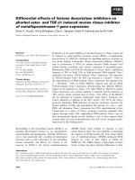
TNBS induced significant inflammation within the proxi-
mal and distal regions of the colon although the extent of
inflammation was greater in the distal region (Figure 1A).
Histone acetylation in inflamed and non-inflamed regions
of the colon in the rat TNBS model of colitis
TNBS induced a significant increase in pan histone 4
acetylation in the distal (592 ± 54% vs 135 ± 24 Sham
operated animals, p < 0.05) and the proximal regions of
the colon (315 ± 39% vs 125 ± 19% sham operated ani-
mals, p < 0.05) with the inflamed distal region showing
a greater increase (Figure 1B).
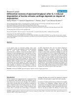
Acetylation of lysine (K) residues 8 and 12 were signif-
icantly increased in both the inflamed distal (K8: 818 ±
111 vs 138 ± 19%; K12: 741 ± 64 vs 121 ± 34%, both
p < 0.05) and less-inflamed proximal (K8: 546 ± 50 vs
100 ± 21%; K12: 533 ± 69 vs 100 ± 26%, both p < 0.05)
regions following TNBS treatment (Figure 2). However,
the effect was significantly greater in the inflamed tissue
than in the less-inflamed tissue for both K8 (818 ± 111
vs 546 ± 50%, p < 0.05) and K12 (741 ± 64 vs 533 ±
69%, p < 0.05).
In contrast, there was no significant induction of K5
or K16 induction by TNBS in the inflamed distal region
(Figure 2). Moreover, K5 (255 ± 39 vs 100 ± 15% Sham
operated animals, p < 0.05) and K16 (300 ± 63 vs 100 ±
29% Sham operated animals, p < 0.05) acetylation was
enhanced in the non-inflamed proximal region.
Tsaprouni et al. Journal of Inflammation 2011, 8:1
/>Page 3 of 12
Localisation of acetylated histones 4 and 3 in DSS-treated
animal models
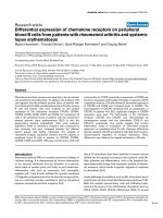
Acetylation of both histones 4 and 3 was evident in
non-DSS treated rats but this was enhanced in all
inflamed areas, regardless of distinct positions in the
colon, of both for Lewis rats (H4: 222 ± 31 DSS t reated
vs 100 ± 31% non-DSS treated animals, p < 0.05; H3
292 ± 40 DSS treated vs 100 ± 13% non-DSS treated
animals, p < 0.05 ) and Spraque-Dawley rats (H4: 1 87 ±
30 DSS treated vs 100 ± 21% non-DSS treated animals,
p < 0.05; H3 361 ± 36 DSS treated vs 100 ± 15% non-
DSS treated animals, p < 0.05) (Figure 3). Similar results
were obtained from Sprague-Dawley DSS-treated cells.
Localisation of acetylated histones 4 and 3 in Peyer’s
patches
We also investiga ted whether DSS-treatment would have
an effect on histone acetylation in the Peyer’ s p atches
found in the small intestine. Acetylate d histones are indi-
cated by the brown colour in the micrographs. Pan acety-
lated H3 was situated in the mantle zone of Peyer’ s
patches in DSS-treated Lewis and Sprague-Dawley rats in
contrast to the more uniformed staining for acetylated
histone 4 througho ut the surface of Peyer’s patches (Fig-
ure 3D).
Specificity of histone 4 lysine acetylation in Peyer’s
patches after DSS treatment
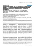
DSS induced acetylation of histone 4 lysines K5, K8,
K12 and K16 in both rat strains (Figure 4). However, a
greater induction was seen on K8 in both Lewis (414 ±
51 DSS treated vs 100 ± 23% non-DSS treated animals)
and Sprague-Dawley rats (1275 ± 123 DSS treated vs
100 ± 18% non-DSS treated animals). Similar results
were seen with K12 in both Lewis (703 ± 64 DSS trea-
ted vs 100 ± 14% non-DSS treated animals) and Spra-
gue-Dawley rats (1117 ± 113 DSS treated vs 100 ± 27%
non-DSS treated animals). K5 acetylation in Lewis rats
(346 ± 17 DSS t reated vs 100 ± 12% non-DSS treated
animals) and Sprague-Dawley rats (263 ± 22 DSS treated
vs 100 ± 16% non-DSS treated animals) was also
induced albeit to a lesser extent. Our findings were
similar for K16 acetylation in both Lewis (235 ± 43 DSS
treated vs 100 ± 22% non-DSS treated animals) and
Sprague-Dawley rats (321 ± 24 DSS treated vs 100 ±
26% non-DSS treated animals).
Distal colon
Proximal colon
Sham TNBS
Sham
Sham
TNBS
TNBS
Prox
Distal
0
200
400
600
800
% of control
Proximal
Region
Distal Regio
n
*
*
S
h
a
mTNB
S
AB
2cm
distal to
the
caecum
3cm
proximal
to the
anus
pan H4 acetylation
β-actin
Pan
acetyl H4
Figure 1 Acetylation on histone 4 in the trinitrobenzen e sulfonic acid (TNBS) rat mo del of inflammation. A: Sham (saline treated)
operated and TNBS treated rat large intestine. Rats were Sham or TNBS treated for 7 days before sacrifice. Well-advanced inflammation is
apparent in the colon of the TNBS rat model. B: Pan acetylation on histone 4 (H4). The Sham model was saline-treated and therefore less
inflamed (control). Results were obtained by Western blotting. The ratio of the density of histone H4 bands over b-actin control bands was
calculated. In order to evaluate changes in density from different Western blotting experiments control densitometry was denoted as 100% and
differences were accounted as increase percentage of the control. Representative examples of bands obtained are also illustrated. Columns
represent the densitometric evaluation of three independent experiments (mean ± SEM). (*p < 0.05 vs Sham proximal or Sham distal
respectively).
Tsaprouni et al. Journal of Inflammation 2011, 8:1
/>Page 4 of 12
Histone acetylation in Crohn’s disease
Acetylation on H4 was slightly induced in the non-
inflamed ileum of Crohn’ s disease patients. In contrast,
H4 acetylation was significantly elevated in the inflamed
regions (472 ± 88 vs 100 ± 34% control, p < 0.05) (Fig-
ure 5A). Peyer’s patches from Crohn’ s disease patients
also showed a significant increase in pan H4 acetylation
(382 ± 29%) compared to the control non-inflamed tis-
sue (100 ± 34%, p < 0.05) (Figure 5A). Levels of acety-
lated K5 were not significantly upregulated compared to
control (Figure 5). More specifically, K8 acetylation was
significantly induced compared to control samples in
the i nflamed regions (527 ± 44% vs 100 ± 25% control
tissue, p < 0.05) and the non-inflamed CD samples (527 ±
44% vs 195 ± 42% non-inflamed CD, p < 0.05). In Peyer’s
patches from CD patients, K8 was significantly upregu-
lated compared to control (488 ± 52% vs 100 ± 25% con-
trol tissue, p < 0.05) (Figure 5).
Enhanced acetylation on K12 was detected in inflamed
regions of CD compared to control (442 ± 54% vs 100 ±
29% control tissue, p < 0.05) and non-inflamed CD tis-
sue (4 42 ± 54% vs 223 ± 38% non-inflame d IBD tissue,
p < 0.05). Similarly, enhanced acetylation on K12 was
detected in Peyer’s patches compared to control (429 ±
65% vs 100 ± 29% control tissue, p < 0.05). Acetylation
on lysine 12 was not significantly increased in non-
inflamed tissue compared to control. No changes in
lysine 16 acetylation were observed in either inflamed or
non-inflamed tissue from Crohn’ s disease patients. In
the Peyer’s patches, however, a significant elevation of
acetylation on K16 was observed (Figure 5).
Discussion
Our results show that acetylation of histone H4 was sig-
nificantl y elevat ed in the inflamed mucosa in the TNBS
model of colitis particularly on lysine residues (K) 8 and
H4K12
0
200
400
600
800
1000
% of control
Proximal
Region
Distal Regio
n
*
*
H4K16
0
100
200
300
400
% of control
Proximal
Region
Distal Regio
n
*
H4K8
0
400
800
1200
% of control
Proximal
Region
Distal Region
*
*
H4K5
0
100
200
300
400
% of control
Proximal
Region
Distal Region
*
S
h
a
mTNB
S
Sham TNBS
Sham TNBS
Sham TNBS
A
C
B
D
β-actin
β-actin
β-actin
β-actin
Figure 2 Acetylation on histone 4 (H4) specific lysine residues 5 (K5) (A), 8 (K8) (B), 12 (K12) (C) and 16 (K16) (D) in a Sham (control)
and trinitrobenzene sulfonic acid (TNBS) rat model of colitis. Results were obtained by Western blotting. In order to evaluate changes in
density from different western blotting experiments control densitometry was denoted as 100% and differences were accounted as increase
percentage of the control. Representative examples of bands obtained are also illustrated. Columns represent the densitometric evaluation of
three independent experiments (mean ± SEM). (*p < 0.05 vs Sham proximal or Sham distal respectively).
Tsaprouni et al. Journal of Inflammation 2011, 8:1
/>Page 5 of 12
12 in contrast to non-inflamed tissue. In addition, acety-
lated H 4 was localised to inflamed tissue and to PP in
DSS-treated rat models. Within the PP, H3 acetylation
was detected in the mantle zone whereas H4 acetylation
was seen in both the periphery and the germinal centre.
Finally, acetylation of H4 was significantly increased in
inflamed biopsies and PP from patients with CD.
Enhanced acetylation of H4K5 and K16 was seen in the
PP. Acetylation of K5 and K16 was localized to the
mantle zone whereas acetylation of K8 and K12 was
localized to both the mantle zone and the germinal cen-
ter (data not shown).The diversity of IBD and the diffi-
culty in successfully distinguishing between Ulcerative
colitis and Crohn’ s disease underlined the criteria for
E-actin
Lewis
Acetylated Histone 3
Acetylated Histone 4
sham shamDSS DSS
S-D
*
Lewis
Rats
Sprague-Dawley
Rats
*
Ac H4
0
100
200
300
400
500
% of control
Sprague-Dawley
Rats
Lewis
Rats
*
Ac H3
0
100
200
300
% of control
A
B
C
Histone H3
Histone H4
D
*
sham sham
D
SS
D
SS
Figure 3 Acetylation on histones 3 (H3) and 4 (H4) in Lewis and Sprague-Dawley dextran sulfate sodium (5% DSS) treated rats. Tissue
samples were obtained from the sigmoid colon of the animals. A: Representative bands of H4 and H3 acetylation as obtained by Western
blotting. b-actin levels were measured to ensure equal protein loading. The results are representative of three independent experiments. B, C:
Graphical analysis of data Lanes represent: (1) non-DSS treated Lewis rats (control), (2) DSS-treated Lewis rats, (3) non-DSS treated Sprague-
Dawley rats (control) (4) DSS-treated Sprague-Dawley rats. Columns represent the mean ± SEM of three independent experiments (*p < 0.05).
D: Histone 3 (H3) and histone 4 (H4) localisation in Peyer’s patches of dextran sulfate sodium (DSS) treated Lewis rats. H3 is acetylated mainly in
the mantle zone and H4 is acetylated throughout the surface of Peyer’s patches to both mantle zone and germinal centre cells. In Peyer’s
patches of untreated animals no acetylation on either histone 3 or 4 was apparent. Micrographs are representative of two individual experiments
for each strain. Isotype controls show no staining.
Tsaprouni et al. Journal of Inflammation 2011, 8:1
/>Page 6 of 12
employing two different animal models for studying his-
tone acetylation (TNBS and DSS) associated with
Crohn’s disease and Ulcerative colitis respectively [30].
Although in many ca ses it is not clear whether cyto-
kines ar e the cause or the result of the under lying dis-
ease p rocess there is little question that their presence
can have profound effects upon gut epithelial cell func-
tion and that pro-inflammatory cytokines are key factors
in the pathogenesis of Crohn’s disease (CD). Activation
of nuclear factor kappa B (NF-B), which is involved in
pro-inflammatory cytokine gene transcription, is
increased in the intestinal mucosa o f CD patients [31].
Modulation of histone acetylation is involved in tran-
scriptional regulation, associated with the NF-B
pathway [32-34]. Importantly, eith er a lack or an excess
of NF-B can lead to IBD. As enhanced intestinal
epithelial permeability may cause IBD by itself, NF-B
deficiency could underline epithelial barrier function
directly by deregulating the expression of proteins
involved in cellular adhesion. Alternatively, NF-B fail-
ure could break the barrier in directly by comp romising
the survival of epithelial cells [35]. This might explain
the complex molecular mode of action of butyrate in
IBD, where for example reports show that butyrate inhi-
bits NF-B activation and increases IBb levels in vitro
in intestinal epithelial cell lines [36]. In gain of function
mutations in the Nod2 gene, there is an induction of
TH1 and IL-17 secreting T helper response that
Sham DSS
H4K5
H4K5
H4K8
Lewis
S
-D
H4K12
0
500
1000
1500
% of control
H4K16
0
100
200
300
400
% of control
H4K8
0
500
1000
1500
% of control
H4K5
0
100
200
300
400
% of control
Sham
Sham
Sham ShamSham
Sham Sham
Sham
DSS
DSS
DSS
DSS
DSS
DSS
DSS
DSS
Lewis
L
ewis
L
ewis
LewisS-D
S
-D
S
-D
S-D
A
B
C
D
E
*
*
#
#
*
*
*
*
β-actin
H4K8
H4K12
H4K12
H4K16
H4K16
β-actin
Sham
DSS
Figure 4 Acetylation on histone 4 (H4) specific lysine residues 5 (K5), 8 (K8), 12 (K12) and 16 (K16) in Lewis and Sprague-Dawley
dextran sulfate sodium (5% DSS). A: Representative bands of H4K5, K8, K12 and K16 acetylation. Lanes for Lewis rats represent: non-DSS
treated (control) and DSS-treated. Likewise representative bands are illustrated for the Sprague-Dawley rats. Graphical representation of Western
blotting data. H4 acetylation of K5 (B),K8(C), K12 (D) and K16 (E). Columns represent the mean ± SEM (bar) of three independent experiments.
Tsaprouni et al. Journal of Inflammation 2011, 8:1
/>Page 7 of 12
promotes tissue damage and Crohn’sdisease[37].On
the other hand, loss-of-function mutations compromise
NF-B activation and TH1 driven colitis [35].
A number of articles demonstrate that ace tylation of
histone H4 plays a primary role in the structural
changes that mediate enhanced binding of transcription
factors to their recognition sites within nucleosomes
[38]. In primary airway smooth muscle cells, TNF-a
induced histone 4 acetylation and this induction was
attenuated by pre-treatment of cells with a glucocorti-
coid [39]. Finally, variations in global levels of histone
marks in different grades, morphologic types, and phe-
notype classes of invasive breast cancer have been
reported to be clinically significant [40]. The use of
sodium butyrate, a histone deacetylase inhibitor, in the
treatment of IBD lead to the hypothesis that in addition
Control
Non-
Inflam.
Inflam.
Peyer
’
s
Patches
0
200
400
600
% of control
*
*
H4K5
0
100
200
300
% of control
Control
Non-
Inflamed
Peyer’s
Patches
A
B
H4K16
0
100
200
300
400
% of control
*
H4K12
0
200
400
600
% of control
#
*
*
H4K8
0
200
400
600
800
% of control
#
*
*
Crohn’s Disease
Control Non-
Inflamed
Inflamed Peyer’s
Patches
Crohn’s Disease
C
D
E
panAcH4
Inflamed
β-actin
panAcH4
H4K5
H4K8
H4K12
H4K16
Figure 5 Acetylation on histone 4 (H4) and H4 lysine residues in Crohn’ sdisease. Columns represent the mean ± SEM of three
independent experiments. Four biopsies were pooled to obtain sufficient protein for one experiment (50 μg of protein) (*p < 0.05 vs control).
Pan acetylation on H4 in Crohn’s disease (A). Acetylation on histone 4 (H4) specific lysine residues 5 (K5) (B),K8(C), K12 (D), and 16 (E), in non-
inflamed, inflamed tissue and Peyer’s patches of Crohn’s disease patients. Results were obtained by Western blotting. Columns represent the
mean ± SEM of three independent experiments. (*p < 0.05 vs control, #p < 0.005 vs non-inflamed CD). Representative images of the bands
obtained are illustrated.
Tsaprouni et al. Journal of Inflammation 2011, 8:1
/>Page 8 of 12
to its anti-proliferative action, an effect on histone acety-
lation could be associated with its therapeutic effects.
For example, in human umbilical vein endothelial cells
(HUVEC), induction of tissue-type plasminogen activa-
tor (t-PA) transcription by butyrate and Trichostatin A
was preceded by histone 4 acetylation [41]. Recent evi-
dence reve aled that butyrate decreases pro-inflammatory
cytokineexpressionviainhibitionofNF-B activation
and IBa degradation [14,18,42] while it has also been
demonstrated that NF-B induction of inflammatory
gene expression is associated with histone acetylation
[28,34] and indee d with p65 acetylation [43].With the
importance of H4 acetylation having been studied and
described in other disease models, experiments were
carried out in to in vestigate whether acetylated histone
4 activity was altered in inflamed and non-inflamed tis-
sue of a TNBS model of colitis. We observed differences
in histone 4 acetylation levels between inflamed and
non-inflamed tissue particularly with respect to K8 and
K12 acetylation. This specificity towards lysine acetyla-
tion could be explained by the selective recruitment of
transcriptional co-activators containi ng HAT activity by
transcription factors such as NF-B [44,4 5]. Although
tempting to suggest a cause-and-effect model it is
unclear whether increased inflammation leads directly to
increased histone acetylation in vivo at specific gene
promoters. Further studies will be needed to address
this in IBD but preliminary evidence suggests that this
may be the case for the GM-CSF promoter in alveolar
macrophages from smokers [46]. Also another interest-
ing study investigating the effect of pro-inflammatory
cytokines in intestinal alkaline phosphatase (IAP) gene
expression comes to further support the possible role of
histone acetylation in intestinal inflammation. The
authors report both histones 3 and 4 we re hyperacety-
lated in HT-29 cells when they were stimulated with
TNF-a or IL-1b concluding that both pro-inflammatory
cytokines affect sodium butyrate-induced activation of
the IAP gene likely via deacetylation of its promoter
region [47].
Macroscopic analysis of tissue from both Lewis and
Sprague-Dawley rats treated with 5% DSS revealed areas
of severe inflammation . However, Peyer’ s patches did
not sho w any signs of inflammation agreeing wit h pre-
vious results showing that the DSS model resembles
ulcerative colitis with inflammatio n present in the des-
cending and sigmoid colon and the rectum but is not
apparent along the wall of the small intestine where
Peyer’s patches are situated. In the DSS model, acetyla-
tion of histones 4 and 3 was upregulated in both Lewis
and Sprague-Dawley rats. Comparison of acetylated
levels between histones 3 and 4 revealed that while both
were acetylated, the latter reached significantly higher
levels. Similarly, in Peyer’s patches of t he DSS model,
histone 4 acetylation was g reater than that of histone 3.
Immunohistochemical investigation of Peyer’s patches
revealed a distinct pattern of histone acetylation. Acety-
lation on H3 was only detected in t he mantle zone of
Peyer’s patches, whilst acetylated H4 occurred in both
the periphery and the germinal centre of Peyer’ s
patches. Therefore, it was concluded that acetylation on
H3 could possibly be cell specific, whereas H4 is gener-
ally induced in all cell types present in Peyer’s patches
(T-cells, B-cells, dendritic cells and macrophages)
although this needs to be formally assessed (possibly by
counter staining). These data indicate an increase in his-
tone acetyla tion during gut inflammation. In support, a
number of reports show differential H3 acetylation pat-
terns between TH1 and TH2 cells [48,49].
Acetylation of K8 and K12 is associated with the upre-
gulation o f inflammatory genes [28]. In the DSS model
of colitis, H4 K8 and K12 were highly acetylated in the
Sprague- Dawley rats. These findings were in agreement
with previous results document ed in vitro [50]. Interest -
ingly, in the Lewis rats, only K12 acetylation was
strongly induced. This difference could be attributed to
genetic variances between the two rat strains, as dis-
cussed by other groups [51,52].
ThepresentstudywasconcludedbymeasuringH4
acetylation in Crohn’s disease patient biopsies. As with
the TNBS model, Peyer’s patches, non-inflamed and
inflamed biopsies were assessed. Levels of acetylated H4
were most prominent in the inflamed biopsies, followed
by those in Peyer’ s patches albeit to a lesser extent.
Acetylation was also detectable in the non-inflame d
mucosa of Crohn’s disease patients. The results for acet-
ylation on H4 lysines in Crohn’s disease were very simi-
lar t o those obtained in the TNBS treated animals. K5
and K16 were only slightly acetylated in all samples,
with the inflamed and non-infla med samples presenting
no significant difference in acetylation. Peyer’s patches
showed the highest levels of K5 and K16 acetylation.
Finally, in biopsies of inflamed bowel and in Peyer’ s
patches
of Crohn’s disease patients, K8 and K12 were
both s ignificantly acetylated. Acety lation on lysine r esi-
dues in the non-inflamed biopsies was only slightly
upregulated. The results suggested that although pan
acetylation on H4 in the Peyer’s patch es is probably not
cell specific, it is possible that acetylation of its specific
lysine residues is cell type dependent. This could also
explain the significant increase in K8 and K12 acetyla-
tion revealed by Western blotting. An increased Treg
number in Peyer’ s patches indicates that they have a
very important niche in the peripheral gut, where new
encounters with antigens are very critical. In this
respect, it seems natural that Treg are more numerous
in Peyer’s patches as it is in the gut that antigens to
cross the intestinal barrier are to be processed and exert
Tsaprouni et al. Journal of Inflammation 2011, 8:1
/>Page 9 of 12
their effect, and thus it is an area where essential anti-
genic surveillance is taking place [53].
Site specific histone acetylation and deacetylation have
been associated in more re cent years with a number of
different functions such as nucleosome assembly, het-
erochromatin silencing, transcription and ge ne repres-
sion [54]. The human chromatin assembly factor 1
(CAF-1) co mplex co-purifies with histone H4 modified
at sites that are indicative of recent synthesis. Acetyla-
tion is observed at K5, K8 and/or K12 but not at K16
[55]. In yeast H4K16 appears to be critica l for the silen-
cing information regulator protein (Sir) binding because
the interacti on betw een full length Sir3 and an H4 pep-
tide in vitro is abolished by acetylation of lysine 16 but
not other lysines [56]. Another example of site specific
lysine acetylation involves the SMRT mammalian co-
repressor. SMRT preferentially binds to the unacetylated
histone 4 tail and its binding is d ependent on deacety-
lated H4K5 [57]. Finally, another example of the e ffect
of specific lysine residue acetylation in gene function is
the observation that with the coding region of ERG11,
an active gene, deacetylases Hos2 and Rpd3 redundantly
deacetylate all lysines in histone 4 and H4 tails except
for H4K16, which is deacetylated primarily by Hos2
[58]. Precise patterns of acetylation at promoter s, there-
fore, may be recognized by particular transcription fac-
tors because specific combinations of hypoacetylated
residue s at genes correlate with spec ific expression pro-
files over a variety of conditions [54].
Paradoxicall y, HDAC inhibit ors are used in the treat-
ment of IBD. This may reflect either an anti-proliferative
effect seen with h igh, non-specific doses of HDAC inhi-
bitors or an effect on the acetylation status of non-
histone proteins e.g. tubulin and transcription factors
such as NF-B and GATA [20,59,60]. Recent reports,
however, show that administration of an HDAC inhibi-
tor in vivo increased Foxp3 gene expression, as well as
the production and the suppr essive function of regula-
tory T cells (Treg cells). It has been shown that H DAC
inhibition therapy in vivo enhanced Treg-mediated sup-
pression of a homeostatic proliferation and decreased
IBD through Treg-dependent effects [61]. These results
may, at least in part, reflect the activation of regulatory
T-cells involved in active NF-B suppression (and
incr eased histone acetylation) of inflammation primarily
induced in the Peyer’s patches [62].
The results presented here are indicative of the impor-
tance of histone 4 acetylation in the expression of
inflammatory genes in inflammatory diseases, such as
IBD. Whether this is causal or downstream to activation
of inflammation is unclear but suggests that HAT inhi-
bitors may be useful in treatment. Deacetylase inhibitors
in vivo, such as Belinostat (PXD101) and Phenylbutyrate,
are currently used in clinical trials. However, most
clinical trials have not had much success either due to
the disease being stable or due to adverse effects of the
drug [63]. The mechanism might be better understood
when the target proteins (histone or non-histone) of
these compounds are identified.
The present preliminary studies aim to provide further
understanding in the role that histone acety lation plays
in the re gulation o f inflammation. Future studies should
examine the activity of specific HATs and HDACs in
individual immune and resident cells types. It is, there-
fore, possible to speculate that further understanding of
the role of histone modifications in IBD may lead to
new therapeutic strategies in the treatment of IBD and
explain the therapeutic utility of current treatment.
Acknowledgements
This work was funded by the University of Bedfordshire and GlaxoSmithKline
(UK).
Author details
1
Airways Disease Section, National Heart & Lung Institute, Imperial College
London, Dovehouse Street, London, SW3 6LY, UK.
2
School of Health and
Biosciences, University of East London, Stratford Campus, Romford Road,
London, E15 4LZ, UK.
3
Gastroeintestinal Laboratory, Rayne Institute, St.
Thomas Hospital, London, SE1 7EH, UK.
Authors’ contributions
LGT performed all experiments and drafted the manuscript. KI participated
in the histone extraction methods. JJP provided clinical and animal samples.
IMA participated in the design and coordination of the study and to
manuscript writing. NP participated in the design and coordination of the
study. All authors read and approved the final manuscript.
Competing interests
The authors declare that they have no competing interests.
Received: 12 April 2010 Accepted: 27 January 2011
Published: 27 January 2011
References
1. Baumgart DC, Carding SR: Inflammatory bowel disease: cause and
immunobiology. Lancet 2007, 369(9573):1627-1640.
2. D’Haens G, Daperno M: Advances in biologic therapy for ulcerative colitis
and Crohn’s disease. Curr Gastroenterol 2006, 8(6):506-512.
3. Travis S: Advances in therapeutic approaches to ulcerative colitis and
Crohn’s disease. Curr Gastroenterol 2005, 7(6):475-484.
4. Neurath M, Fuss I, Strober W: TNBS-colitis. Int Rev Immunol 2000, 19:51-62.
5. Fujno K, Takami Y, dela Fuente SG, Ludwig KA, Mantyh CR: Inhibition of
the vanilloid receptor subtype-1 attenuates TNBS-colitis. Jof
Gastrointestinal Surg 2004, 8(7):842-848.
6. Dohi T, Fujihashi K, Rennert PD, Iwatani K, Kiyoto H, McGhee JR: Hapten-
induced colitis is associated with colonic patch hypertrophy and T
Helper cell-2-type responses. J Exp Med 1999, 189:1169-1179.
7. Alex P, Zachos NC, Nguyen T, Gonzales L, Chen TE, Conklin LS, Centola M,
Li X: Distinct cytokine patterns identified from multiplex profiles of
murine DSS and TNBS-induced colitis. Inflammatory Bowel Diseases 2008,
15(3):341-352.
8. Zaph C, Troy AE, Taylor BC, Berman-Booty LD, Guild KJ, Du Y, Yost EA,
Gruber AD, May MJ, Greten FR, Eckmann L, Karin M, Artis D: Epithelial-cell-
intrinsic IKK-β expression regulates intestinal immune homeostasis.
Nature 2007, 446:552-556.
9. Nenci A, Becker C, Wullaert A, Gareus R, van Loo G, Danese S, Huth M,
Nikolaev A, Neufert C, Madison B, Gumucio D, Neurath MF, Pasparakis M:
Epithelial NEMO links innate immunity to chronic intestinal
inflammation. Nature 2007, 446:557-561.
Tsaprouni et al. Journal of Inflammation 2011, 8:1
/>Page 10 of 12
10. Murano M, Maemura K, Hirata I, Toshina K, Nishikawa T, Hamamoto N,
Sasaki S, Saitoh O, Katsu K: Therapeutic effect of intracolonically
administered nuclear factor kappa B (p65) antisense oligonucleotide on
mouse dextran sulphate sodium (DSS)-induced colitis. Clin Exp Immunol
2000, 120:51-58.
11. Herfarth H, Brand K, Rath HC, Rogler G, Scholmerich J, Falk W: Nuclear
factor-kappa B activity and intestinal inflammation in dextran sulphate
sodium (DSS)-induced colitis in mice is suppressed by gliotoxin. Clin Exp
Immunol 2000, 120:59-65.
12. Fitzpatrick LR, Wang J, Le T: In vitro and in vivo effects of gliotoxin, a
fungal metabolite: efficacy against dextran sodium sulfate-induced
colitis in rats. Dig Dis Sci 2000, 45:2327-2336.
13. Rahman I, Marwick J, Kirkham P: Redox modulation of chromatin
remodeling: impact on histone acetylation and deacetylation, NF-κB and
pro-inflammatory gene expression. Biochem Pharmacol 2004,
68(6):1255-1267.
14. Place RF, Noonan EJ, Giardina C: HDAC inhibition prevents NF-κB activation
by suppressing proteasome activity: Down-regulation of proteasome
subunit expression stabilizes IκBα. Biochem Pharmacol 2005, 70:394-406.
15. Siavoshian S, Segain JP, Kornprobst M, Bonnet C, Cherbut C, Galmiche JP,
Blottiere HM: Butyrate and trichostatin A effects on the proliferation/
differentiation of human intestinal epithelial cells: induction of cyclin D3
and p21 expression. Gut 2000, 46:507-514.
16. Glauben R, Batra A, Fedke I, Zeitz M, Lehr HA, Leoni F, Mascagni P,
Fantuzzi G, Dinarello CA, Siegmund B: Histone hyperacetylation is
associated with amelioration of experimental colitis in mice. J Immunol
2006, 176:5015-5022.
17. Roy CC, Kien CL, Bouthillier L, Levy E: Short-chain fatty acids: ready for
prime time? Nutr Clin Pract 2006, 21(4):351-366.
18. Park JS, Lee EJ, Lee JC, Kim WK, Kim HS: Anti-inflammatory effects of short
chain fatty acids in IFN-gamma-stimulated RAW 264.7 murine
macrophage cells: involvement of NF-kappaB and ERK signalling
pathways. Int Immunopharmacol 2007, 7(1):70-77.
19. Xu WS, Parmigiani RB, Marks PA: Histone deacetylase inhibitors: molecular
mechanisms of action. Oncogene 2007, 26(37):5541-5552.
20. Bhavsar P, Ahmad T, Adcock IM: The role of histone deacetylases in
asthma and allergic diseases. J Allergy Clin Immunol 2008, 121(3):580-584.
21. Matthews JG, Ito K, Barnes PJ, Adcock IM: Defective glucocorticoid
receptor nuclear translocation and altered histone acetylation patterns
in glucocorticoid-resistant patients. J Allergy Clin Immunol 2004,
113:1100-1108.
22. Li L, Leung DYM, Strand MJ, Golevs E:
ATF2 impairs glucocorticoid
receptor
mediated transactivation in human CD8
+
T cells. Blood 2007,
110(5):1570-1577.
23. Fraga MF, Ballestar E, Villar-Garea A, Boix-Chornet M, Espada J, Schotta G,
Bonaldi T, Haydon C, Ropero S, Petrie K, Iyer NG, Pérez-Rosado A, Calvo E,
Lopez JA, Cano A, Calasanz MJ, Colomer D, Piris MA, Ahn N, Imhof A,
Caldas C, Jenuwein T, Esteller M: Loss of acetylation at Lys16 and
trimethylation at Lys20 of histone H4 is a common hallmark of human
cancer. Nat Gen 2005, 37(4):391-400.
24. Morris GP, Beck PL, Herridge MS, Depew WT, Szewczuk MR, Wallace JL:
Hapten-induced models of chronic inflammation and ulceration in the
rat colon. Gastroenterol 1989, 96:795-803.
25. Vowinkel T, Kalogeris TJ, Mori M, Krieglstein CF, Granger DN: Impact of
dextran sulfate sodium load on the severity of inflammation in
experimental colitis. Dig Dis Sci 2004, 49(4):556-564.
26. Ellis RD, Goodlad JR, Limb GA, Powell JJ, Thompson RP, Punchard NA:
Activation of nuclear factor kappa B in Crohn’s disease. Inflamm Res
1998, 47:440-445.
27. Ito K, Barnes PJ, Adcock IM: Histone acetylation and histone
deacetylation. Methods in Molecular Medicine. Asthma: Mechanisms and
Protocols 2000, 44:309-319.
28. Ito K, Barnes PJ, Adcock IM: Glucocorticoid receptor recruitment of
histone deacetylase 2 inhibits interleukin-1β-induced histone H4
acetylation on lysines 8 and 12. Mol Cell Biol 2000, 20(18):6891-6903.
29. Ito K, Caramori G, Lim S, Oats T, Chung KF, Barnes PJ, Adcock IM:
Expression and activity of histone deacetylases in human asthmatic
airways. Am J Respir Crit Care Med 2002, 166(3):392-396.
30. Neurath MF, Finotto S, Fuss I, Boirivant M, Galle PR, Strober W: Regulation
of T-cell apoptosis in inflammatory bowel disease: to die or not to die,
that is the mucosal question. Trends Immunol 2001, 22:21-26.
31. Schreiber S: Inflammatory bowel disease: Immunologic concepts. Hepato-
Gastroenterol 2000, 47:15-28.
32. Adcock IM, Cosio B, Tsaprouni L, Barnes PJ, Ito K: Redox regulation of
histone deacetylases and glucocorticoid-mediated inhibition of the
inflammatory response. Antioxid Redox Signal 2005, 7(1-2):144-152.
33. Ito K, Yamamura S, E ssilfie-Quaye S, Cosio B, Ito M, Barnes PJ,
Adcock IM: Histone deacetylase 2-mediated deacetylation of the
glucocorticoid receptor enables NF-kappa B suppression. JExpMed
2006, 203(1):7-13.
34. Ashburner BP, Westerheide SD, Baldwin AS Jr: The p65 (RelA) subunit of
NF-κB interacts with the histone deacetylase (HDAC) corepressors
HDAC1 and HDAC2 to negatively regulate gene expression. Molecular
and Cellular Biology 2001, 21:7065-7077.
35. Ben-Neriah Y, Schmidt-Supprian M: Epithelial NF-kappaB maintains host
gut microflora homeostasis. Nat
Immunol 2007, 8(5):479-481.
36. Hamer HM, Jonkers D, Venema K, Vanhoutvin S, Troost FJ, Brummer RJ:
Review article: the role of butyrate on colonic function. Aliment
Pharmacol Ther 2008, 27(2):104-119.
37. Maeda S, Hsu LC, Liu H, Bankston LA, Iimura M, Kagnoff MF, Eckmann L,
Karin M: Nod2 mutation in Crohn’s disease potentiates NF-kappaB
activity and IL-1beta processing. Science 2005, 307(5710):734-738.
38. Vettese-Dadey M, Grant PA, Hebbes TH, Crane-Robinson C, Allis CD,
Workman JL: Acetylation of histone H4 plays a primary role in enhancing
transcription factor binding to nucleosomal DNA in vitro. EMBO J 1996,
15(10):2508-2518.
39. Nie M, Knox AJ, Pang L: Beta-2 Adrenoreceptor agonists, like
glucocorticoids repress eotaxin gene transcription by selective inhibition
of histone H4 acetylation. J Immunol 2005, 175:478-486.
40. Elsheikh SE, Green AR, Rakha EA, Powe DG, Ahmed RA, Collins HM, Soria D,
Garibaldi JM, Paish CE, Ammar AA, Grainge MJ, Ball GR, Abdelghany MK,
Martinez-Pomares L, Heery DM, Ellis IO: Global Histone Modifications in
Breast Cancer Correlate with Tumor Phenotypes, Prognostic Factors, and
Patient Outcome. Cancer Res 2009, 69(9):3802-3809.
41. Arts J, Lansink M, Grimbergen J, Toet KH, Kooistra T: Stimulation of tissue-
type plasminogen activator gene expression by sodium butyrate and
trichostatin A in human endothelial cells involves histone acetylation.
Biochem J 1995, 310:171-176.
42. Song M, Xia B, Li J: Effects of topical treatment of sodium butyrate and
5-aminosalicylic acid on expression of trefoil factor 3, interleukin 1beta,
and nuclear factor kappaB in trinitrobenzene sulphonic acid induced
colitis in rats. Postgrad Med J 2006, 82(964):130-135.
43. Chen L, Fischle W, Verdin E, Greene WC: Duration of nuclear NF-kappaB
action regulated by reversible acetylation. Science 2001, 293:1653-1657.
44. Smoak KA, Cidlowski JA: Mechanisms of glucocorticoid receptor signalling
during inflammation. Mechs Age Dev 2004, 125:697-706.
45. Hoffmann A, Natoli G, Ghosh S: Transcriptional regulation via the NF-κB
signalling module. Oncogene 2006, 25:6706-6716.
46. Ito K, Lim S, Caramori G, Chung KF, Barnes PJ, Adcock IM: Cigarette
smoking reduces histone deacetylase 2 expression, enhances cytokine
expression, and inhibits glucocorticoid actions in alveolar macrophages.
FASEB J 2001, 15:1110-1112.
47. Malo MS, Biswas S, Abedrapo MA, Yeh L, Chen A, Hodin RA: The pro-
inflammatory cytokines, IL-1β and TNF-α, inhibit intestinal alkaline
phosphatase gene expression. DNA and Cell Biology 2006, 25(12) :684-695.
48. Wang L, Kametani Y, Katano I, Habu S: T-cell
specific enhancement of
histone H3 acetylation in 5’ flanking region of the IL-2 gene. Biochem
and Bipophys Commun 2005, 331(2):589-594.
49. Letting DL, Rakowski C, Weiss MJ, Blobel GA: Formation of a Tissue-
Specific Histone Acetylation Pattern by the Hematopoietic Transcription
Factor GATA-1. Mol and Cell Biol 2003, 23(4):1334-1340.
50. Wako T, Houben A, Furushima-Shimogawara R, Belyaev ND, Fukui K:
Centromere-specific acetylation of histone H4 in barley detected
through three-dimensional microscopy. Plant Mol Biol 2003, 51(4):533-541.
51. Quary S, Bizat N, Altairac S, Menetrat H, Mittoux V, Conde F, Hantraye P,
Brouillet E: Major strain differences in response to chronic systemic
administration of the mitochondrial toxin 3-nitropropionic acid in rats:
implications of neuroprotection studies. Neuroscience 2000, 97:521-530.
52. Jurado F, Bellon JM, Golitsin A, Gimero MJ, Pascual G, Bujan J: Role of
macrophages in myocardial apoptosis following cardiac transplant.
Influence of immunosuppressive treatment. Histology and Histopathology
1999, 14:1033-1043.
Tsaprouni et al. Journal of Inflammation 2011, 8:1
/>Page 11 of 12
53. Ejsing-Duun M, Josephsen J, Aasted B, Buschard K, Hansen AK: Dietary
Gluten Reduces the Number of Intestinal Regulatory T Cells in Mice.
Scandinavian Journal of Immunol 2008, 67:553-559.
54. Shahbazian MD, Grunstein M: Functions of site-specific histone
acetylation and deacetylation. Annual Review of Biochem 2007, 76:75-100.
55. Verreault A, Kaufman PD, Kobayashi R, Stillman B: Nucleosome assembly
by a complex of CAF-1 and acetylated histones H3/H4. Cell 1996,
87:95-104.
56. Liou GG, Tanny JC, Kruger RG, Walz T, Moazed D: Asssembly of the SIR
complex and its regulation by O-acetyl-ADP-ribose, a product of NAD-
dependent histone deacetylation. Cell 2005, 121:515-527.
57. Hartman HB, Yu J, Alenghat T, Ishizuka T, Lazar MA: The histone binding
code of nuclear receptor co-repressors matches the substrate specificity
of histone deacetylase 3. EMBO Rep 2005, 6:445-451.
58. Wang A, Kurdistani SK, Grunstein M: Requirement of Hos2 histone
deacetylase for gene activity in yeast. Science 2002, 298(5597):1412-1414.
59. Adcock IM, Caramori G: Cross talk between pro-inflammatory
transcription factors and glucocorticoids. Immunol and Cell Biol 2001,
79:376-384.
60. Dokmanovic M, Clarke C, Marks PA: Histone deacetylase inhibitors:
Overview and perspectives. Mol Cancer Res 2007, 5(10):981-989.
61. Tao R, de Zoeten EF, Özkaynak E, Chen C, Wang L, Porrett PM, Li B,
Turka LA, Olson EN, Greene MI, Wells AD, Hancock WW: Deacetylase
inhibition promotes the generation and function of regulatory T cells.
Nature Medicine 2007, 13(11):1299-1307.
62. Tsuji NM, Mizumachi K, Kurisaki J: Interleukin-10-secreting Peyer’s patch
cells are responsible for active suppression in low-dose oral tolerance.
Immunology 2001, 103(4):458-464.
63. Ma X, Ezzeldin HH, Diasio RB: Histone deacetylase inhibitors: Current
status and overview of recent clinical trials. Drugs 2009, 69(14):1911-1934.
doi:10.1186/1476-9255-8-1
Cite this article as: Tsaprouni et al.: Differential patterns of histone
acetylation in inflammatory bowel diseases. Journal of Inflammation 2011
8:1.
Submit your next manuscript to BioMed Central
and take full advantage of:
• Convenient online submission
• Thorough peer review
• No space constraints or color figure charges
• Immediate publication on acceptance
• Inclusion in PubMed, CAS, Scopus and Google Scholar
• Research which is freely available for redistribution
Submit your manuscript at
www.biomedcentral.com/submit
Tsaprouni et al. Journal of Inflammation 2011, 8:1
/>Page 12 of 12