Báo cáo y học: "Meconium pseudocyst secondary to ileum volvulus perforation without peritoneal calcification: a case report" pps
Bạn đang xem bản rút gọn của tài liệu. Xem và tải ngay bản đầy đủ của tài liệu tại đây (528.82 KB, 5 trang )
CAS E REP O R T Open Access
Meconium pseudocyst secondary to ileum
volvulus perforation without peritoneal
calcification: a case report
Esther Valladares
*
, David Rodríguez, Antonio Vela, Sergi Cabré, Josep Maria Lailla
Abstract
Introduction: A case of giant meconiu m pseudocyst secondary to ileum volvulus perforation is presented.
Conventional radiographic features of meconium peritonitis with secondary meconium pseudocyst formation are
well described. Our case is unusual in comparison to other cases reported in the literature and needs to be
reported because the meconium pseudocyst presented without the typical ultrasound fe atures (calcifications,
polyhydramnios and ascites) and was initially identif ied as an abdominal mass.
Case presentation: We describe the case of a 29-year-old Caucasian woman in her third tri mester of pregna ncy,
in which an abdominal mass was detected in the fetus. The newborn was diagnosed in the early neonatal perio d
with meconium pseudocyst secondary to ileum volvulus perforation.
Conclusions: The prenatal appearance of a meconium pseudocyst can be complemented by other signs of bowel
obstruction (if present) such as polyhydramnios and fetal bowel dilatation. This is an original case report of interest
to all clinicians in the perinatology and fetal ultrasound field. We consider that the utility of this case is the
recognition that a meconium pseudocyst might appear without the typical ultrasound features and should be
considered as a differential diagnosis when an echogenic intra-abdominal cyst is seen.
Introduction
Intra-uterine intestinal perforation causes a sterile
inflammatory reaction of the peritoneum known as
meconium peritonitis.
The ultrasound diagnosis of meconium peritonitis
should be considered in the presence of a fetal intra-
abdominal hyper-echoic mass, particularly if associated
with ascites and polyhydramnios. Meconium cysts
usually contain characteristic punctate echogenic calcifi-
cations as well.
With technical advances in imaging and increasing use
of high-resolution ultrasonic equipment, a significant
number can now be diagnosed prenatally. Magnetic
resonance imaging may also be a valuable diagnostic
tool.
Meconium pseudocyst secondary to ileum volvulus
perforation is a n uncommon cause of fetal abdominal
mass. We report an unusual case of meconium
pseudocyst presenting without the typical features iden-
tified on ultrasound examination.
Case presentation
A 29-year-old Caucasian woman with a 32.3 week, twin
bicorial biamniotic pregnancy was admitted to the
Emergency Service with threat of preterm labor. Tocoly-
sis with atosiban and fet al lung maturation pattern were
provided.
Social and medical history were remarkable for gesta-
tional diabetes and a previous evaluation for primary
sterility through laparoscopy and hysteroscopy, but were
otherwise non-contributory.
The first day of hospitalization (32.3 weeks), third tri-
mester fetal ultrasound was performed. An abdominal
mass o ccupying the entire left hemiabdomen with
mixed echogenicity was identified in the first fetus
(cephalic presentation) (Figures 1 and 2). No calcifica-
tions were observed. The fetus’s stomach and amniotic
fluid volume were normal. Neuroblastoma or meconium
pseudocyst were suspected. The first fetus had abnormal
* Correspondence:
Department of Obstetrics and Gynaecology, Hospital Sant Joan de Déu,
Esplugues de Llobregat, 08950, Barcelona, Spain
Valladares et al. Journal of Medical Case Reports 2010, 4:292
/>JOURNAL OF MEDICAL
CASE REPORTS
© 2010 Valladares et al; licensee BioMed Central Ltd. This is an Open Access article distributed under the terms of the Creative
Commons Attribution License ( which permits unrestricted use, distribution, and
reproduction in any medium, provided the original work i s properly cited.
umbilical artery and normal middle cerebral artery Dop-
pler studies. The second fetus (transverse situation) had
no appare nt pathology and normal Doppler studies. Pre-
vious ultrasound examinations of both fetuses before
32 weeks were normal.
Fetal magnetic resonance imaging (performed at 32.4
weeks) identified a 72 × 58 mm, heterogeneous, mesen-
teric mass without necrosis causing significant distortion
of the small intestine to the left. There were no patholo-
gic find ings in the rest of abdominal structures (Figures
3 and 4). There were no calcifications, ascites, polyhy-
dramnios or bowel loop dilatation.
At 32.6 weeks of gestation, uterine contractions and
cervical ripening began. Urgent cesarean section was
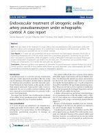
Figure 1 Ultrasound image: 32.3 weeks of gestation. Transverse
scan image thorough the fetal abdomen identifying a mass
occupying the entire left hemiabdomen (meconium pseudocyst),
with mixed echogenicity. No calcifications were observed.
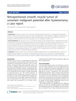
Figure 2 Ultrasound image: 32.3 weeks of gestation.
Longitudinal scan image thorough the fetal abdomen identifying
a mass occupying the entire left hemiabdomen (meconium
pseudocyst), with mixed echogenicity. No calcifications were
observed.
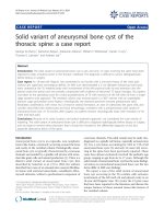
Figure 3 Magnetic resonance imaging: 32.4 weeks of gestation.
Longitudinal magnetic resonance image of the fetus demonstrating
the meconium pseudocyst; a 72 × 58 mm, heterogeneous,
mesenteric mass without necrosis causing significant distortion of
the small intestine to the left. No calcification or ascites were
observed.
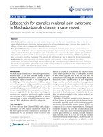
Figure 4 Magnetic resonance imaging: 32.4 weeks of gestation.
Transverse magnetic resonance image of the fetus demonstrating
the meconium pseudocyst; a 72 × 58 mm, heterogeneous,
mesenteric mass without necrosis causing significant distortion of
the small intestine to the left. No calcification or ascites were
observed.
Valladares et al. Journal of Medical Case Reports 2010, 4:292
/>Page 2 of 5
performed due to preterm labor associated with fetal
malposition (transverse situation). Birth weights were
1980 g (fetus 1) and 2060 g (fetus 2).
Laparotomy and bowel resection were performed
within the first day following delivery. During surgery a
10 cm, volvulated, necrotic portion of small intestine
was identified, at 10 cm from ileocecal valve. Small
bowel volvulus resection, termi no-terminal anastomosis,
and appendectomy were performed.
Pathological anatomy reports revealed distal ileum
vascular congestion, in testinal wall bleeding and areas of
acute inflammation.
The final diagnosis was a perforated ileum volvulus
and secondary meconium pseudocyst.
Bowel obstruction was suspected at three days follow-
ing the initial surgical intervention. A second laparot-
omy identified a segment of obstructed bowel. This was
resected and a termino-terminal re-anastomosis was
performed. Sweat chloride test for cystic fibrosis was
negative.
Due to the newborn’s torpid post-operative course and
lack of gastrointestinal tolerance, an exploratory laparot-
omy was performed 51 days after birth. Intra-opera-
tively, a stenosis of the re-anastomosis was observed.
Resection of a 5 cm section of bowel including ileocecal
valve, as well as ileostomy and colostomy were
performed.
The newborn remained hospitalized receiving total
parenteral nutritio n and with secretory diarrhea due to
short bowel syndrome, and died during the seventh
month of life.
Discussion
The differential diagnosis of a sonographically visualized
intra-abdominal cyst in a fetus is extensive, and includes
intestinal duplications cyst, mesenteric cysts, choledo-
chal cyst, meconium pseudocyst, congenital cyst of the
pancreas, renal cyst, obstructive uropathy, urachal cyst,
ovarian cyst, ureterocele and tumorous lesions such a s
cystic sacroccocygeal teraromas.
Fetal tumors comprise 0.5% to 2% of all childhood
neoplasms. Extra-cranial teratomas, neuroblastomas,
soft-tissue and intra-cranial tumors are the most com-
mon (85% of all tumors). The remaining 15% are made
up of renal tumors, liver tumors, retinoblastoma and
other less common processes that can mimic a tumor,
such as meconium peritonitis (cystic type) [1].
Meconium peritonitis is a sterile chemical peritonitis
caused by meconium extruding into the peritoneal cav-
ity through a small bowel perforation in utero. The esti-
mated prevalence is about 0.29 per 10,000 live births
and the mortality ranges from 11% to 50%. It usually
appears in the neonatal period with abdominal
distension, vomiting, acidosis and intra-abdominal
calcifications.
Perfora tion occurs most commonly in the ileum prox-
imal to an obstruction, but this cannot always be
demonstrated. The obstruction can be caused by atresia,
stenosis, volvulus, internal bowel hernia, Meckel’s diver-
ticulum, meconium ileus, or peritoneal bands. Intest inal
stenosis or atresia and meconium ileus account for 65%
of the cases. Adhesions between loops of intestine an d
omentum act to contain the meconium collection
extruded into the peritoneal cavity, creating a cystic
mass that can be visualized on ultrasound. The reactio n
may alternatively result in the formation of a solid non-
cystic mass with calcium deposits sealing off the per-
foration [2]. When t he formation of this apparently
solid abdominal mass occ urs, an accurate diagnosis
between an abdominal tumor and meconium collection
may be challenging.
In a review of 12 cases of meconium perit onitis, intra-
peritoneal calcifications were present in 60% of the
patients with cystic fibrosis and 100% of patients with-
out cystic fibrosis [3]. The authors postulate t hat pan-
creatic enzymes, which are in a low concentration in
80% of patients with cystic fibr osis, may be necessary
for the calcifications to occur. Our case showed no evi-
dence of cystic fibrosis. It is possible that the ultrasound
was performed soon after the creation of the pseudocyst
and before the calcification could be visible sonographi-
cally. Calcifications can develop within days, but may
need several weeks to be visible sonographically [3].
Cystic fibrosis is the most common fatal autosomal
recessive disease a mong Caucasian population, with a
frequency of one in 2000 to 3000 live births. The sweat
chloride test remains the primary test for the diagnosis
of this disease; the DNA testing is used for confirmation
of patients with intermediate sweat chloride results. The
sweat testing is performed by the collection of sweat
with pilocarpine iontophoresis, and chemical determina-
tion of the chloride concentration [4]. Meconium ileus
is the presenting problem in 10 to 20 percent of new-
borns with cystic fibrosis, and is virtually pathognomo-
nic of the disease. Volvulus in fetal life is suggestive of
cystic fibrosis; episodes of small bowel obstruction may
also occur in older children and adults.
Depending on when the bowel perforation occurs dur-
ing development and the severity of the inflammatory
reaction induced by the meconium extruded into the
peritoneal cavity, three differen t types of meconium
peritonitis can be described according to the ultrasound
findings [5]. The fibroadhesive type is the most frequent
and is characterized by an intense fibroblastic reaction
causing the f ormation of fibrotic membranes which are
adherent to the intestinal wall and cover the perforation.
Valladares et al. Journal of Medical Case Reports 2010, 4:292
/>Page 3 of 5
Ultrasound reveals the presence of diffuse punctiforme
hyper-echogenic lesions around the peritoneal cavity.
Intra-abdominal calcifications are not usually observed.
Ascitis, hydramnios or bowel loop dilatation are also
characteristic. The perforation may not be visualized as
it often seals spontaneously. The cystic type, as found in
the present case, is formed by a meconium collection
surrounded by fibrotic membranes (pseudocyst).
Through ultrasound imaging the pseudocyst appears as
a large meconiu m-fill ed cyst lined by a thick membrane
containing multiple calcium deposits and plaques. The
cystic type is usually formed secondary to a prenatal vol-
vulus with perforation [6]. The last category is the gen-
eralized type, and is the consequence of a peri-natal
perforation with meconium spread throughout the
abdominal cavity.
One s tudy [7] has described the relationship between
ultrasound findings and the post-natal course of meco-
nium peritonitis. A tot al of 69 cases were divided into
four grades according to their ultrasound features.
Grade 0, isolated intra-abdominal calcifications; grade 1,
intra-abdominal calcifications and ascites or pseudocyst
or bowel dilatation; grade 2, two associated findings;
grade 3, a ll sonographic features. The authors found an
increasing need for neonatal surgery with higher grades
of the sonographic classification [7]. Another study also
found a correlation between ultrasound features and
clinical implications [8]. Persistent ascites, pseudocyst or
dilated bowel loop were reported to be the most sensi-
tive predictors of post-natal surgery (92%, P < 0.022) [9].
Meconium pseudocysts are often accompanied by
polyhydramnios [10]. It is often the consequence of
associated bowel atresia or extrinsic mechanical obstruc-
tion of the bowel due to mass effect. A large fetal intra-
abdominal mass may additionally cause fetal lung imma-
turity; however, percutaneous drainage of these cysts
may cause leakage of the meconium into the amniotic
fluid.
The MR appearance of meconium pseudocysts have
been described in the literature [11,12].
With one exception, all cases of meconium pseudocyst
were associated with bowel dilatation or free intra-
abdominal flu id [13]. In another case [14], the meconium
pseudocyst was associated with dilated bowel and ascites,
but had no calcifications in a newborn with a normal
sweat test. A separate study describes 11 cases of meco-
nium peritonitis [15]. In one case from this study which
was si milar to ours, the only ultrasound finding was a
meconium pseudocyst. In nine o ther cases, the meco-
nium pseudocyst was associated with polyhydramnios,
ascites or dilated bowel loops. In the remaining case, fetal
ascites was the only ultrasound finding.
Treatment for meconium pseudocyst usually consists
of surgical resection, although definitive procedures in
the early neonatal period are usually difficult. Conse-
quently, many patients require more than one surgical
intervention. Some authors recommend immediate cyst
drainage and decompression through paracentesis fol-
lowing birth with delayed defi nitive resection [16]. The
prognosis was poor in the past, but has improved due to
the development of newer surgical techniques. Eckoldt
[15] demonstrated a successful management with
patient survival i n nine out of 11 cases. In cases with
underlying atresia, temporary diversion enterostomy
with planned secondary reconstruction at two to three
weeks showed good results. For large meconium pseu-
docysts, a two-stage approach with cyst decortication
and temporary enterostomy, followed by elective rever-
sal is the gold standard.
Conclusions
Meconium peritonitis is an uncommon fetal and neona-
talconditionanditshouldbeconsideredinthediffer-
ential diagnosis when an echogenic intra-abdominal
mass is observed. The prenatal appearance can be
accompanied by signs of bowel obstruction, such as
polyhydramnios and bowel dilatation. Generalized
hydrops increases the severity of this disease.
Surgery should be performed as soon as possible after
delivery and initial resuscitation although immediate
decompression paracentesis may result in a rapid
improvement in the overall state of the newborn while
preparation for surgery i s underway. A two stage-
approach with temporary enterostomy and delayed
reversal is the best choice.
Our case is unusual in comparison to other sonogra-
phically described prenatal cases due to the large size of
the pseudocyst, the absence of ascites, bowel dilatation,
or polyhydramnios, as well as a lack of abdominal calci-
fications in a newborn without cystic fibrosis.
The c linical utility of this case is the recognition that
meconium pseudocyst may present without typical
ultrasound features, and should be considered in the dif-
ferential diagnosis of an abdominal mass. This will facili-
tate delivery of appropriate treatment as soon as
possible after birth.
Consent
Written informed consent was obtained from the patient
for both her case and the case of her child for publica-
tion of this case report and any accompanying images.
A copy of the written consent is available for review by
the Editor-in-Chief of this journal.
Authors’ contributions
EV collected the clinical case, wrote the manuscript, and conducted the
literature search. DR collected previous similar clinical cases from the
literature, drafted the manuscript, and attended the discussion. AV visited
Valladares et al. Journal of Medical Case Reports 2010, 4:292
/>Page 4 of 5
the patient, made the ultrasound diagnosis, and gave advice on the
literature search. SC developed the article concept, provided ultrasound
images and contributed to writing the introduction. JML provided general
supervision and analyzed and interpreted the patient data. All authors have
read and approved the final manuscript
Competing interests
The authors declare that they have no competing interests.
Received: 22 October 2009 Accepted: 31 August 2010
Published: 31 August 2010
References
1. Albert A, Cruz O, Muntaner A, Vela A, Badosa J, Castañón M, Morales L:
Congenital solid tumors. A thirteen year review. Cir Pediatr 2004,
17(3):133-136.
2. Konge JC, de Chazal R, MacFadyen U, Taylor DJ: Antenatal diagnosis and
management of meconium peritonitis: a case report and review of the
literature. Ultrasound Obstet Gynecol 1995, 6:66-69.
3. Finkel LI, Slovis TL: Meconium peritonitis, intraperitoneal calcifications
and cystic fibrosis. Pediatr Radiol 1982, 12(2):92-93.
4. Denning CR, Huang NN, Cuasay LR, Shwachman H, Tocci P, Warwick WJ,
Gibson LE: Cooperative study comparing three methods of performing
sweat tests to diagnose cystic fibrosis. Pediatr 1980, 66:752.
5. Garel C: Imagerie d’une péritonite méconiale à présentation clinique
pseudotumorale. J Radiol 1997, 78:1288-1290.
6. Foster MA, Nyberg DA, Mahoney BS: Meconium peritonitis: prenatal
sonographic findings and clinical significance. Radiology 1987,
165:661-665.
7. Zangheri G, Andreani M, Ciriello E, Urban G, Incerti M, Vergani P: Fetal
intra-abdominal calcifications from meconium peritonitis: sonographic
predictors of postnatal surgery. Prenat Diagn 2007, 27(10):960-963.
8. Tseng JJ, Chou MM, Ho ES: Meconium peritonitis in utero: prenatal
sonographic findings and clinical implications. J Chin Med Assoc 2003,
66(6):355-359.
9. Shyu MK, Shih JC, Lee CN, Hwa HL, Chow SN, Hsieh FJ: Correlation of
prenatal ultrasound and postnatal outcome in meconium peritonitis.
Fetal Diagn Ther 2003, 18(4):255-261.
10. Olnick HM, Hatcher MB: Meconium peritonitis. JAMA 1953, 152:582-584.
11. Saguintaah M, Couture A, Veyrac C, Baud C, Quere MP: MRI of the fetal
gastrointestinal tract. Pediatr Radiol 2002, 32:395-404.
12. Veyrac C, Couture A, Saguintaah M, Baud C: MRI of the fetal GI tract
abnormalities. Abdom Imaging 2004, 29:411-420.
13. Simonosky V, Lisy J: Meconium pseudocyst secondary to ileal atresia
complicated by volvulus:antenatal MR demonstration. Pediatr Radiol 2007,
37:305-309.
14. Yang WT, Ho SW, Metreweli C: Case Report: Antenatal Sonographic
Diagnosis of meconium Peritonitis and Subseqüent Evolving Meconium
Pseudocyst Formation Without Peritoneal Calcification. Clin Radiol 1997,
52:477-479.
15. Eckoldt F, Heling KS, Woderich R, Kraft S, Bollmann R, Mau H: Meconium
peritonitis and pseudo-cyst formation: prenatal diagnosis and post-natal
course. Prenat Diagn 2003, 23:904-908.
16. Tanaka K, Hashizume K, Kawarasaki H, Iwanaka T, Tsuchida Y: Elective
surgery for cystic meconium peritonitis: report of two cases. J Pediatr
Surg 1993, 28(7):960-961.
doi:10.1186/1752-1947-4-292
Cite this article as: Valladares et al.: Meconium pseudocyst secondary to
ileum volvulus perforation without peritoneal calcification: a case
report. Journal of Medical Case Reports 2010 4:292.
Submit your next manuscript to BioMed Central
and take full advantage of:
• Convenient online submission
• Thorough peer review
• No space constraints or color figure charges
• Immediate publication on acceptance
• Inclusion in PubMed, CAS, Scopus and Google Scholar
• Research which is freely available for redistribution
Submit your manuscript at
www.biomedcentral.com/submit
Valladares et al. Journal of Medical Case Reports 2010, 4:292
/>Page 5 of 5