Báo cáo y học: " HIV-1 neutralization by monoclonal antibody against conserved region 2 and patterns of epitope exposure on the surface of native viruses" pot
Bạn đang xem bản rút gọn của tài liệu. Xem và tải ngay bản đầy đủ của tài liệu tại đây (663.88 KB, 8 trang )
BioMed Central
Page 1 of 8
(page number not for citation purposes)
Journal of Immune Based Therapies
and Vaccines
Open Access
Original research
HIV-1 neutralization by monoclonal antibody against conserved
region 2 and patterns of epitope exposure on the surface of native
viruses
Apichai Sreepian
1
, Jongruk Permmongkol
2
, Wannee Kantakamalakul
1
,
Sontana Siritantikorn
1
, Nattaya Tanlieng
1
and Ruengpung Sutthent*
1
Address:
1
Department of Microbiology, Faculty of Medicine Siriraj Hospital, Mahidol University, Bangkok, Thailand and
2
Faculty of Medical
Technology, Mahidol University, Bangkok, Thailand
Email: Apichai Sreepian - ; Jongruk Permmongkol - ;
Wannee Kantakamalakul - ; Sontana Siritantikorn - ; Nattaya Tanlieng - ;
Ruengpung Sutthent* -
* Corresponding author
Abstract
Background: Conserved neutralizing epitopes are considered to be a key role for eliciting broadly
neutralizing antibody (NAb). Previously, two conserved neutralizing epitopes of HIV-1 CRF01_AE
envelope were identified at amino acid 93-112 of the C1 (C1E) and at 218-239 of the C2 (C2E)
regions. To access the potency of antibody directed against conserved epitopes, a monoclonal
antibody (MAb) specific to the C2E region was developed and characterized.
Methods: The immunogenicity of two epitopes was examined by immunizing BALB/c mice with
the matching synthetic peptides. One MAb, C2EB5, directed against peptide C2E, was generated
by conventional methods, while C1E1 and C1E2 peptides induced slight antibody response in mice.
The neutralizing activity of MAb C2EB5 was examined using a peripheral blood mononuclear cell
(PBMC) based method and various HIV-1 subtypes including A, B, C, D, and CRF01_AE; C2EB5
was compared with other known neutralizing MAbs (4E10, 447-52D) and with sCD4. The
exposure of the C2 epitope on native virus was investigated using virus capture by these MAbs.
Results: The MAb C2EB5 demonstrated cross-neutralization against various HIV-1 subtypes. The
overall potency of MAb C2EB5 against 5 subtypes was ranked in the following order: subtype C>
CRF01_AE> subtype D> subtype A> subtype B. The epitope exposure for MAb C2EB5 was also
correlated with the neutralization properties of each subtype.
Conclusion: This study demonstrates the cross-clade neutralizing activity of a MAb directed
against an epitope located in the C2 region of the HIV-1 env and highlights differences in the
exposure of antigenic epitopes on the surface of various HIV-1 subtypes. The epitope for this newly
identified neutralizing MAb made against a subtype CRF01_AE peptide is particularly exposed in
subtype C viral isolates.
Published: 12 October 2009
Journal of Immune Based Therapies and Vaccines 2009, 7:5 doi:10.1186/1476-8518-7-5
Received: 25 September 2008
Accepted: 12 October 2009
This article is available from: />© 2009 Sreepian et al; licensee BioMed Central Ltd.
This is an Open Access article distributed under the terms of the Creative Commons Attribution License ( />),
which permits unrestricted use, distribution, and reproduction in any medium, provided the original work is properly cited.
Journal of Immune Based Therapies and Vaccines 2009, 7:5 />Page 2 of 8
(page number not for citation purposes)
Background
The great variability HIV-1 antigenic epitopes has been
considered to be a major mechanism used by the virus to
evade the host immune response. To elicit broadly neu-
tralizing antibody (NAb) against HIV-1, one or more con-
served epitopes should be recognized to overcome the
extensive antigenic diversity. However, there are few con-
served epitopes on the envelope protein that are accessible
for specific antibody binding and neutralization. These
epitopes have been hidden either by glycosylation or con-
formational masking [1,2]. The major targets of HIV-1
neutralizing antibodies are located on the surface gp120,
whose diverse antigenic epitopes mediate receptor and co-
receptor binding [3,4], and on the transmembrane gp41,
which causes membrane fusion and allows the virus to
gain entry into host cells [5]. A previous report has shown
that one-third of neutralizing specificities of subtypes B
and C neutralizing antibodies in polyclonal sera recognize
the CD4 binding site (CD4b) and gp41 epitopes, while
two-thirds of the antibodies were estimated to be directed
against unidentified epitopes [6].
Three monoclonal antibodies (2G12, IgG1b12, 447-52D)
directed against gp120 and three MAbs against gp41
(MAbs: 2F5, 4E10, Z13) have been extensively described
in their neutralizing activities. Of the anti-gp120 MAbs,
2G12 recognizes a unique epitope in a carbohydrate-rich
region on the outer domain involving the C3-V4 region
[7,8], whereas IgG1b12 binds to the CD4 binding site and
447-52D recognizes the V3 loop of gp120 [9]. The anti-
gp41 MAbs, 2F5, 4E10 and Z13 bind to the same contin-
uous membrane proximal region of gp41. 2F5 is mapped
to the conserved sequence ELDKWA [10], whereas 4E10
and Z13 recognize an epitope involving the sequence
NWF(D/N)IT, which is located on the C-terminus of the
2F5 epitope [11,12]. There have been several MAbs devel-
oped against various conserved epitopes that show some
neutralization, such as 17b and 48d. The MAbs 17b and
48d recognize a cluster of gp120 epitopes that are cen-
tered on the β 19 strand and partially overlap the co-recep-
tor binding site [13,14]. While many of the known HIV
Env MAbs are specific for conserved regions, several
reports have demonstrated that some variable amino acid
patterns lead to NAb resistance [15,16].
The emergence of circulating recombinant forms (CRFs)
has been recognized and it is thought that they will make
the HIV-1 epidemic more complex. This may have serious
issues for the future of antiretroviral therapy and vaccine
development. At least 32 circulating recombinant forms
have been reported in HIV-1 group M [17]. CRF01_AE, a
hybrid of subtype A (gag) and E (env), is an important
HIV-1 recombinant form prevalent in Asia. Since we dem-
onstrated some conserved neutralizable epitopes, which
are located on amino acids 93-112 (C1 region) and 218-
239 (C2 region) of HIV-1 CRF01_AE primary isolates in
previous study [18], we have attempted to test the immu-
nogenicity of these conserved epitopes and potencies of
these induced MAbs. Toward that aim, we immunized
BALB/c mice with peptides corresponding to these
epitopes and MAbs specific to these epitopes were pro-
duced. A monoclonal antibody directed against peptide
C2E (amino acids 218-239) was produced and the neu-
tralization pattern for this C2EB5 MAb has revealed a
cross-neutralizing activity and the presence of antigenic
epitopes for this site on the surface of native viruses. The
antigenic portion of this epitope appears to be particularly
exposed in subtype C envelopes.
Methods
Monoclonal antibodies 4E10 and 447-52D and soluble
CD4 (sCD4) [19]
Two human MAbs (4E10 and 447-52D) and sCD4 were
kindly gifted from the National Institute for Biological
Standards and Control (NIBSC, UK) whereas MAb C2EB5
was produced in this study [20]. The MAb 447-52D recog-
nizes GPGR motif at amino acids 312-315 on the tip of V3
loop whereas MAb 4E10 recognizes NWFDIT located at
amino acid position 671-676 in gp41. Soluble CD4
(sCD4) is an entry inhibitor devised as a decoy for the
HIV-1 gp120 protein. These MAbs and sCD4 were aliquot
and stored at -20°C.
Primary isolates and T-cell line adapted (TCLA) strains of
HIV-1
Five HIV-1 CRF01_AE primary isolates were obtained
from National HIV Repository and Bioinformatic Center
(Thailand) (NHRBC). These viruses with prefix MENO
were collected from HIV-1 seropositive cases from the
northern part of Thailand through National serosurveil-
lance in the year 2002, including MENO12 (AY243187),
MENO23 (AY243194), MENO24 (AY243195), MENO31
(AY243202) and MENO43 (AY243213). HIV-1 TCLA
strains including, 2 subtype A (92RW009 and VI191), 4
subtype B (QH0692, SF162, IIIB and MN), 2 subtype C
(92BR025 and DU174), 1 subtype D (92UG024) and 2
CRF01_AE (NP1525 and NPO3) were obtained from
National Institute for Biological Standards and Control
(NIBSC, UK). These viruses were thawed from liquid
nitrogen and co-cultivated with PHA-stimulated donor
PBMCs in IL-2 medium. The viral multiplications were
followed up by measuring p24 level (Vironostika HIV-1
Antigen, bioMerieux). The value of 50% tissue culture
infectious dose (TCID
50
) for each virus stock, both pri-
mary isolates and TCLA strains, was titrated on PHA-stim-
ulated PBMCs and the value of TCID
50
was calculated by
Spearman-Karber method.
Journal of Immune Based Therapies and Vaccines 2009, 7:5 />Page 3 of 8
(page number not for citation purposes)
Peptides corresponding to conserved neutralizable
epitopes on C1 and C2 regions of gp120 (HIV-1 CRF01_AE)
The designation of peptides corresponding C1 (C1E1 and
C1E2) and C2 regions (C2E) has been described previ-
ously [18]. The peptides were designed from alignment of
env nucleotide sequences (C2-V4) obtained from 43 HIV-
1 CRF01_AE primary isolates (GenBank under accession
number AF373037
-AF373043, AY005164-AY005179 and
AF322195
-AF322214) [18]. The amino acid sequence of
peptide C1E1 (amino acids 93-112 of C1 region) is ENF-
NMWKNN
MVEQMQEDVIS whereas amino acid
sequence of peptide C1E2 is different from C1E1 at posi-
tion 101, where an N residue is changed to a K, as under-
lined in Table 1. Peptide C2E, 22-mer peptide containing
DPIPIHYCTPAGYAILKCNDKN, is located at residues
218-239 of the C2 region. The activities of these peptides
have been investigated in previous studies by inhibiting
the neutralizing activities of sera from long-term non-pro-
gressors (LTNPs) infected with HIV-1 CRF01_AE [18].
Immunization and monoclonal antibody production
Six to eight week old female BALB/c mice (from National
Laboratory Animal Center, Thailand) were immunized
with synthetic peptides by intraperitoneal injection proto-
cols. Two groups of mice (2 mice/group) were primed
with 100 μg peptide/100 μl complete Freund's adjuvant
(Sigma, USA). Two weeks later, the first group was intra-
peritoneally boosted with 100 μg peptide/100 μl incom-
plete Freund's adjuvant (IFA) (Sigma, USA) whereas the
second group was boosted with 200 μg peptide/100 μl
IFA. For control groups, the mice were immunized with
normal saline instead of peptide utilized the same prepa-
ration of peptide immunizations. All mice were bled and
sacrificed after boosting 3 days, and then all sera was kept
frozen. The spleenocytes were separated immediately to
hybridize with myeloma cells Ag8.653 by using 41.3%
polyethylene glycol (Sigma, USA) as fusion reagent [20].
The hybridoma cells were cultured in HAT medium, RPMI
1640 medium supplemented with hypoxanthine-ami-
nopterin-thymidine (Sigma, USA) and 20% FBS, for a
week before transferring to HT medium, medium without
aminopterin, until the colonies of hybridoma cells were
grown. Initially, the hybridoma cells were diluted in
round-bottom 96-well plate by limiting dilutions to
obtain 1-10 cells per well and cultured for 5-7 days. The
supernatant from each well was screened for antibody by
peptide ELISA to identify the antibody producing clones.
Then, they were subcloned by limiting dilutions (< 1.0
cell per well) twice and antibody positive clones were
selected. Monoclonal IgG was purified by passing culture
supernatant through ProPur™ protein G spin column
(Nunc, Denmark) according to the manufacturer's
instruction. IgG purity was determined by SDS-PAGE and
Western blot with goat anti-mouse conjugated with HRP
(Invitrogen, USA).
Detection of antibody responses in BALB/c mice and
antibody produced in hybridoma cell lines by peptide
ELISA
The flat-bottom 96-well plates were coated with 100 μl of
peptide (5 μg/ml carbonate buffer, pH 9.6) for overnight
at 4°C. Following washing steps, the plates were blocked
with blocking buffer (5% skimmed milk, 0.3% Tween20
in PBS) for 1 h. The plates were washed again before incu-
bating with 100 μl of sera from BALB/c mice (for detect Ab
response in BALB/c mice) or 100 μl of culture superna-
tants (for detect Ab production in hybridoma cell culture)
for 1 h at 37°C. After washing steps, 100 μl of HRP-conju-
gated goat-anti mouse IgG (Invitrogen, USA) was added
and the plates were allowed to incubate for 1 h at 37°C.
Then, 100 μl of TMB substrate (Zymed, USA) was added
and the reaction was stopped with 100 μl of 1 M H
2
SO
4
.
The absorbance was measured at wavelength 450 nm. The
cutoff is defined as the mean value of absorption of serum
samples from mice immunized with normal saline or that
of fresh culture medium.
HIV-1 neutralization assay
Neutralization test was assayed by a method based on
PBMC infection and reduction of p24 gag protein in cul-
ture fluids, as described previously [18]. Briefly, 75 μl of
virus supernatant (30 TCID
50
) was pre-incubated with
equal volume of serially diluted MAb or sCD4 at 37°C for
1 h. After that, 75 μl of PHA-stimulated PBMCs (1.34 ×
10
6
cells/ml) was added and allowed to incubate at 37°C,
5% CO
2
for 18 h. The infected cells were then washed
twice and re-suspended in 400 μl of IL-2 medium before
transferring 200 μl of cell suspension into duplicated well
of round-bottom 96-well plate. The replication of the
virus in supernatant was followed up by measuring p24
antigen on day 4. For virus control, it was performed by
incubating the virus supernatant with HIV seronegative
sera. To calculate percent neutralizing activity, p24 level of
virus control was subtracted with p24 level of virus con-
taining each dilution of MAb or sCD4 before being
divided by p24 level of virus control and then multiplied
by 100. The toxicity of MAb C2EB5 on PBMCs was
assayed by adding various concentration of C2EB5 into
50,000 PBMCs (10
6
cells/ml) overnight. The viable
PBMCs were counted after washing and stained with vital
dye (typan blue).
Determination of antigenic exposures on the surface of
intact, native viruses
This procedure was modified from the protocol, which
has been described previously [21,22]. Briefly, the flat-
bottom 96-well plates were coated directly with 100 μl of
each MAb or sCD4 (10 μg/ml carbonate buffer, pH 9.6).
Following washing steps, the plates were blocked with
blocking solution and incubated with 100 μl of virus
supernatant (100 ng p24 antigen/ml) for 1 h. After wash-
Journal of Immune Based Therapies and Vaccines 2009, 7:5 />Page 4 of 8
(page number not for citation purposes)
Table 1: Alignment of amino acid sequences of the Env glycoprotein gp120 at position 218 to 239.
Clade Accession
no.
218 219 220 221 222 223 224 225 226 227 228 229 230 231 232 233 234 235 236 237 238 239 240
C2E DEPIPIHYCTPAGYAILKCNDKN
NP1525 AE AAW57720 K
MENO23 AE AY621208
MENO43 AE AY621222
92BR025 C AAB61124
A N.T
DU174 C DQ411853
A NNK
SF162 B P19550
E A F K
QH0692 B AY669730
F A F N.T
IIIB B AB037858
E A F N.T
MN B P05877
E A F K
VI191 A ABY26917
E A F R E
92RW009 A AY669700
F N A F K K
92UG024 D AAT67532
E A F N.M
Dots and dashes indicate similarity of amino acids, respectively.
Journal of Immune Based Therapies and Vaccines 2009, 7:5 />Page 5 of 8
(page number not for citation purposes)
ing steps to remove unbound viruses, 250 μl of 1% Triton
X-100 was added to remove the contents of each well for
measuring p24 antigen. The control well was performed
by adding 100 μl of IL-2 medium and influenza virus (10
TCID50) instead of HIV-1 supernatant.
Statistic analysis
All statistical analyses were performed on non-parametric
analysis by program SPSS version 1.5. The association
between antigenic exposures and HIV-1 subtypes were
determined by Wilcoxon Signed-Rank test. The difference
of antigenic epitope exposure of MAb C2EB5 and neutral-
izing activity was determined by Mann-Whitney U test.
Results
C1E1, C1E2 and C2E peptide immunogenicity
Previously, we have investigated the activity of epitopes
located at amino acids 93-112 of the C1 and 218-239 of
the C2 regions as conserved neutralizable epitopes in HIV-
1 CRF01_AE primary isolates by using peptides from
C1E1, C1E2 and C2E to inhibit neutralizing activities of
sera from HIV-1 CRF01_AE infected LTNPs [18]. The
results led to the hypothesis that antibodies directed
against theses epitopes should be broadly neutralizing
antibodies. These peptides were used to immunize BALB/
c mice. There were low titer antibody responses, as meas-
ured by peptide ELISA, in sera from mice immunized with
peptides C1E1 and C1E2. However, peptide C2E induced
antibody responses in BALB/c mice to a higher titer than
that of the C1E1 and C1E2 peptides. The response to the
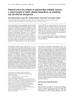
C2E peptide occurred in a dose dependent manner (Fig-
ure 1). The mouse antisera from C2E immunization neu-
tralized the HIV-1 CRF01_AE laboratory strain NP03 at a
1:30 dilution (data not shown). We therefore proceeded
to produce monoclonal antibody from C2E immunized
mice.
Neutralizing activity of MAbs directed against peptide C2E
To explore the neutralizing activity of MAbs directed
against peptide C2E, a murine MAb specific to this pep-
tide was produced. The MAb clone B5 with the greatest
neutralizing activity against NPO3 (CRF01_AE) HIV-1
strain was selected for further study and named MAb
C2EB5. This MAb C2EB5 did not show any cross-reaction
with peptides C1E1 and C1E2 by the ELISA method. The
neutralizing activity of MAb C2EB5 was investigated
against 14 isolates of HIV-1 from various subtypes includ-
ing, subtype A; 92RW009 and VI191, subtype B; MN, IIIB,
QH0692, and SF162, subtype C; 92BR025 and DU174,
subtype D; 92UG024 and CRF01_AE; NPO3, CM244,
NP1525, MENO23, MENO43. HIV-1 isolates used in this
neutralization study were selected based on C2 amino
acid similarity (CRF01_AE) and difference (other sub-
types) to explore the cross-reactivity of the monoclonal
antibody (Table 1). The results revealed that MAb C2EB5
neutralized subtype A, B, C, D and CRF01_AE with mean
IC
50
± SD 32.00 ± 6.92, > 50, 24.94 ± 21.11, 29.78 and
21.81 ± 6.71 μg/ml, respectively (Table 2). The cellular
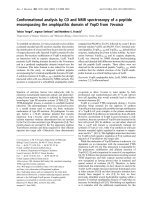
toxicity of MAb C2EB5 was demonstrated at a concentra-
tion > 50 μg/ml, as shown in Figure 2.
The exposure of antigenic epitopes on the surface of
intact, native HIV-1
The MAb C2EB5 was also characterized by performing
virus capture to assess the exposure of the antigenic
epitopes on the virion; data for C2EB5 was compared with
capture data for the 4E10 and 447-52D MAbs and for
sCD4. We initially assessed MAb binding to native, intact
viruses by a virus binding ELISA [21]. We localized various
epitopes by coating MAbs and sCD4 onto flatted-bottom
96-well plates, and adding the native viruses, without
ionic detergent treatment, for attachment. The exposures
of antigenic epitopes to these MAbs were compared. The
virus binding activity (± SD) of HIV-1 subtypes A, B, C, D
and CRF01_AE against MAb C2EB5 were 11.59 ± 0.36,
5.36 ± 3.39, 35.65 ± 3.56, 31.54 and 29.89 ± 5.18 pg/ml,
respectively. HIV-1 subtype B virions showed the lowest
amount of antibody capture by MAb C2EB5, and this was
significantly lower than that observed for other subtypes
(p < 0.05). This observation was correlated with low neu-
tralizing activity of the C2EB5 MAb against HIV-1 subtype
Dose response curve of antibody production in immunized miceFigure 1
Dose response curve of antibody production in
immunized mice. Sera from 5 mice per group were col-
lected 1 week after the last immunization and tested by
ELISA for the presence of specific antibodies. The peptides
from C1E1 and C1E2 represented amino acids 93-112 of the
C1 region, whereas peptides C2E represented amino acids
218-240 of the C2 region. Antibody titers of sera from mice
immunized with C1E1, C1E2, and C2E at 50, 100, and 200
μg, are expressed as the log
2
values of reciprocal endpoint
titers. The control group was injected with normal saline.
The immunization by peptides C1E1 and C1E2 induced a
poor antibody response, whereas peptide C2E induced a
robust antibody response, in a dose dependent manner.
Journal of Immune Based Therapies and Vaccines 2009, 7:5 />Page 6 of 8
(page number not for citation purposes)
B isolates. HIV-1 subtype C demonstrated high antigenic
epitope exposure to MAb C2EB5, which also correlated
well with the neutralizing activity of the C2EB5 MAb
against subtype C isolates. However, this comparison is
based upon analysis of a small number of samples inves-
tigated in this study.
Discussion
The antigenic diversity of HIV-1, particularly within the
Env glycoprotein, is a major tool used by the virus as an
immune evasion strategy, and this poses a major obstacle
for the development of an effective HIV-1 vaccine. There-
fore, a major focus of vaccine developers has been the dif-
ficulty in the elicitation of a broadly neutralizing antibody
response. This effort has been directed towards a limited
number of conserved epitopes on the envelope glycopro-
tein of HIV-1 primary isolates. Previously, we attempted
to define these conserved neutralizable epitopes in
CRF01_AE primary isolates from Thailand. We defined
locations of various conserved epitopes and utilized these
data to design synthetic peptides. We found that synthetic
peptides representing amino acids 93-112 (C1E1 and
C1E2) of C1 and 218-239 (C2E) of C2 regions could
absorb NAbs in sera collected from Thai long-term non-
progressors (LTNPs). Presence of these NAbs in sera of
these subjects implies that these amino acids are associ-
ated with particular properties of neutralizable epitopes.
Recently, these data were re-examined by creating a mon-
oclonal antibody directed against peptide C2E to investi-
gate its neutralization property.
Unfortunately, conserved neutralizable epitopes appear
to be poorly immunogenic and Abs against them are
rarely produced in infected subjects [23,24]. The peptides
C1E1, C1E2 and C2E were also described to be poorly
antigenic. Our previous study demonstrated that these
peptides were bound at low titers by sera from HIV-1
infected individuals [18]. We found that peptide C2E
could induce an antibody response in BALB/c mice
whereas peptides C1E (C1E1 and C1E2) failed to do so.
Table 2: Neutralization and virus capture (epitope exposure) of 14 HIV-1 isolates by monoclonal antibodies C2EB5, 4E10, 447-52D,
and sCD4.
HIV-1 strain Subtype IC
50
(μg/ml) Virus binding activity
3
(p24 antigen (pg/ml))
C2EB5
1
447-52D 4E10 sCD4 C2EB5 447-52D sCD4
92RW009 A 36.89 22.5 17.5 8.7 11.33 7.26 6.8
VI191 A 27.1 15 13.75 8.7 11.84 17.78 7.33
QH0692 B > 50 10.13 > 25 10 3.37 28.59 5.2
SF162 B > 50 3.7 1.25 7.5 9.72 35.16 8.74
MN B > 50 6.25 13.75 9.89 6.25 20.6 5.4
IIIB B > 50 8.06 > 25 4.2 2.08 27.69 22.8
92BR025 C 10.01 15 9 10 38.16 5.08 29.44
DU174 C 19.87 1.5 1.25 10 33.13 24.19 32.74
92UG024 D 29.78 21.25 20 6.25 31.54 5.41 29.27
NPO3 AE 12.8 > 25 6.47 3.79 32.47 13.2 29.73
CM244 AE 17.5 21.5 7.5 10 35.55 15.17 28.71
NP1525 AE 12.7 > 25 15 1.87 30.1 2.51 18.68
MENO23
2
AE 28.35 > 25 15 10 29.5 4.84 4.66
MENO43
2
AE 27.72 > 25 17.3 10 21.62 0 11.23
1
The neutralization activity of C2EB5 at concentrations more than 50 μg/ml was not reported because of cellular toxicity observed above this
concentration.
2
HIV-1 primary isolate
3
The influenza virus was used as negative control virus in each test.
Journal of Immune Based Therapies and Vaccines 2009, 7:5 />Page 7 of 8
(page number not for citation purposes)
The differences amongst these peptides to induce anti-
body responses in BALB/c mice might be due to the fact
that amino acid substitutions in peptide C2E results in the
presence of highly immunogenic amino acids (His, Lys,
Ala, Leu, Asp and Arg) within this epitope. These amino
acids occur at a greater frequency than in the C1E pep-
tides. The C2E (218-239) epitope is located around a β-
turn near the loop α domain and C1E (93-112) spans the
coil region located in the inner domain of gp120. These
positions within C1E might be difficult for antibodies to
recognize. However, previously, we found that there were
NAbs against these epitopes in the sera of HIV-1 LTNPs
[18]. The C1E and C2E epitopes might be less potent in
vitro due to their lacking of conformational structure,
combined epitopes, and allelic representations [25-27].
Indeed, the epitopes around amino acids 93-112 and 218-
239 have been previously described, including epitopes at
amino acids 90-100 of C1 and 222-231 of C2. Several
MAbs against these epitopes have also been produced
[23,24]. The MAb against 222-231 was reported to be
reactive with a denatured form of gp120 [24], whereas we
demonstrated that our MAb against amino acids 218-239
could neutralize native viruses albeit at high concentra-
tions of MAb. This might be due to location of this
epitope at the inner domain of gp120. While the MAb
C2EB5 showed poor neutraliztion against the subtype B
pseudovirus SF162 in the TZM-bl pseudovirus neutraliza-
tion assay (data not shown) [28,29], it will be interesting
to further test the breadth of this MAb, especially against
subtype C isolates in this assay. A low IC50 against SF162
was also observed in the PBMC-based assay [IC50>50 μg/
ml] (Table 2)
The reason that MAb C2EB5 was able to neutralize HIV-1
subtype C comparable to CRF01_AE may be due to the
homology within the C2 amino acids (218-239) for these
2 subtypes (except at only one position at 227, Table 1).
In contrast, HIV-1 subtype B contains 3-4 amino acid
(position 218, 227, 231, 238) differences from CRF01_AE
C2 amino acid. Interestingly, subtype A and D also have
3-4 amino acid (position 218, 227, 231, 237 or 238) dif-
ferences from CRF01_AE, but they could be neutralized by
MAb C2EB5 potently. The C2 (218-239) epitopes of sub-
types A and D might be more exposed than subtype B
epitopes because of shorter variable loops, such as V1-V4,
or perhaps a lack of glycosylation sites that shield the con-
served C2 neutralizable epitopes [30]. The neutralization
resistance of HIV-1 subtype B against MAb C2EB5 was
likely due to a reduced exposure of this epitope on the sur-
face of this HIV-1 subtype B. However, this study is pre-
liminary and further experiments will be required to
confirm these observations.
Conclusion
This is the first such study utilizing amino acid sequences
of HIV-1 CRF01_AE primary isolates to design MAb. This
MAb, in addition to neutralizing CRF01_AE, also cross-
neutralizes other subtypes, particularly subtype C, which
accounts for the largest population of HIV-1 infection in
the world. As described above, high concentration of MAb
C2EB5 was required to neutralize subtype B. However, it
is our hope that MAbs directed against conserved regions
are an alternative way to develop an effective vaccine
against HIV. Accordingly, these data may facilitate our
understanding of essential characteristics to design an
immunogen to induce broadly neutralizing antibodies;
this information may assist in the development of an
effective HIV-1 vaccine.
Abbreviations
CRF: circulating recombinant form; gp: glycoprotein;
HRP: horse radish peroxidase; IFA: incomplete Freund's
adjuvant; IL-2: interleukin-2; LTNP: long-term non-pro-
gressor; MAb: monoclonal antibody; NAb: neutralizing
antibody; PBMC: peripheral blood mononuclear cell;
PHA: phytohemagglutinin; sCD4: soluble CD4; SD:
standard deviation; TCID
50
: 50% tissue culture infectious
dose; TCLA: T-cell line adapted.
Competing interests
The authors declare that they have no competing interests.
Authors' contributions
AS participated in the design of the study, determined
immunogenicities, performed MAb, investigated neutral-
izing activities, analyzed data and drafted the manuscript.
JP participated in determining immunogenicities and per-
forming MAb. WK and SS participated in the design of this
study and were responsible for data analysis. NT prepared
virus primary isolates and TCLA strains. RS conceived of
the study, participated in the design of this study, ana-
Cellular toxicity of the C2EB5 MAb tested at various concen-tration on PBMCsFigure 2
Cellular toxicity of the C2EB5 MAb tested at various
concentration on PBMCs. Some toxic effects for the
PBMC target cells were observed above concentrations > 50
ug/ml.
0 1.56 3.13 6.25 12.5 25 50 100 200
ʅ
Journal of Immune Based Therapies and Vaccines 2009, 7:5 />Page 8 of 8
(page number not for citation purposes)
lyzed data and drafted the manuscript. All authors have
read and approved the final manuscript.
Acknowledgements
We thank Dr. Susan Zolla-Pazner, Dr. Hermann Katinger, National Insti-
tute for Biological Standards and Control (NIBSC), and National HIV
Repository and Bioinformatic Center (Thailand) for providing monoclonal
antibodies 447-52D, 4E10, sCD4, and HIV-1 isolates, respectively. We also
thank Dr. David Montefiori and Dr. Victoria Polonis for supporting the
TZM-bl neutralization assay. This study was supported from the Thailand
Research Fund through the Royal Golden Jubilee Ph.D. Program (Grant No.
PHD/0100/2546) to Apichai Sreepian and Prof. Dr. Ruengpung Sutthent.
References
1. Poignard P, Saphire EO, Parren PW, Burton DR: Gp120: biological
aspects of structural features. Annu Rev Immunol 2001,
19:253-274.
2. Wei X, Decker JM, Wang S, Hui H, Kappes JC, Wu X, Salazar-
Gonzalez JF, Salazar MG, Kilby JM, Saag MS, Komarova NL, Nowak
MA, Hahn BH, Kwong PD, Shaw GM: Antibody neutralization
and escape by HIV-1. Nature 2003, 422:307-312.
3. Chan DC, Kim PS: HIV entry and its inhibition. Cell 1998,
93:681-684.
4. Jones PL, Korte T, Blumenthal R: Conformational changes in cell
surface HIV-1 envelope glycoproteins are triggered by coop-
eration between cell surface CD4 and co-receptor. J Biol Chem
1998, 273:404-409.
5. Este JA, Telenti A: HIV entry inhibitors. Lancet 2007, 370:81-88.
6. Binley JM, Lybarger EA, Crooks ET, Seaman MS, Gray E, Davis KL,
Decker JM, Wycuff D, Harris L, Hawkins N, Wood B, Nathe C, Rich-
man D, Tomaras GD, Bibollet-Ruche F, Robinson JE, Morris L, Shaw
GM, Montefiori DC, Mascola JR: Profiling the specificity of neu-
tralizing antibodies in a large panel of plasmas from patients
chronically infected with human immunodeficiency virus
type 1 subtypes B and C. J Virol 2008, 82:11651-8.
7. Trkola A, Pomales A, Yuan H, Korber B, Maddon PJ, Allaway GP, Kat-
inger H, Barbas CF 3rd, Burton DR, Ho DD, Moore JP: Cross-clade
neutralization of primary isolates of human immunodefi-
ciency virus type 1 by human monoclonal antibodies and
tetrameric CD4-IgG. J Virol 1995, 69:6609-6617.
8. Trkola A, Purtscher M, Muster T, Ballaun C, Buchacher A, Sullivan N,
Srinivasan K, Sodroski J, Moore JP, Katinger H: Human monoclonal
antibody 2G12 defines a distinctive neutralization epitope on
the gp120 glycoprotein of human immunodeficiency virus
type 1. J Virol 1996, 70:1100-1108.
9. Zwick MB, Parren PW, Saphire EO, Church S, Wang M, Scott JK,
Dawson PE, Wilson IA, Burton DR: Molecular features of the
broadly neutralizing immunoglobulin G1 b12 required for
recognition of human immunodeficiency virus type 1 gp120.
J Virol 2003, 77:5863-5876.
10. Muster T, Steindl F, Purtscher M, Trkola A, Klima A, Himmler G,
Ruker F, Katinger H: A conserved neutralizing epitope on gp41
of human immunodeficiency virus type 1. J virol 1993,
67:6642-6647.
11. Buchacher A, Predl R, Strutzenberger K, Steinfellner W, Trkola A,
Purtscher M, Gruber G, Tauer C, Steindl F, Jungbauer : Generation
of human monoclonal antibodies against HIV-1 proteins;
electrofusion and Epstein-Barr virus transformation for peri-
oheral blood lymphocyte immortalization. AIDS Res Hum
Retrvir 1994, 10:359-369.
12. Zwick MB, Labrijn AF, Wang M, Spenlehauer C, Saphire EO, Binley
JM, Moore JP, Stiegler G, Katinger H, Burton DR, Parren PW:
Broadly neutralizing antibodies targeted to the membrane-
proximal external region of human immunodeficiency virus
type 1 glycoprotein gp41. J Virol 2001, 75:10892-10905.
13. Rizzuto C, Sodroski J: Fine definition of a conserved CCR5-
binding region on the human immunodeficiency virus type 1
glycoprotein 120. AIDS Res Hum Retrovir 2000, 16:741-749.
14. Wu L, Gerard NP, Wyatt R, Choe H, Parolin C, Ruffing N, Borsetti
A, Cardoso AA, Desjardin E, Newman W, Gerard C, Sodroski J:
CD4-induced interaction of primary HIV-1 gp120 glycopro-
teins with the chemokine receptor CCR-5. Nature 1996,
384:179-183.
15. Mo H, Stamatatos L, IP JE, Barbas CF, Parren PW, Burton DR, Moore
JP, Ho DD: Human immunodeficiency virus type 1 mutants
that escape neutralization by human monoclonal antibody
IgG1b12. J Virol 1997, 71:6869-6874.
16. Purtscher M, Trkola A, Grassauer A, Schulz PM, Klima A, Dopper S,
Gruber G, Buchacher A, Muster T, Katinger H: Restricted anti-
genic variability of the epitope recognized by neutralizing
gp41 antibody 2F5. AIDS 1996, 10:587-593.
17. HIV-1 subtype and circulating form (CRF) reference
sequences 2005 [ />PENDIUM/2005/partI/leitner.pdf].
18. Sreepian A, Srisurapanon S, Horthongkham N, Tunsupasawasdikul S,
Kaoriangudom S, Khusmith S, Sutthent R: Conserved neutralizing
epitopes of HIV type 1 CRF01_AE against primary isolates in
long-term nonprogressors. AIDS Res Hum Retroviruses 2004,
20:531-542.
19. Binley JM, Wrin T, Korber B, Zwick MB, Wang M, Chappey C, Stiegler
G, Kunert R, Zolla-Pazner S, Katinger H, Petropoulos CJ, Burton DR:
Comprehensive cross-clade neutralization analysis of a panel
of anti-human immunodeficiency virus type 1 monoclonal
antibodies. J Virol 2004, 78:13232-13252.
20. Fuller SA, Takahashi M, Hurrell JGR: Preparation of monoclonal
antibodies. In Current protocols in molecular biology Edited by:
Ausubel FM, Brent R, Kingston RE, Moore DD, Seidman JG, Smith JA,
Struhl K. New York: John Wiley and Sons; 1991:11.01-11.11.5.
21. Nyambi PN, Mbah HA, Burda S, Williams C, Gorny MK, Nadas A,
Zolla-Pazner S: Conserved and exposed epitopes on intact,
native, primary human immunodeficiency virus type 1 viri-
ons of group M. J Virol 2000, 74:7096-7107.
22. Kusk P, Holmback K, Lindhardt BO, Hulgaard EF, Bugge TH: Map-
ping of two new human B-cell epitopes on HIV-1 gp120. AIDS
1992, 6:1451-1456.
23. Phogat S, Wyatt R: Rational modifications of HIV-1 envelope
glycoproteins for immunogen design. Cur Pharm Design 2007,
13:213-227.
24. Burton DR, Desrosiers RC, Doms RW, Koff WC, Kwong PD, Moore
JP, Nabel GJ, Sodroski J, Wilson IA, Wyatt RT: HIV vaccine design
and the neutralizing antibody problem. Nat Med 2004,
5:233-236.
25. Draenert R, Allen TM, Liu Y, Wrin T, Chappey C, Verrill CL, Sirera
G, Eldridge RL, Lahaie MP, Ruiz L, Clotet B, Petropoulos CJ, Walker
BD, Martinez-Picado J: Constraints on HIV-1 evolution and
immunodominance revealed in monozygotic adult twins
infected with the same virus. J Exp Med 2006, 203:529-539.
26. Zhang PF, Cham F, Dong M, Choudhary A, Bouma P, Zhang Z, Shao
Y, Feng YR, Wang L, Mathy N, Voss G, Broder CC, Quinnan GV Jr:
Extensively cross-reactive anti-HIV-1 neutralizing antibodies
induced by gp140 immunization. Proc Natl Acad Sci USA 2007,
104:10193-10198.
27. Choudhry V, Zhang M-Y, Sidrov IA, Bouma P, Cham F, Choudhary A,
Rybak SM, Fouts T, Montefiori DC, Broder CC, Quinnan GV Jr, Dim-
itrov DS: Cross-reactive HIV-1 neutralizing monoclonal anti-
bodies selected by screening of an immune human phage
library against an envelope glycoprotein (gp140) isolate from
a patient (R2) with broadly HIV-1 neutralizing antibodies.
Virology 2007, 363:79-90.
28. Li M, Gao F, Mascola JR, Stamaattos L, Polonis VR, Koutsoukos M,
Voss G, Goepfert P, Gilbert P, Greene KM, Bilska M, Kothe DL, Sala-
zar-Gonzalez JF, Wei X, Decker JM, Hahn BH, Montefiori DC:
Human immunodeficiency virus type 1 env clones from
acute and early subtype B infections for standardized Assess-
ments of vaccine-elicited neutralizing antibodies. J Virol 2005,
79:10108-25.
29. Brown BK, Wieczorek L, Sanders-Buell E, Borges AR, Robb ML, Birx
DL, Michael NL, McCutchan FE, Polonis VR: Cross-clade neutrali-
zation patterns among HIV-1 strains from the six major
clades of the pandemic evaluated and compared in two dif-
ferent models. Virology 2008, 375:529-38.
30. Moore JP, Sattentau Q, Wyatt R, Sodroski J: Probing the structure
of the human immunodeficiency virus surface glycoprotein
gp120 with a panel of monoclonal antibodies. J Virol 1994,
68:469-484.