Báo cáo y học: "Demonstration of a novel technique to quantitatively assess inflammatory mediators and cells in rat knee joints" pps
Bạn đang xem bản rút gọn của tài liệu. Xem và tải ngay bản đầy đủ của tài liệu tại đây (457.76 KB, 8 trang )
BioMed Central
Page 1 of 8
(page number not for citation purposes)
Journal of Inflammation
Open Access
Research
Demonstration of a novel technique to quantitatively assess
inflammatory mediators and cells in rat knee joints
Nicola J Barton*
1
, David A Stevens
2
, Jane P Hughes
2
, Adriano G Rossi
3
,
Iain P Chessell
2
, Alison J Reeve
2
and Daniel S McQueen
1
Address:
1
Division of Neuroscience, University of Edinburgh, Medical College, 1 George Sq, Edinburgh, EH8 9JZ, UK,
2
Neurology CEDD,
GlaxoSmithKline R&D Ltd, Harlow, Essex CM19 5AW, UK and
3
MRC Centre for Inflammation Research, The Queens Medical Research Institute,
University of Edinburgh, EH16 4TJ, UK
Email: Nicola J Barton* - ; David A Stevens - ; Jane P Hughes - ;
Adriano G Rossi - ; Iain P Chessell - ; Alison J Reeve - ;
Daniel S McQueen -
* Corresponding author
Abstract
Background: The inflammation that accompanies the pain and swelling associated with osteo- and
rheumatoid arthritis is mediated by complex interactions of inflammatory mediators. Cytokines
play a pivotal role in orchestrating many of these processes, including inflammatory cell
recruitment, adhesion and activation. In addition, prostaglandins are secreted into the synovial
cavity and are involved in perpetuation of local inflammation, vasodilatation and vasoconstriction,
and also with bone resorption. Pre-clinical models have been developed in order to correlate to
the human disease and principle among these is the adjuvant-induced arthritis model in the rat.
Methods: We have developed a technique to quantitatively assess the contents of synovial fluid
samples from rat joints. Two needles joined together are inserted into the knee joint of
anaesthetised rats and connected to a Watson-Marlow perfusion pump. Sterile saline is infused and
withdrawn at 100 µl min
-1
until a 250 µl sample is collected.
Results: Our results demonstrate up to 125 fold increases in synovial IL1α and IL1β
concentrations, approximately 30 fold increases in levels of IL6 and IL10 and a 200–300 fold
elevation in synovial concentrations of TNFα during FCA-induced experimental arthritis. Finally,
this novel technique has demonstrated a dose-response relationship between FCA and the total
cell counts of synovial perfusates.
Conclusion: In summary, this new technique provides a robust method for quantifying
inflammatory mediators and cells from the synovial cavity itself, thereby detailing the inflammatory
processes from within the capsule and excluding those processes occurring in other tissues
surrounding the entire articulation.
Published: 13 June 2007
Journal of Inflammation 2007, 4:13 doi:10.1186/1476-9255-4-13
Received: 19 December 2006
Accepted: 13 June 2007
This article is available from: />© 2007 Barton et al; licensee BioMed Central Ltd.
This is an Open Access article distributed under the terms of the Creative Commons Attribution License ( />),
which permits unrestricted use, distribution, and reproduction in any medium, provided the original work is properly cited.
Journal of Inflammation 2007, 4:13 />Page 2 of 8
(page number not for citation purposes)
1. Background
Inflammatory joint diseases such as rheumatoid arthritis
(RA) are regulated by complex interactions involving
many mediators, such as prostanoids and cytokines. The
infiltration of cells into the synovial tissue and joint space
is another key characteristic of synovitis, which combined
with release of these mediators and degradative enzymes,
eventually leads to cartilage and bone destruction (for
reviews see [1]).
Measuring the levels of these mediators of inflammation
in the synovial fluid from patients can provide informa-
tion about the underlying pathophysiology of joint dis-
ease [2], for example the level of severity and current
activity [3-5] as well as inter-individual variations in dis-
ease [6] and effectiveness of drug-treatments (for review
see [7]). Furthermore changes occurring in the synovial
fluid can be used as biomarkers of disease; this has already
been demonstrated in RA patients with plasma levels of
inflammatory proteins [8,9].
Human joint fluid samples have been taken and analysed
for inflammatory mediator content from both healthy
volunteers and patients with joint diseases. These studies
revealed the importance of particular cytokines, including
Tumour Necrosis Factor (TNF)α, Interleukin (IL) 1β, and
IL6, which are now targets for disease-modifying anti-
rheumatic drugs (DMARDs; for review see [10,11]). Fur-
thermore increases in virtually all the prostanoids have
been detected from these samples [12,13], but notably
Prostaglandin E
2
(PGE
2
), which has been associated with
erosion of bone and cartilage in RA [14-17].
Although studies have investigated the fluid taken from
joints, most research has focused on the inflammatory
mediators within the synovial membrane, rather than
those released into the intra-articular space. One reason
for this is the technical difficulty of trying to assess
cytokine levels in such a viscous material as synovial fluid.
Several studies have assessed cytokine gene expression lev-
els in the synovial membrane, rather than the actual pro-
tein content, both in human clinical samples [18,19] and
in animal models of arthritis [20-22]. In addition, PGE
synthase, the enzyme responsible for the conversion of
cyclooxygenase-derived PGH
2
to PGE
2
has been detected
in synovial tissues of patients with RA [23].
The early time course of release of key mediators cannot
be determined using human synovial fluid samples, as
patients rarely report to the clinic until the disease has
progressed and is causing chronic pain and swelling [24].
Even then, repeated sampling from individuals is difficult,
and most patients are prescribed drugs, to improve their
symptoms and quality of life, which interfere with inflam-
matory regulatory processes and cytokine expression.
Therefore by using animal models of disease, the early
events of inflammation can be elucidated, and the effects
of drugs on inflammatory markers can be measured under
controlled conditions.
Rat adjuvant-induced unilateral arthritis is a well estab-
lished RA disease model. [25-27] and use of this model
has gone a long way in aiding the understanding of the
time-course of the pathology in clinical RA. The model
closely mimics the pathology of human RA, including his-
topathological changes, cell infiltration, hypersensitivity
and swelling of the affected joint [28-30]. Previous studies
in animal models of joint inflammation have investigated
the time course of cytokine protein or gene expression
using homogenates of whole rat joints or paws post mor-
tem [20-22,31-33]. A major limitation of these studies is
that such sampling always includes bone, synovial tissue,
synovial fluid and surrounding muscles and connective
tissue, which will not allow the origin of any analytes to
be determined. Others have surgically dissected and lav-
aged knee joints in order to collect the synovial fluid from
dead animals [34-36]. However, this does not allow for
acute repeated sampling from the same animal over a
period of up to a day to determine the affect of drugs on
the levels of inflammatory mediators, or the acute effect of
an inflammatory insult on inflammatory processes in the
synovial cavity, a significant benefit of the perfusion
method described here. A further study used an in vivo
microdialysis procedure to determine the levels of inflam-
matory mediators in the synovial fluid of rats with adju-
vant induced polyarthritis [37]. However, the apparatus
used for this had limitations, for example the molecular
weight cut-off of the microdialysis membrane was 50 kD,
and therefore potentially underestimated the levels of
IL1β in the joints. Furthermore, this limits the molecules
that could be assessed by this method, which is in contrast
to the present method, in which there is no limit to the
size of molecules collected. The perfusion technique
described in the present study also allows for the collec-
tion of cells from the joint space. As yet, no studies appear
to have been carried out by perfusing saline through the
intact joint space and collecting samples of cells and
mediators from intact anaesthetised animals. The primary
aim of this study was to develop a perfusion method to
sample only the synovial fluid. A secondary aim was to
study the effects of a joint insult on the intra-articular
cytokine concentrations and cell infiltrate levels associ-
ated with adjuvant-induced arthritis in the joint space
were also measured, as these are known key mediators in
human RA conditions.
2. Methods
Experiments were performed in accordance with Home
Office regulations and within UK animal welfare guide-
lines, and received Local Ethics Committee approval.
Journal of Inflammation 2007, 4:13 />Page 3 of 8
(page number not for citation purposes)
Male Wistar rats (Charles River, UK; initial weight ranges
240–290 g) were used. Rats were housed four to a cage in
a 12-h light: dark environment and were given free access
to standard animal feed and water for the duration of the
study.
2.1 Arthritis induction
Briefly, rats (8) were transiently anaesthetised using 3%
halothane in oxygen. The left knee was injected with 150
µl of Freund's Complete Adjuvant (FCA; 1 mg ml
-1
Myco-
bacterium tuberculosis, Sigma, UK; i.art). A further 3 rats
received a higher dose of FCA (500 µg), in order to assess
the effect of adjuvant dose on inflammatory cell recruit-
ment and mediator release into the joint space (100 µl; 5
mg ml-1 Mycobacterium tuberculosis, MAFF, UK; i.art).
Only 3 rats were used for this part of the study, as it was
designed as a pilot study to determine whether differences
in the number of inflammatory cells and mediators
present in the knee joint were evident between normal
animals and those injected with the two doses of adjuvant
using this new technique. The right joints were untreated.
Animals were then allowed to recover from the anaesthe-
sia.
2.2 Perfusion of joint space and analysis of samples
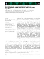
2.2.1. The perfusion needles
A needle perfusion system was constructed by binding a
25- and a 23-gauge needle together using epoxy putty,
with the bevels of the needles positioned on the outside
edges facing away from one another (see Figure 1). The
tips of the needles were set 1–1.5 mm apart.
2.2.2. Perfusion of knee joints
Rats were anaesthetised with urethane (ethyl carbamate;
0.6 ml 100 g
-1
body weight; 25% w v
-1
solution; single i.p.
injection). Once fully anaesthetised the animal was laid
on its back on an automated heating blanket (Harvard
Apparatus Limited, UK) and its core body temperature
maintained at 37°C via a thermistor probe positioned in
the rectum.
The limbs of the rat were flexed over a 20 ml glass vial,
with the patella facing directly upwards for insertion of
the perfusion needles, and the limb was secured in place
with tape. The 23-gauge needle was connected to a
Watson-Marlow roller pump via silicone rubber perfusion
tubing (internal diameter 1 mm, external diameter 4.2
mm, Watson Marlow, UK). Sterile saline was infused at a
constant rate of 100 µl min
-1
. After infusion of 100 µl of
vehicle (sterile saline), the outflow tubing was connected
to the 25-gauge needle, to minimise pressure build-up
within the joint space. Fluid was infused and withdrawn
at a constant rate until a 250 µl basal sample was collected
in a 1.5 ml centrifuge tube. Samples were immediately fro-
zen at -20°C.
2.2.3 Cytokine assay of joint samples
Luminex assay
Samples from the studies investigating the effects of
anaesthetic on joint cytokine levels (n = 10) and the dif-
ferences between normal (n = 10), high dose FCA-injected
(n = 3) and low dose FCA- injected joints (n = 8) were ana-
lysed using a multi-cytokine bead array detection system
capable of detecting rat IL1α, IL1β, IL2, IL4, IL6, IL10,
Interferon (IFN) γ, Granulocyte Macrophage-Colony
Stimulating Factor (GM-CSF) and TNFα, according to the
manufacturers instructions (Bio-Rad cytokine rat 9-plex,
Biorad, USA). Briefly, a monoclonal antibody directed
against the desired analyte was covalently coupled to dyed
5.5 µm polystyrene beads (2.5 × 10
6
beads ml
-1
cytokine
-
1
). The conjugated beads were exposed to 50 µl of sample
or standard solutions containing a known amount of
cytokine, in a 96-well filter plate and incubated overnight
at 4°C, protected from light. After a series of washes and
vacuum filtration to remove unbound protein, a bioti-
nylated detection antibody specific for a different epitope
on the analyte was added to the reaction. After incubation,
the unbound antibody was removed; the reaction mixture
was detected by the addition of streptavidin-phycoeryth-
rin (streptavidin-PE), which binds to the biotinylated
detection antibodies. Following a further series of washes
and vacuum filtration, the beads were re-suspended in
200 µl 5% BSA in PBS; the plate was stored at 4°C in the
dark until analysis. The reaction mixture was read using a
Luminex Data Collector in a Luminex 100 flow cytometer
(Luminex, USA). The minimum detection limit of the
assay was 2 pg ml
-1
for each mediator measured. Any val-
ues lower than these levels were classed as 0 for the pur-
poses of this study.
The perfusion needles and the perfusion system managing inflow and outflow from the knee joint spaceFigure 1
The perfusion needles and the perfusion system managing
inflow and outflow from the knee joint space. A Watson-
Marlow pump controlled the rate of saline infusion and sam-
ple extraction (100 µl min
-1
) from the joint. After the knee
was secured to prevent movement of the limb, needles were
inserted into the knee joint through the patella tendon.
Journal of Inflammation 2007, 4:13 />Page 4 of 8
(page number not for citation purposes)
Luminex data analysis
Excel data files were generated containing individual bead
numbers and the associated median fluorescence intensi-
ties. Standard curves were plotted to calculate the relative
amount of each cytokine in samples, using the aliquoted
serial dilutions of a positive control solution for calibra-
tion. Unknown sample cytokine concentrations were cal-
culated from the curve.
ELISA assay
The levels of TNFα and ILβ in samples from studies inves-
tigating the effects of the needles (n = 6), and leakage of
infusion from the joint cavity (n = 2) were measured using
commercially available ELISA kits that specifically recog-
nize the rat cytokines (BioSource International,
Camarillo, USA) according to the manufacturer's instruc-
tions. Briefly, 100 µl aliquots of sample were pipetted into
the wells of a microtiter plate pre-coated with an antibody
specific for rat IL-1β or TNFα and incubated for 3 h at
room temperature. After washing, a different biotinylated
anti-rat IL-1β or TNFα antibody was added and incubated
at ambient temperature for 1 h. Streptavidin-peroxidase
was added and incubated for 30 min. After a third incuba-
tion and washing to remove all unbound enzyme, colour
was developed by addition of stabilized chromogen
(tetramethylbenzidine), a stop solution added and the
intensity of the coloured product quantified spectropho-
tometrically at 450 nm. The minimum detection limit of
the assay was 2 pg ml
-1
.
2.3 Study design
2.3.1 Anaesthetic effects
In order to determine what effect anaesthetic agents had
on inflammatory mediators in joints, control experiments
were carried out. Firstly, five naive rats were anaesthetised
with urethane (ethyl carbamate; 0.6 ml 100 g
-1
body
weight; 25% w v
-1
solution; single i.p. injection), and five
further rats with sodium pentobarbital (1 ml kg
-1
body
weight; 60 mg ml
-1
solution; single i.p. injection main-
tained with i.v. 375 µl hr
-1
20 mg ml
-1
solution of pento-
barbital). No other procedures were carried out for 7
hours, at which point perfusion needles were inserted into
both knee joints and a 250 µl sample collected. The sam-
ple was frozen immediately at -20°C, and later assayed
using the Luminex assay.
2.3.2 Needle effects
In order to determine what effects inserting the perfusion
needles had on synovial cytokine concentrations, an
experiment was carried out in which six animals were
anaesthetised with urethane (as described above), and the
perfusion needles inserted into both knee joints and held
in position for 7 hours, at which time a 250 µl sample was
collected. The sample was frozen immediately at -20°C,
and later assayed using an ELISA.
2.3.3 Perfusion effects on the concentration of analyte
Two naïve rats were anaesthetised with urethane (as
described above) and a basal sample taken immediately.
Then 1000 pg recombinant rat IL1β (Bioclone, USA) in
100 µl was infused over 1 min. A second sample was
taken1 hour later; this was repeated hourly until 7 hours
post-IL1β infusion. The samples were frozen and later
assayed for IL1β content using an ELISA, to determine if
the sample contained the same amount of IL1β that was
initially infused.
2.3.4 Cytokine levels in normal and FCA-injected joints
Basal samples from ipsilateral and contralateral joints of
10 normal animals were compared with basal samples
from 8 rats which had received i.art low dose FCA (150
µg) and 3 that were injected with i.art high dose FCA (500
µg) 14 days earlier. Samples (250 µl) were collected and
frozen for later testing with the Luminex bead array.
2.3.5 Total cell counts
Joint perfusion samples were collected from ten naïve rat
knee joints, eight 150 µg FCA-injected ipsilateral and con-
tralateral joints and three 500 µg FCA-injected ipsilateral
and contralateral joints. Undiluted samples were viewed
by light microscopy in a haemocytometer. If red blood
cells were present, or a high number of inflammatory
cells, samples were diluted in saline, with added Zap-
poglobin, as per the manufacturer's instructions (1 drop
per 20 ml).
2.4 Data Analysis
Data were collected and analysed using Microsoft Excel
and Graphpad Prism software. Results are expressed as
mean ± standard error of the mean (SEM) where appropri-
ate.
Statistics
The Mann-Whitney U (non-parametric) test was used to
analyse differences between groups, which were not nor-
mally distributed, or in which the sample size was small.
To determine differences between the means of more than
two groups a non-parametric one-way analysis of variance
(Kruskal-Wallis) test was performed and a post-hoc test
(Dunn's) undertaken if the test was significant. In all cases
the null hypothesis was rejected at P < 0.05.
3. Results
3.1 Anaesthetic effects
Samples from naïve animals (n = 5) which received no
treatment during 7 hours of urethane anaesthesia, showed
a slight trend for increased levels of cytokines, but the
increases were not statistically significant for IL1α, IL1β,
IL2, IL4, IL6, IL10, GM-CSF, IFNγ, or TNFα compared
with samples taken from rats immediately after adminis-
tration of anaesthetic (n = 10; P > 0.05, Mann Whitney)
Journal of Inflammation 2007, 4:13 />Page 5 of 8
(page number not for citation purposes)
see Table 1. However, in contrast, animals anaesthetised
with pentobarbital (n = 5), had significantly higher levels
of GM-CSF and TNFα (P < 0.05, Mann Whitney) after 7
hours, in comparison with naïve joints, see Table 1.
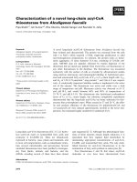
3. 2 Needle effects
Samples taken from knee joints in which the perfusion
needles had been in place for 7 hours while the animal
was anaesthetised with urethane (n = 6) showed increased
levels of TNFα, as measured by ELISA, but these were not
statistically significant from basal samples from the same
rats immediately after needle insertion (P > 0.05, Mann
Whitney). IL1β levels in two joints increased to approxi-
mately 40 pg ml
-1
over this time period, see Figure 2.
3.3 Perfusion effects on the concentration of analyte
Two joint perfusions were carried out to determine if any
of the infused solution leaked from the joint space prior
to withdrawal of samples. Recombinant rat IL1β (1000
pg), a cytokine known to be detectable by ELISA, was
infused into the joint, along with saline, and samples were
collected hourly. In both cases the full amount (1000 pg)
administered was recovered in the first two samples. How-
ever, a greater amount of IL1β was recovered compared to
the initial dose administered; Table 2 shows the results.
3.4 Levels of cytokines in normal and FCA-injected joints
Fourteen days after rats received 150 µg or 500 µg FCA
i.art (n = 8 and 3 respectively), the ipsilateral joint con-
tained significantly higher levels of IL1α, IL1β, IL6 and
TNFα compared with samples from naïve joints (n = 10),
as measured by the Luminex assay (P < 0.05, Two-way
ANOVA; see Figure 3a). The contralateral joints of rats
injected with 500 µg FCA also contained significantly
higher levels of IL1α, IL1β, IL6 and TNFα (P < 0.05, Two-
way ANOVA; see Figure 3b).
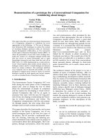
3.5 Total cell counts
Total inflammatory cell counts from normal animals (n =
5) and those injected with FCA (n = 8) 14 days prior to
sampling are shown in Figure 4. Normal joints had no
cells detectable, whereas all others samples had measura-
ble levels. However, only the 500 µg FCA ipsilateral (n =
3) joints proved to have a significantly greater number of
cells than normal joints (4.8 ± 0.06 × 10
6
cells ml
-1
; P <
0.05, Mann Whitney). A dose-response relationship was
demonstrated by the total cell count in both ipsilateral
and contralateral joints.
4. Discussion
The main aim of this study was to develop a method for
sampling synovial fluid from the knee joint of anaesthe-
tized rats. The technique was firstly validated by assessing
whether any inflammatory response was evoked by the
experimental set up, including the anaesthetic or the nee-
dles themselves; the efficiency of the system was investi-
gated, i.e. whether any infused solution leaked from the
joint space prior to sample extraction. Once the above fac-
tors had been assessed, they were taken into consideration
when comparing samples from naïve and adjuvant-
injected inflamed joints. Finally, the novel perfusion tech-
nique was used to quantify inflammatory cell numbers
within the rat synovial cavity. This technique proved to be
reliable and consistent when perfusing the joint cavity,
and regular volumes of sample were easily collected.
There were no problems with measuring protein content
due to high sample viscosity, and this technique is there-
fore a valuable addition to protocols which use homoge-
nates of entire joints to assess inflammatory mediator
content.
Levels of (a) TNFα and (b) IL1β from joints immediately after needle insertion (basal), and 7 hours laterFigure 2
Levels of (a) TNFα and (b) IL1β from joints immediately after
needle insertion (basal), and 7 hours later. Cytokines were
assayed using an ELISA, and although there was an apparent
increase in TNFα concentrations, to approximately 300 pg
ml
-1
in two samples, this was not statistically significant (P >
0.05, Mann Whitney). The horizontal lines on the graphs rep-
resent the median values in each group.
Table 1: The effect of anaesthetic on basal levels of cytokines in the joint.
IL1α IL1β IL2 IL4 IL6 IL10 GM-CSF IFNγ TNFα
Basal (n = 10)
Mean ± SEM
0.8 ± 0.5 1.0 ± 1.0 0.5 ± 0.5 0.2 ± 0.2 2.5 ± 2.5 1.5 ± 1.0 0 ± 0 0.1 ± 0.1 0.2 ± 0.2
Urethane (n = 5)
Mean ± SEM
2.6 ± 1.1 0 ± 0 0 ± 0 0 ± 0 0 ± 0 0 ± 0 1.0 ± 0.6 0.3 ± 0.3 36.6 ± 20.9
Pentobarbital (n = 5)
Mean ± SEM
1.4 ± 0.7 0.2 ± 0.2 0.1 ± 0.1 0 ± 0 0 ± 0 6.2 ± 6.2 1.7 ± 0.4* 0.1 ± 0.1 44.2 ± 21.6*
Levels of nine cytokines in rats anaesthetised for 7 hours with either urethane or pentobarbital, in comparison with samples taken immediately after
urethane anaesthesia (basal). Mann Whitney test were performed to determine differences between each anaesthetic and basal levels. Pentobarbital
anaesthesia resulted in a significant elevation of GM-CSF and TNFα levels; statistical significance P < 0.05 indicated by *.
Journal of Inflammation 2007, 4:13 />Page 6 of 8
(page number not for citation purposes)
It was established that the choice of anaesthetic may play
a role in initiating an inflammatory response within the
knee joints. Urethane, a hypnotic anaesthetic agent com-
monly used for laboratory animals, resulted in very little
change in any of the mediators measured over a 7 hour
period. In contrast, pentobarbital (pentobarbitone), a
short-acting anaesthetic which must be maintained by i.v
infusion, therefore requiring further surgical preparation
of the animal, induced increases in GM-CSF and TNFα
after continuous administration during the day, perhaps a
result of the surgery of the implanted cannulae. It was
therefore decided to use urethane for experiments, given
that it provides an extended period of anaesthesia with
minimal physiological changes [38], without the need for
invasive surgical preparation. Furthermore, pentobarbital
can cause respiratory depression in rats, whereas urethane
causes minimal cardiopulmonary disturbances [38,39].
Once it was established that urethane anaesthesia had no
adverse effects on the system, it was necessary to evaluate
any inflammatory component as a result of the perfusion
needles themselves, over a sustained time period of 7
hours. It was noted that a few rats developed increased
TNFα or IL1β levels as a result of the needles being main-
tained within the joint. However, the change occurred in
only 20% of animals, and was not significant; moreover,
The effects of 150 µg (low dose; n = 5) and 500 µg (high dose; n = 3) FCA on total cell count from joint perfusatesFigure 4
The effects of 150 µg (low dose; n = 5) and 500 µg (high
dose; n = 3) FCA on total cell count from joint perfusates.
Naïve joints contained no cells (0), whereas all other joints
contained increased levels, although only high dose ipsilateral
joints proved to have significantly raised levels (P < 0.05,
Mann Whitney); statistical significance donated by *.
Table 2: Perfusion effects on the concentration of analyte.
Animal 1 Animal 2
IL1β concentration (pg ml
-1
; 250 µl) Amount of IL1β (pg) IL1β concentration (pg ml
-1
; 250 µl) Amount of IL1β
(pg)
1 hour 2000 500 2000 500
2 hour 2000 500 2000 500
3 hour 200 50 356 89
4 hour 544 136 216 54
5 hour 350 87.5 210 52.5
6 hour 458 114.5 200 50
7 hour 318 79.5
Total (pg) 1467.5 1245.5
IL1β concentrations in each 250 µl sample collected, up to 7 hours post-infusion of IL1β (1000 pg). The amount of IL1β protein in each sample was
calculated and summed, to show that little or no leakage from the joint space occurred. In fact, more IL1β was present than was injected, in both
cases as a result of de novo release of endogenous IL1β protein.
Levels of IL1α, IL1β, IL6, IL10 and TNFα in (a) ipsilateral and (b) contralateral joints of normal rats and those injected with low (150 µg; n = 8) and high (500 µg; n = 3) dose FCA 14 days earlierFigure 3
Levels of IL1α, IL1β, IL6, IL10 and TNFα in (a) ipsilateral and
(b) contralateral joints of normal rats and those injected with
low (150 µg; n = 8) and high (500 µg; n = 3) dose FCA 14
days earlier. There were negligible levels of any of the media-
tors measure in naïve joints (n = 10), but a significant
increase in the expression of IL1α, IL1β, IL6 and TNFα was
seen in all ipsilateral inflamed joints and in contralateral joints
of rats injected with the high dose FCA (P < 0.05, Two-way
ANOVA; compared with normal joints); statistical signifi-
cance represented by *.
Journal of Inflammation 2007, 4:13 />Page 7 of 8
(page number not for citation purposes)
the increases in the two mediators did not occur in the
same animals.
This study has demonstrated that very little, if any, solu-
tion infused into the joint is lost into the surrounding tis-
sue, and can be recovered in full through the effusion
tubes. This was confirmed by injection of Evans blue dye
into the joint cavity and later dissection of the tissue (data
not shown here). Furthermore, there was an increased
quantity of IL1β detected in the perfusate collected.
Although this study was not designed to show the effects
of the protein on the joint, the 1 ng dose of IL1β adminis-
tered resulted in de novo release of natural IL1β, as shown
by the fact that elevated levels of IL1 were detected, in
addition to the 1 ng dose.
Adjuvant-induced arthritis is a widely used model of
inflammatory joint disease, and will be the primary sub-
ject of future studies applying this novel perfusion
method. It was therefore important that samples collected
in this way could detect differences between cytokine lev-
els in naïve joints and FCA-treated joints. Levels of all
cytokines measured in this study (IL1α, IL1β, IL6, IL10
and TNFα) showed dramatic increases 14 days after an
initial inflammatory insult to the joint, including high
and low doses of FCA. Furthermore, the contralateral joint
of rats injected with the high dose of FCA also had higher
levels of all cytokines measured, illustrating the contralat-
eral effect also noted in the inflammatory cell count study.
Finally, this study investigated the total number of white
blood cells present in the joint washout samples. Not sur-
prisingly it was observed that FCA-injected joints con-
tained higher levels than normal rat knee joints, as
previously shown [40]. However, of particular interest are
the cell counts in contralateral, non-injected limbs. Con-
tralateral effects arising from a unilateral insult is a well
documented phenomenon. In general, contralateral
changes in behaviour, magnitude of biochemical fluctua-
tions or histopathological lesions are less than those
observed on the ipsilateral side (for review see [41]). Total
cell count data from this study are in agreement with this
finding, and although the lower dose of FCA used here
does not elicit behavioural signs of inflammation or
hypersensitivity in the contralateral joint, there is evi-
dence of infiltration of inflammatory cells.
5. Conclusion
In summary, we have demonstrated the use of a novel
method for sampling synovial fluid and washing out the
joint cavity to collect the "inflammatory soup", and have
performed assays to measure levels of cytokines during
adjuvant-induced arthritis. This method has the advan-
tage of enabling the contents of synovial fluid to be inves-
tigated alone, without the contamination of the
surrounding tissue. We have also revealed its value in
measuring cellular components of inflammation. In con-
clusion, as this new method of joint perfusion uses anaes-
thetised animals, acute effects of anti-inflammatory drugs
or novel compounds could be investigated, thus improv-
ing the knowledge of how novel drug targets are affecting
the inflammatory process.
Competing interests
The author(s) declare that they have no competing inter-
ests.
Authors' contributions
NJB planned and carried out all in vivo studies, in vitro
assays, data interpretation, statistical analysis and compi-
lation of the manuscript. DAS and JPH assisted with the
Luminex assay use and data collection, then read and
edited the manuscript after completion. AGR assisted with
the total inflammatory cell count studies and reviewed
and edited the article. IPC, AJR and DSM contributed
intellectually to the experimental designs, as well as to
structural and editorial aspects of the paper. All authors
read and approved the final manuscript.
Acknowledgements
We would like to thank GlaxoSmithKline for funding these studies and my
PhD studentship.
References
1. Sweeney SE, Firestein GS: Rheumatoid arthritis: regulation of
synovial inflammation. Int J Biochem Cell Biol 2004, 36:372-378.
2. Kubota E, Kubota T, Matsumoto J, Shibata T, Murakami KI: Synovial
fluid cytokines and proteinases as markers of temporoman-
dibular joint disease. J Oral Maxillofac Surg 1998, 56:192-198.
3. Alstergren P, Ernberg M, Kvarnstrom M, Kopp S: Interleukin-1beta
in synovial fluid from the arthritic temporomandibular joint
and its relation to pain, mobility, and anterior open bite. J
Oral Maxillofac Surg 1998, 56:1059-65; discussion 1066.
4. Chang H, Israel H: Analysis of inflammatory mediators in tem-
poromandibular joint synovial fluid lavage samples of symp-
tomatic patients and asymptomatic controls. J Oral Maxillofac
Surg 2005, 63:761-765.
5. Rooney M, Symons JA, Duff GW: Interleukin 1 beta in synovial
fluid is related to local disease activity in rheumatoid arthri-
tis. Rheumatol Int 1990, 10:217-219.
6. Ulfgren AK, Grondal L, Lindblad S, Khademi M, Johnell O, Klareskog
L, Andersson U: Interindividual and intra-articular variation of
proinflammatory cytokines in patients with rheumatoid
arthritis: potential implications for treatment. Ann Rheum Dis
2000, 59:439-447.
7. Barrera P, Boerbooms AM, van de Putte LB, van der Meer JW:
Effects of antirheumatic agents on cytokines. Semin Arthritis
Rheum 1996, 25:234-253.
8. Eastgate JA, Symons JA, Wood NC, Grinlinton FM, di Giovine FS, Duff
GW: Correlation of plasma interleukin 1 levels with disease
activity in rheumatoid arthritis. 1988, 2:706-709.
9. Houssiau FA, Devogelaer JP, Van Damme J, de Deuxchaisnes CN, Van
Snick J: Interleukin-6 in synovial fluid and serum of patients
with rheumatoid arthritis and other inflammatory
arthritides. Arthritis Rheum 1988, 31:784-788.
10. Christodoulou C, Choy EH: Joint inflammation and cytokine
inhibition in rheumatoid arthritis. Clin Exp Med 2006, 6:13-19.
11. Zwerina J, Redlich K, Schett G, Smolen JS: Pathogenesis of rheu-
matoid arthritis: targeting cytokines. Ann N Y Acad Sci 2005,
1051:716-729.
12. Egg D: Concentrations of prostaglandins D2, E2, F2 alpha, 6-
keto-F1 alpha and thromboxane B2 in synovial fluid from
Publish with BioMed Central and every
scientist can read your work free of charge
"BioMed Central will be the most significant development for
disseminating the results of biomedical research in our lifetime."
Sir Paul Nurse, Cancer Research UK
Your research papers will be:
available free of charge to the entire biomedical community
peer reviewed and published immediately upon acceptance
cited in PubMed and archived on PubMed Central
yours — you keep the copyright
Submit your manuscript here:
/>BioMedcentral
Journal of Inflammation 2007, 4:13 />Page 8 of 8
(page number not for citation purposes)
patients with inflammatory joint disorders and osteoarthri-
tis. Z Rheumatol 1984, 43:89-96.
13. Trang LE, Granstrom E, Lovgren O: Levels of prostaglandins F2
alpha and E2 and thromboxane B2 in joint fluid in rheuma-
toid arthritis. Scand J Rheumatol 1977, 6:151-154.
14. Dayer JM, Krane SM, Russell RG, Robinson DR: Production of col-
lagenase and prostaglandins by isolated adherent rheuma-
toid synovial cells. Proc Natl Acad Sci U S A 1976, 73:945-949.
15. Fulkerson JP, Damiano P: Effect of prostaglandin E2 on adult pig
articular cartilage slices in culture. Clin Orthop Relat Res
1983:266-269.
16. Robinson DR, Smith H, McGuire MB, Levine L: Prostaglandin syn-
thesis by rheumatoid synovium and its stimulation by colch-
icine. Prostaglandins 1975, 10:67-85.
17. Robinson DR, Tashjian AH Jr., Levine L: Prostaglandin-stimulated
bone resorption by rheumatoid synovia. A possible mecha-
nism for bone destruction in rheumatoid arthritis. J Clin Invest
1975, 56:1181-1188.
18. Firestein GS, Alvaro-Gracia JM, Maki R: Quantitative analysis of
cytokine gene expression in rheumatoid arthritis. J Immunol
1990, 144:3347-3353.
19. Wagner S, Fritz P, Einsele H, Sell S, Saal JG: Evaluation of synovial
cytokine patterns in rheumatoid arthritis and osteoarthritis
by quantitative reverse transcription polymerase chain reac-
tion. Rheumatol Int 1997, 16:191-196.
20. Patten C, Bush K, Rioja I, Morgan R, Wooley P, Trill J, Life P: Char-
acterization of pristane-induced arthritis, a murine model of
chronic disease: response to antirheumatic agents, expres-
sion of joint cytokines, and immunopathology. Arthritis Rheum
2004, 50:3334-3345.
21. Rioja I, Bush KA, Buckton JB, Dickson MC, Life PF: Joint cytokine
quantification in two rodent arthritis models: kinetics of
expression, correlation of mRNA and protein levels and
response to prednisolone treatment. Clin Exp Immunol 2004,
137:65-73.
22. Thornton S, Duwel LE, Boivin GP, Ma Y, Hirsch R: Association of
the course of collagen-induced arthritis with distinct pat-
terns of cytokine and chemokine messenger RNA expres-
sion. Arthritis Rheum 1999, 42:1109-1118.
23. Westman M, Korotkova M, af Klint E, Stark A, Audoly LP, Klareskog
L, Ulfgren AK, Jakobsson PJ: Expression of microsomal prostag-
landin E synthase 1 in rheumatoid arthritis synovium. Arthritis
Rheum 2004, 50:1774-1780.
24. Cohen SB, Katsikis PD, Chu CQ, Thomssen H, Webb LM, Maini RN,
Londei M, Feldmann M: High level of interleukin-10 production
by the activated T cell population within the rheumatoid
synovial membrane. Arthritis Rheum 1995, 38:946-952.
25. Bileviciute I, Lundeberg T, Ekblom A, Theodorsson E: Bilateral
changes of substance P-, neurokinin A-, calcitonin gene-
related peptide- and neuropeptide Y-like immunoreactivity
in rat knee joint synovial fluid during acute monoarthritis.
Neurosci Lett 1993, 153:37-40.
26. Billingham ME: Mechanisms and Models of Rheumatoid Arthri-
tis. Edited by: Pettipher ER. London, Academic Press; 1995:389.
27. Mapp PI, Terenghi G, Walsh DA, Chen ST, Cruwys SC, Garrett N,
Kidd BL, Polak JM, Blake DR: Monoarthritis in the rat knee
induces bilateral and time-dependent changes in substance P
and calcitonin gene-related peptide immunoreactivity in the
spinal cord. Neuroscience 1993, 57:1091-1096.
28. Donaldson LF, Seckl JR, McQueen DS: A discrete adjuvant-
induced monoarthritis in the rat: effects of adjuvant dose. J
Neurosci Methods 1993, 49:5-10.
29. Pelegri C, Franch A, Castellote C, Castell M: Immunohistochemi-
cal changes in synovial tissue during the course of adjuvant
arthritis. J Rheumatol 1995, 22:124-132.
30. Wilson AW, Medhurst SJ, Dixon CI, Bontoft NC, Winyard LA, Brack-
enborough KT, Alba JD, Clarke CJ, Gunthorpe MJ, Hicks GA: An ani-
mal model of chronic inflammatory pain: Pharmacological
and temporal differentiation from acute models. European
Journal of Pain 2006, 10:537-549.
31. Magari K, Miyata S, Nishigaki F, Ohkubo Y, Mutoh S, Goto T: Differ-
ential effects of FK506 and methotrexate on inflammatory
cytokine levels in rat adjuvant-induced arthritis. J Rheumatol
2003, 30:2193-2200.
32. Marinova-Mutafchieva L, Williams RO, Mason LJ, Mauri C, Feldmann
M, Maini RN: Dynamics of proinflammatory cytokine expres-
sion in the joints of mice with collagen-induced arthritis
(CIA). Clin Exp Immunol 1997, 107:507-512.
33. Smith-Oliver T, Noel LS, Stimpson SS, Yarnall DP, Connolly KM: Ele-
vated levels of TNF in the joints of adjuvant arthritic rats.
Cytokine 1993, 5:298-304.
34. Keeble JE Curtis, B, Mallaghan, FA & Brain, SD: The role of sensory
nerves in joint inflammation: studies using TRPV1 knockout
mice. pA2 online 2004, 2:43P.
35. Singh HN, Blancuzzi V, Greenwood S, Skiles JW, O'Byrne EM: Syno-
vial fluid levels of tumor necrosis factor-alpha in the inflamed
rat knee: modulation by dexamethasone and inhibitors of
matrix metalloproteinase and phosphodiesterase. Inflamm
Res 1997, 46 Suppl 2:S153-4.
36. Vale ML, Benevides VM, Sachs D, Brito GA, da Rocha FA, Poole S,
Ferreira SH, Cunha FQ, Ribeiro RA: Antihyperalgesic effect of
pentoxifylline on experimental inflammatory pain. Br J Phar-
macol 2004, 143:833-844.
37. Liu SH, Wong CS, Chang DM: Increase Monocyte chemoattract-
ant protein-1 in knee joints of rats with adjuvant-induced
arthritis: in vivo microdialysis. The journal of rheumatology 2005,
32:2205-2211.
38. Sapru HN, Krieger AJ: Cardiovascular and respiratory effects of
some anesthetics in the decerebrate rat. Eur J Pharmacol 1979,
53:151-158.
39. Wixson SK, White WJ, Hughes HC Jr., Lang CM, Marshall WK: The
effects of pentobarbital, fentanyl-droperidol, ketamine-xyla-
zine and ketamine-diazepam on arterial blood pH, blood
gases, mean arterial blood pressure and heart rate in adult
male rats. Lab Anim Sci 1987, 37:736-742.
40. Santos L, Tipping PG: Attenuation of adjuvant arthritis in rats
by treatment with oxygen radical scavengers. Immunol Cell Biol
1994, 72:406-414.
41. Shenker N, Haigh R, Roberts E, Mapp P, Harris N, Blake D: A review
of contralateral responses to a unilateral inflammatory
lesion. Rheumatology (Oxford) 2003, 42:1279-1286.