Báo cáo y học: "Gender-based reciprocal expression of transforming growth factor-β1 and the inducible nitric oxide synthase in a rat model of cyclophosphamide-induced cystitis" ppsx
Bạn đang xem bản rút gọn của tài liệu. Xem và tải ngay bản đầy đủ của tài liệu tại đây (4.23 MB, 13 trang )
BioMed Central
Page 1 of 13
(page number not for citation purposes)
Journal of Inflammation
Open Access
Research
Gender-based reciprocal expression of transforming growth
factor-β1 and the inducible nitric oxide synthase in a rat model of
cyclophosphamide-induced cystitis
Pradeep Tyagi
1
, Vikas Tyagi
2
, Naoki Yoshimura
2
, Erich Witteemer
2
,
Derek Barclay
3
, Patricia A Loughran
3
, Ruben Zamora
3
and
Yoram Vodovotz*
3,4
Address:
1
Department of Urology, William Beaumont Hospital, MI 48073, USA,
2
Department of Urology, University of Pittsburgh, PA 15213,
USA,
3
Department of Surgery, University of Pittsburgh, PA 15213, USA and
4
Center for Inflammation and Regenerative Modeling, McGowan
Institute for Regenerative Medicine, University of Pittsburgh, PA 15219, USA
Email: Pradeep Tyagi - ; Vikas Tyagi - ; Naoki Yoshimura - ;
Erich Witteemer - ; Derek Barclay - ; Patricia A Loughran - ;
Ruben Zamora - ; Yoram Vodovotz* -
* Corresponding author
Abstract
Background: The pluripotent cytokine transforming growth factor-β1 (TGF-β1) is the central
regulator of inducible Nitric Oxide Synthase (iNOS) that is responsible for nitric oxide (NO)
production in inflammatory settings. Previous studies have implicated a role for NO, presumably
derived from iNOS, in cyclophosphamide (CYP)-induced cystitis in the bladder. TGF-β1 is
produced in latent form and requires dissociation from the latency-associated peptide (LAP) to act
as primary anti-inflammatory and pro-healing modulator following tissue injury in the upper urinary
tract. Since the role of TGF-β1 in lower urinary tract inflammation is currently unknown, and since
gender-based differences exist in the setting of interstitial cystitis (IC), the present study examined
the relationship between TGF-β1 and iNOS/NO in the pathogenesis of CYP-induced cystitis in
both male and female rats.
Methods: Sprague-Dawley rats, 4 months of age, of either gender were given 150 mg/kg CYP
intraperitoneally. Urinary and bladder tissue TGF-β1 and NO reaction products (NO
2
-
/NO
3
-
) were
quantified as a function of time following CYP. Expression of active and latent TGF-β1 as well as
iNOS in harvested bladder tissue was assessed by immunohistochemistry.
Results: Female rats had significantly higher levels of NO
2
-
/NO
3
-
in urine even at baseline as
compared to male rats (p < 0.001), whereas there was no gender based significant difference in
urine levels of active or latent TGF-β1 prior to CYP injection. Inflammatory and cytotoxic changes
were induced by CYP in the bladder of both sexes that were accompanied by differences in the
urine levels of NO
2
-
/NO
3
-
and TGF-β1. Male rats responded to CYP with significantly lower levels
of NO
2
-
/NO
3
-
and significantly higher levels of TGF-β1 in urine (p < 0.05) as compared to females
at all time points after CYP. The urine levels of NO
2
-
/NO
3
-
after CYP were inversely correlated to
latent and active TGF-β1 (Pearson coefficient of -0.72 and -0.69 in females and -0.89 and -0.76 in
males, respectively; p < 0.01). Bladder tissue of male rats exhibited significantly higher levels of both
Published: 19 August 2009
Journal of Inflammation 2009, 6:23 doi:10.1186/1476-9255-6-23
Received: 31 March 2009
Accepted: 19 August 2009
This article is available from: />© 2009 Tyagi et al; licensee BioMed Central Ltd.
This is an Open Access article distributed under the terms of the Creative Commons Attribution License ( />),
which permits unrestricted use, distribution, and reproduction in any medium, provided the original work is properly cited.
Journal of Inflammation 2009, 6:23 />Page 2 of 13
(page number not for citation purposes)
latent and active TGF-β1 (p < 0.01) compared to female rats after CYP. TGF-β1 and iNOS protein
was mostly localized in the urothelium.
Conclusion: The results of this study suggest that there exists an inverse relationship between the
expression of TGF-β1 and iNOS/NO
2
-
/NO
3
-
in CYP-inflamed bladder. The gender of the animal
appears to magnify the differences in urine levels of TGF-β1 and NO
2
-
/NO
3
-
in this inflammatory
setting. These results support the hypothesis that TGF-β1 can suppress iNOS expression
associated with bladder inflammation and reduce systemic levels of NO
2
-
/NO
3
-
, and further suggest
that this feature of TGF-β1 can be harnessed for therapy and diagnosis of interstitial cystitis.
Background
Cyclophosphamide is an oxazaphosphorine DNA alkylat-
ing agent, known for its anti-neoplastic and immunosup-
pressant properties, that is used clinically for malignancy,
bone marrow transplantation, and multiple sclerosis. A
prominent side effect of CYP is hemorrhagic cystitis [1,2].
It has been proposed that acrolein, a phase I metabolic
product of CYP, is the causative agent of the edema, ulcer-
ation, and hemorrhage evident upon direct contact with
bladder lumen [3]. This ability of CYP to cause cystitis has
been utilized to simulate interstitial cystitis (IC) in pre-
clinical studies [4].
A recent study from our laboratory suggested that changes
in the cytokine milieu of the bladder after CYP describes a
pro-inflammatory phenotype in this organ, likely due to
the rapid infiltration of innate immune cells. These
inflammatory changes correlate with the abnormal void-
ing and histology characteristic of cyclophosphamide
(CYP)-induced cystitis in rats [4]. Temporal changes in the
levels of pro-inflammatory cytokines and chemokines
such as interleukin IL-1α, IL-1β, IL-6, IL-17, IL-18, and
GRO/KC preceded or concurred with pathological
changes induced by CYP. Studies from other groups dem-
onstrate that various inflammatory cytokines seem to
mediate the pathogenesis of CYP-induced cystitis through
the induction of high levels of iNOS and NO production
as well as cyclooxygenase-derived prostaglandins [5-8].
Clinical studies based on tissue biopsies from patients
with IC suggest an elevated expression of both iNOS and
TGF-β1 in the urothelium as compared to patients with
kidney stone or benign hematuria [9,10].
TGF-β1 is expressed by inflammatory cells such as neu-
trophils and eosinophils, as well as by cells in the epithe-
lium, fibroblasts, and smooth muscle cells [11-13]. These
cells express three isoforms of TGF-β, namely TGF-β1,
TGF-β2, and TGF-β3, with TGF-β1 being the most abun-
dant [14]. Though TGF-β1 has both pro- and anti-inflam-
matory effects [11-13], studies have shown this cytokine
to primarily suppress inflammation and promote healing
following tissue injury in the upper urinary tract [14,15].
The numerous biological functions of all TGF-β's require
an initial bioactivation, in which the dimeric TGF-β pre-
cursor is cleaved intracellularly to yield the active TGF-β
dimer, which subsequently remains associated with the
remaining portion of its own pro-form, the latency-asso-
ciated peptide (LAP). This latent TGF-β complex is
secreted, and may bind to other proteins such as latent
TGF-β binding proteins (LTBP) or α2-macroglobulin
[16,17]. Bioactive TGF-β1 is a potent suppressor of iNOS
expression and enzymatic activity [18].
The excessive production of TGF-β1 can promote tissue
fibrosis in a number of diseases including liver cirrhosis,
pulmonary fibrosis, and fibrotic kidney [19]. Coinciden-
tally, a significant degree of fibrosis is also frequently
noticed in the bladder of chronic IC patients on cysto-
scopic exam, the reasons for which remain elusive [20-
22]. Experimental IC is also induced in rats by acrolein, a
metabolite of CYP excreted into the urine from the kidney
[3]. This animal model exhibits gender-based differences
in the observed pathology [23-25], a feature also seen in
human IC [26]. A study on ovariectomized rats revealed
an increased severity of histological changes induced by
CYP that were ameliorated by estrogen replacement [25].
A similar gender disparity in human lower urinary tract
diseases is exemplified by significantly higher levels of IL-
1α and IL-1RA in urine of healthy females that seem to
provide prophylaxis against upper and lower urinary tract
infection [26]. Steroid hormones released from the ovary
can induce expression of IL-1RA and slow down the pro-
gression of renal diseases [27].
We hypothesized that urine levels of TGF-β1 are not spe-
cific for nephropathy, but can also reflect the state of the
acrolein-injured bladder. Given the interplay of regulatory
influences operating in the production of NO, TGF-β1
and other pro-inflammatory cytokines in bladder inflam-
mation, we sought to define the time-dependent changes
in the urinary levels of NO-derived oxidation products as
well as TGF-β1 in a rat model of CYP-induced cystitis. We
also sought to determine if there are gender-specific pat-
terns of iNOS and TGF-β1 expression in this animal
model. We further sought to determine the expression
and cellular localization of active and latent TGF-β1 as
well as that of iNOS in the bladder. Our findings demon-
strate lower levels of iNOS and NO reaction products, and
Journal of Inflammation 2009, 6:23 />Page 3 of 13
(page number not for citation purposes)
concomitantly higher levels of TGF-β1, in male vs. female
rats. We discuss the possible relevance of these findings to
the pathology and possible diagnosis and treatment of
human IC.
Methods
All animal experimentation described was performed in
accordance with NIH guidelines following approval by
the University of Pittsburgh Institutional Animal Care and
Use Committee (IACUC). Cyclophosphamide was pro-
cured from Sigma-Aldrich (St. Louis, MO). Intraperito-
neal CYP injections [28] were performed in 4-month old
Sprague-Dawley rats of either sex. Urine specimens
obtained from rats kept in metabolic cages during day-
light hours were frozen immediately in liquid nitrogen
and stored at -80°C prior to analysis. Baseline urine sam-
ples were obtained throughout the 12 daylight hours prior
to next day's CYP injection, as well as from vehicle-treated
rats. Bladder tissue was harvested from both CYP- and
vehicle-treated rats. Harvested bladders were split into
two halves. One half was cryopreserved for immunohisto-
chemistry and the other half was frozen immediately for
protein analysis.
Measurement of NO reaction products and TGF-
β
1
Frozen urine samples from each hourly interval were
thawed, and 20 μl of each sample were analyzed. NO was
measured as NO
2
-
/NO
3
-
by the nitrate reductase method
[29] using a commercially available kit (Cayman Chemi-
cal, Ann Arbor, MI) according to manufacturer's protocol.
Fifty μL from each sample were analyzed for active and
latent TGF-β1 in triplicate using a commercial antigen
capture ELISA kit (Quantikine™, R&D Systems, Minneap-
olis, MN). Each sample was assayed both in the absence
and presence of 1 M HCl in order to assess both active and
latent TGF-β1, respectively. Urine levels of NO
2
-
/NO
3
-
and
TGF-β1 were normalized to the respective creatinine con-
centrations and expressed as μmol per mg of creatinine
and pg/mg of creatinine, respectively. At the conclusion of
the study, harvested bladders were homogenized, lysed,
and stored at -80°C. All tissue TGF-β1 values were then
standardized by bladder weight and expressed as μg per
bladder.
Immunostaining and Confocal Microscopy of iNOS, active
TGF-
β
1, and latent TGF-
β
1
Bladders were fixed in formalin and frozen with TBS tissue
freezing medium (Pacific Southwest Lab Equipment Inc.,
CA) prior to sectioning to a sample thickness of 8
microns. Tissue was permeabilized with 0.2% Triton X-
100-PBS for 15 min, followed by a 1 h block in 2% BSA-
PBS. Tissue sections were incubated in 0.5% BSA-PBS with
5 μg/ml of chicken-anti-TGFβ1 (to assess the expression of
active TGF-β1) and goat-anti human LAP (to assess total/
latent TGF-β1) [30]. Both antibodies were obtained from
R&D Systems. Mouse anti-human iNOS antibody was
obtained from Santa Cruz Biotechnology (Santa Cruz,
CA) and used at a concentration of 2 μg/ml. The primary
antibodies were incubated overnight at 4°C (anti-TGF-β1
and anti-LAP) or at room temperature for 1 h (anti-iNOS).
Anti-LAP antibody was used for the immunodetection of
latent TGF-β1. Following primary antibody incubation,
the sections were washed 3× with 0.5% BSA-PBS and incu-
bated with the appropriate secondary antibodies in 0.5%
BSA-PBS for 1 h at room temperature. Secondary antibod-
ies were as follows: donkey-anti-chicken Cy3 (1:1000,
Jackson ImmunoResearch, West Grove, PA), donkey-anti-
goat Cy5 (1:500, Jackson ImmunoResearch), donkey-anti
mouse Alexa488 (1:500, Invitrogen), Alexa488-phalloi-
din (1:250, Invitrogen, Carlsbad, CA), or Alexa647-phal-
loidin (1:250, Invitrogen). The tissue sections were then
washed 3× with 0.5% BSA-PBS, followed by 3× washes
with PBS. Nuclei were stained for 10 s with Hoechst dye
(1 mg/100 ml bisbenzimide). The slides were rinsed with
PBS and coverslipped with Gelvatol, a water-soluble
mounting media (a mix of 21 g polyvinyl alcohol in 42 ml
glycerol, 52 ml water, a few crystals of sodium azide, and
106 ml 0.2-M Tris buffer, pH = 8.5). The slides were then
visualized with a confocal microscope (Fluoview 1000;
Olympus, Melville, NY).
Statistical Analysis
Values are expressed as mean ± SEM. Analysis of paramet-
ric data among experimental groups of different sex at
baseline and after CYP injection was carried using one
way ANOVA followed by Tukey's multiple comparison
tests for statistical significance. The Pearson correlation
coefficient using two tailed test for significance was used
to check inverse correlation. Significance was considered
at p < 0.05.
Results
Micturition at Baseline and After CYP
Baseline assessment
Cumulative urine volume for a 12-h period a day prior to
CYP injection and on the day of injection was measured
and plotted (Fig. 1A). At baseline, male rats showed a
slightly higher cumulative urine volume (7.67 ± 0.59 ml)
than female rats (5.88 ± 1.88 ml), but the differences were
not statistically significant (ANOVA, Tukey's Multiple
Comparison post-test; p > 0.05; n = 8 rats per group). Both
female and male rats voided urine with similar average
frequency at baseline (Fig. 1A), as measured by the
number of urination events in a single 12-h period (7.8 ±
0.54 for females and 7.57 ± 0.86 for males; n = 8 rats per
group).
Assessment following treatment with CYP
As previously reported by our group, characteristic dys-
functional voiding after CYP injection (150 mg/kg) [4] in
Journal of Inflammation 2009, 6:23 />Page 4 of 13
(page number not for citation purposes)
female rats was also observed in male rats. The cumulative
urine volume voided as well the urination frequency in
rats of both genders drastically increased for the same 12-
h period. The cumulative urine volume increased to 9.65
± 2.34 ml in females and 12.9 ± 1.03 ml in males (Fig.
1A). The rise in cumulative urine volume in female and
male rats after treatment with CYP was significant relative
to baseline values only in female rats (ANOVA, Tukey post
test comparison; *p < 0.05, n = 4 rats per group). The aver-
age 12-h frequency in male rats after CYP was 19 ± 1.5 vs.
20.25 ± 2.6 (n = 4) in females (not statistically signifi-
cant).
In addition, urinary frequency, as measured by urination
events for each hour, showed a dramatic increase during
the time period of 48 h after CYP injection. Female rats
urinated on an average of five times per hour compared to
three times per hour in male rats during this time period.
These results corroborate the previously-reported high
urination frequency after CYP relative to baseline [4].
Occasional microhematuria was also noted in few of the
urine specimens from this time period (data not shown).
Urinary Levels of NO Reaction Products at Baseline and
After CYP Injection
Baseline assessment
Urine levels of the NO oxidation products NO
2
-
/NO
3
-
served as a proxy for the magnitude of NO production in
bladder tissue. The maxima and minima of NO
2
-
/NO
3
-
during the day in control rats were reciprocal to the
maxima and minima of total TGF-β1 at baseline in both
sexes (Pearson correlation coefficient = 0.2 [two tailed p =
0.56; n = 4] for males and 0.19 [p = 0.75; n = 4] for
females; Fig. 1B).
Assessment following treatment with CYP
Our results demonstrated an elevation of NO reaction
products in the urine of CYP-treated rats when compared
to the levels observed in control rats collected at the same
time point of the day. The levels of NO
2
-
/NO
3
-
in the urine
of CYP-treated female rats remained higher as compared
to both CYP-treated and control male rats (ANOVA,
Tukey post test comparison; *p < 0.01, n = 4 rats per
group). Female rats showed the highest levels of NO
2
-
/
NO
3
-
6 h post-CYP, followed by a steady decline to levels
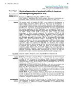
Urinary profile and baseline levels of NO reaction productsFigure 1
Urinary profile and baseline levels of NO reaction products. Panel A:- Effect of CYP on micturition pattern. Cumula-
tive urine volume was measured over the period of 12 daylight hours before and after CYP injection (150 mg/kg) in male and
female rats. In absence of CYP, female rats (empty black dot) voided a cumulative volume of 5.88 ± 1.88 ml compared to
slightly higher volume of 7.67 ± 0.59 ml in male rats (empty black triangle). The mean urinary frequency was 7.8 ± 0.54 in
female rats and 7.57 ± 0.86 in male rats during the 12-h time period at baseline. The cumulative urine volume increased signifi-
cantly to 9.65 ± 2.34 ml in female (solid black dot) and to 12.9 ± 1.03 ml in male rats (solid black triangle) after CYP, relative to
baseline values in female rats (ANOVA, Tukey post hoc test; *p < 0.05). The mean urinary frequency also increased signifi-
cantly after CYP to 19 ± 1.5 and 20.25 ± 2.6 in males and females, respectively. Panel B Urine levels of NO reaction products
at baseline and after CYP. NO
2
-
/NO
3
-
are expressed as μmol/mg creatinine. The measurement of NO
2
-
/NO
3
-
in individual urine
voids from control male rats showed that levels of NO reaction products do not remain constant throughout the day, but are
maximal at the beginning of day and then stabilize for the remainder of the day. Values at baseline in female rats (empty black
dot) did not change over the course of the day. The levels of NO products in urine of CYP treated female rats (solid black dot)
were significantly higher compared to male rats at baseline (empty black triangle) and after CYP injection (solid black triangle)
(ANOVA, Tukey post hoc test; *p < 0.01).
Journal of Inflammation 2009, 6:23 />Page 5 of 13
(page number not for citation purposes)
lower than baseline at 24 h. Treatment of male rats with
CYP was associated with a sharp rise in levels of NO
2
-
/
NO
3
-
at 4 h that remained elevated until 6 h and then pro-
gressively declined to lower values (Fig. 1B).
Levels of TGF-
β
1 in Urine
Baseline assessment
Urine analysis of male (Δ) and female (•) rats before CYP
injection revealed secretion of TGF-β1 in very low
amounts (Fig. 2A). The levels of latent/total and active
forms of TGF-β1 in males were significantly higher than
the respective forms of TGF-β1 in females (*p < 0.001; n
= 8). There was positive correlation between active and
latent forms of TGF-β1 in urine with Pearson's coefficient
of 0.98 (two tailed *p < 0.0001) and 0.87 (two tailed *p
< 0.0001) for female and male rats, respectively. The levels
of active and total TGF-β1 were maintained at similar lev-
els throughout the day in male rats.
Assessment following treatment with CYP
A progressive rise of TGF-β1 was observed in the urine of
male and female rats after CYP injection, starting at 5 h
(Fig. 2B). TGF-β1 levels continued to rise over the 12-h
period of urine collection, reaching a maximum when
experiment was terminated at 24 h. The urine levels of
total TGF-β1 in rats of both sexes rose nearly 100-fold at
24 h relative to their respective baseline values (Fig. 2B),
though this change was significantly higher vs. baseline
values only in male urine (ANOVA, Tukey's Multiple
Comparison post-test; p < 0.01). The levels of total/latent
TGF-β1 in the urine of male rats after CYP were also signif-
icantly higher than the levels of active and total TGF-β1 in
the urine of female rats, both at baseline and after CYP
(ANOVA, Tukey post test comparison; p < 0.01).
Correlation for Urine levels of TGF-
β
1 and NO
2
-
/NO
3
-
The urinary levels of NO metabolites NO
2
-
/NO
3
-
were
inversely correlated to active TGF-β1 and latent TGF-β1 in
both male and female rats (Fig. 3). The Pearson correla-
tion coefficient in female rats was -0.69 (two tailed; *p <
0.03) and -0.72 (two tailed; *p < 0.02) for relationship of
NO
2
-
/NO
3
-
, with active TGF-β1 (Fig. 3A) and latent TGF-
β1 (Fig. 3B), respectively. In male rats, the Pearson corre-
lation coefficient was -0.89 (two tailed; *p < 0.0001) and
-0.76 (two tailed; *p < 0.01) for latent TGF-β1 (Fig. 3D)
and active TGF-β1 (Fig. 3C), respectively.
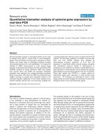
Urine levels of active and latent/total TGF-β1 at baseline and after CYPFigure 2
Urine levels of active and latent/total TGF-β1 at baseline and after CYP. Active and latent/total TGF-β1 values are
reported as pg/mg of creatinine. Panel A Urine levels of TGF-β1 at baseline. In the absence of CYP injection, male and female
rats excreted very low amounts of TGF-β1. Total (empty black triangle) and active (solid black triangle) forms of TGF-
β1 in male urine were significantly higher than total (empty black dot) and active (solid black dot) forms in female urine p
< 0.001 (n = 8). The TGF-β1 levels in male urine were at least 10-fold higher than levels in female urine. Panel B Urine levels
of TGF-β-1 after CYP. Injection of CYP induced time dependent 100-fold increase in urine levels of TGF-β1 in rats of both
sexes relative to the respective baseline values. TGF-β1 levels after CYP were significantly higher than respective baseline val-
ues only in male urine and not in female urine (ANOVA, Tukey's post hoc test; p < 0.01). The levels of total TGF-β1 (empty
black triangle) in male urine after CYP were also significantly higher than the levels of active (solid black dot) and total
(empty black dot) TGF-β1 in female urine both at baseline and after CYP (ANOVA, Tukey post hoc test; p < 0.01). The urine
levels of total TGF-β1 (empty black triangle) were significantly higher than those of active TGF-β1 (solid black triangle) only in
male urine and not in female urine after CYP (*p < 0.01).
Journal of Inflammation 2009, 6:23 />Page 6 of 13
(page number not for citation purposes)
Levels of TGF-
β
1 in Bladder Tissue following CYP injection
We sought to determine if the gender-associated differ-
ences in urinary TGF-β1 levels stemmed from differences
in expression of TGF-β1 in the bladder. Similar to what
was found in urine, female rats at baseline had the lower
levels of both total and active TGF-β1 in bladder tissue as
compared to their male counterparts (Fig. 4). Higher lev-
els of TGF-β1 in urine of male rats were associated with
significantly higher levels of this cytokine in bladder tis-
sue as compared to the tissue levels in the other experi-
mental groups (ANOVA, Tukey post test comparison; *p
< 0.05; Fig. 4). The 100-fold difference in the magnitude
of tissue levels for latent TGF-β1 (panel B) and active TGF-
β1 (panel A) was maintained across all groups. The sub-
stantial levels of latent TGF-β1 in female rats at baseline
and after CYP was accompanied by only minor levels of
active TGF-β1 in bladder tissue (0 3.6 ng; Fig. 4C). In con-
trast, male rats exhibited substantial levels of both active
and latent TGF-β1 at baseline and following treatment
with CYP, with positive Pearson's coefficients of 0.65 and
0.75, respectively (p = 0.24; Fig. 4C).
Immunocytochemical Localization of TGF-
β
1 and iNOS
Having established the presence of gender based differ-
ences in NO
2
-
/NO
3
-
and latent TGF-β1 levels in urine from
control and CYP-treated animals, we next sought to detect
protein expression and localization of iNOS as well as
active and latent TGF-β1. Accordingly, bladders from con-
trol and CYP-treated animals were harvested at 24 h from
the initiation of the experiment, fixed in formalin, and
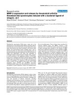
Inverse Relationship between urine TGF-β1 and NO
2
-
/NO
3
-
levelsFigure 3
Inverse Relationship between urine TGF-β1 and NO
2
-
/NO
3
-
levels. Dot matrix plot of NO
2
-
/NO
3
-
in relation to active
and total TGF-β1 in urine of female (Panel A & B) and male (Panel C & D) rats. The different dots (circle, triangle, square and
diamond) represent values of individual rats of each sex at different time points. Mean urinary levels of NO
2
-
/NO
3
-
in female
rats were inversely correlated to mean total TGF-β1 (Panel B) and active TGF-β1 (Panel A), with Pearson correlation coeffi-
cients of -0.72 (two tailed; *p < 0.02) and -0.69 (two tailed; *p < 0.03), respectively. Mean urine levels of NO
2
-
/NO
3
-
in male
rats were inversely correlated to mean total TGF-β1 (Panel D) and active TGF-β1 (Panel C), with Pearson correlation coeffi-
cients of -0.89 (two tailed; *p < 0.0001) and -0.76 (two tailed; *p < 0.01), respectively.
0
400
800
1200
0 200 400 600
0
400
800
1200
0 200 400 600
Female
A
C
Active TGF β
ββ
β1 [pg/mg of Creatinine]
NO
2
-
/NO
3
-
[
μ
μ
μ
μM / mg of Creatinine]
0
400
800
1200
0 200 400 600
B
0
400
800
1200
0 500 1000 1500 2000 2500
Latent TGF β
ββ
β1 [pg/mg of Creatinine]
D
Male
Journal of Inflammation 2009, 6:23 />Page 7 of 13
(page number not for citation purposes)
subjected to immunocytochemistry for iNOS as well as
active and latent TGF-β1 followed by confocal micros-
copy. In bladder tissue sections, active TGF-β1 is repre-
sented by red fluorescence and latent/total TGF-β1
(visualized by immunostaining for LAP) is represented by
blue fluorescence, while green stain represents smooth
muscle actin/phalloidin (Fig. 5). The urothelium region
of sections was marked by a lower expression of actin/
phalloidin. The purple color in the panels (Fig. 5AC) indi-
cates the predominance of blue fluorescence of latent
TGF-β1 over the red fluorescence of active TGF-β1. The
magenta color (Fig. 5D) in the panels indicates overlap of
similar intensity of blue fluorescence from latent TGF-β1
and the red fluorescence of active TGF-β1. In agreement
with the ELISA results in bladder tissue, male CYP-treated
rats exhibited the most intense magenta stain as com-
pared to other groups, indicating higher expression of
active TGF-β1 in the urothelium (Fig. 5D; lumen marked by
white arrow). The expression of active TGF-β1 was much
lower in control male rats (Fig. 5C) and female control
rats (Fig. 5A). The purple color is more evident in controls
of both sexes and in female CYP-treated rats (Figs. 5AB),
suggesting that latent TGF-β1 was elevated and activated
to a moderate degree in these tissues.
We next sought to determine if our emerging impression
of reciprocal expression of iNOS and TGF-β1 in the setting
of CYP-induced bladder inflammation could be con-
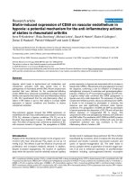
Bladder tissue levels of TGF-β-1 in control and CYP-treated ratsFigure 4
Bladder tissue levels of TGF-β-1 in control and CYP-treated rats. Bladder lysate from different groups were analyzed
for TGF-β1 by ELISA, and levels of TGF-β1 were then standardized by bladder weight and expressed as μg per bladder. Male
rats exhibited the highest expression of TGF-β1 in tissue compared to tissue levels of other groups (ANOVA, Tukey's Multiple
post hoc test; *p < 0.05). Levels of active TGF-β1 (Panel A) in bladder tissue were nearly 100-fold higher than levels of latent
TGF-β1 (Panel B) measured in bladder tissue of all groups. The substantial presence of latent TGF-β1 in female rats at baseline
(open bars) and after CYP (shaded bars) was accompanied by only a minor presence of active TGF-β1 (0 3.6 ng; Panel C). In
contrast, male rats exhibited both active and latent TGF-β1 at baseline and after CYP, with positive Pearson's coefficients of
0.65 and 0.75, respectively but without statistical significance (p = 0.24).
0
10
20
30
40
Active
0.0
0.1
0.2
0.3
0.4
*
*
AB
Latent
0
0.1
0.2
0.3
0.4
0204060
Female
Male
C
Female
Male
Female
Male
Active TGF-
β
β
β
β1
[pg/mg of Creatinine]
Latent TGF-β
ββ
β1 [pg/mg of Creatinine]
TGF-
β
β
β
β1 Bladder Content
[
μ
μ
μ
μg per bladder]
Journal of Inflammation 2009, 6:23 />Page 8 of 13
(page number not for citation purposes)
firmed immunocytochemically at the cellular level. In Fig.
6, iNOS is visualized in green and active TGF-β1 is red.
Except for male CYP-treated rats, the urothelium of other
groups was distinctly red and cells below the lumen were
stained green, indicating predominant iNOS expression
and low active TGF-β1. The male rats showed regions of
equal intensity for red and green fluorescence, just below
the cell layer bordering the lumen. Accordingly, we con-
clude that both iNOS and active TGF-β1 are expressed in
this region, though not co-expressed in the same cells.
This narrowing of tissue regions expressing green and red
fluorescence probably results from more severe tissue
destruction induced by acrolein from CYP in male rats rel-
ative to other groups.
Immunocytochemistry corroborated the urine and tissue
levels of TGF-β1 and NO
2
-
/NO
3
-
. In support of the tissue
ELISA data, bladder tissue from female CYP-treated rats
(Fig. 6B) exhibited the most intense green stain for iNOS
in the urothelium as compared to the other groups. The
immunocytochemical expression of iNOS was much
lower in control male rats (Fig. 6C). The bladders of
Localization of TGF-β1 in rat bladderFigure 5
Localization of TGF-β1 in rat bladder. Control and CYP-treated bladders were harvested at 24 h after CYP injection,
fixed in formalin, and cryopreserved prior to sectioning to a thickness of 8 μm. Bladder sections were stained for TGF-β1 (red
fluorescence) and LAP (blue fluorescence) for the immunodetection of active and latent TGF-β1, respectively. The urothelium
region of sections was marked by a lower degree of green stain for smooth muscle actin/phalloidin. Male CYP-treated rats
exhibited the most intense magenta stain to indicate the substantial presence of active TGF-β1 in urothelium (Panel D; lumen
marked by white arrow), that was much lower in control male rats (Panel C) and nearly absent in female control rats (Panel A).
The purple color emerging from the predominance of blue fluorescence in the overlap with red fluorescence was more prom-
inent in controls of both genders as well as in female rats treated with CYP, but absent in male CYP-treated rats. Magnification
is 60× in all sections and is representative of 4 animals in each group. The experiment is representative of 5 fields per slide.
Journal of Inflammation 2009, 6:23 />Page 9 of 13
(page number not for citation purposes)
female control rats (Fig. 6A) exhibited an elevated expres-
sion of iNOS relative to control male rats.
Discussion
A central observation of our study was the in vivo evidence
for inverse relationship between TGF-β1 and iNOS/NO
synthesis in the setting of bladder inflammation: when
NO
2
-
/NO
3
-
were at their lowest (24 h after CYP injection),
urinary TGF-β1 level reached their peak in both male and
female rats. These results suggest that TGF-β1 is an endog-
enous negative regulator of iNOS and subsequent produc-
tion of NO reaction products, a notion supported in
several biological settings [31-37]. Further support for this
hypothesis comes from our immunostaining studies
showing reciprocal staining of iNOS and TGF-β1, studies
that agree with previous reports on TGF-β1 synthesis by
epithelial and immune cells [12]. Those studies, along
with ours, suggest that the urothelium is the likely source
of TGF-β1 and NO metabolites measured in urine [12].
Prior studies in rat smooth muscle cells suggested that
Co-localization of TGF-β1 and iNOS in rat bladderFigure 6
Co-localization of TGF-β1 and iNOS in rat bladder. The confocal images show iNOS (red stain) and active TGF-β1
(green stain) in bladder sections. iNOS appears to be expressed at low levels in control male (Panel C) and female (Panel A)
rats. In contrast, iNOS immunostaining is increased following treatment with CYP (Panels B and D). The red stain for TGF-β1
was mostly localized in the urothelium region of all the groups. This region was also marked by a lower degree of blue stain for
smooth muscle actin/phalloidin in all the groups. Tissue destruction caused by CYP is prominent in Panel B and D relative to
the normal tissue architecture observed in Panels A and C. The lumen region adjoining the urothelium is indicated by white
arrows. Magnification is 60× in all sections. The experiment is representative of 5 fields per slide.
Journal of Inflammation 2009, 6:23 />Page 10 of 13
(page number not for citation purposes)
bioactive TGF-β1 is a potent suppressor of iNOS expres-
sion and enzymatic activity [18]. Further studies in this
cell type have also shown that TGF-β1 does not directly
inhibit enzymatic activity of iNOS, but rather that this
cytokine both suppresses the induction of iNOS mRNA as
well as increases the degradation of iNOS protein [38,39].
In the present study, we also observed a constitutive, basal
secretion of active TGF-β1 in the urine. The levels of active
TGF-β1 in the urine were generally correlated with the lev-
els of latent/total TGF-β1 in the urine of both males and
females (with the possible exception of the 10-h time
point in untreated male rats), suggesting that there is an
elevation in the expression of total TGF-β1 and that a con-
stant fraction of total TGF-β1 is active in urine regardless
of whether or not the animals were exposed to CYP. We
hypothesize that this active TGF-β1 originates in the blad-
der urothelium due to our data on the presence of TGF-β1
in the bladders of control rats. Likewise, the expression of
iNOS is observed to a low degree in control rat bladder.
The basal secretion of TGF-β1 and NO
2
-
/NO
3
-
seems to
fluctuate slightly throughout the day. Interestingly, we
noted that the maxima and minima of urinary TGF-β1
and NO
2
-
/NO
3
-
occurred at reciprocal time points of each
other, supporting the hypothesis that TGF-β1 is a physio-
logical suppressor of iNOS. The levels of NO
2
-
/NO
3
-
do
not remain constant throughout the day, but progres-
sively fall from maximum levels measured during the
morning hours. The rise of NO
2
-
/NO
3
-
in the morning
before falling to a stable value suggests that some of the
NO
2
-
/NO
3
-
assessed are contributed through the enzy-
matic action of the constitutive NOS enzymes (endothe-
lial NOS and neuronal NOS) as well as potentially from
the stress to the animal from handling and transport to
metabolic cage from animal facility. These results may
indicate an interplay among components of the endocrine
system, especially the hypothalamic-pituitary-adrenal
(HPA) axis that regulates circadian rhythm and stress
response with paracrine signaling in the bladder, a phe-
nomenon previously demonstrated in the aorta [40].
Indeed, given that activation of TGF-β1 is increased in set-
tings of physiological stress, our data may suggest involve-
ment of TGF-β1 in the homeostatic mechanism linked to
HPA axis [41]. It is worth noting that this large diurnal
variation in urinary levels of NO reaction products and
TGF-β1 in control rats argues for the need to sample urine
at multiple time points in studies assessing inflammatory
analytes in urine.
In contrast to earlier studies, we now demonstrate interre-
lated NO
2
-
/NO
3
-
and TGF-β1 levels in individual urine
voids separated by as little as 5 min in CYP-treated rats.
Our results show elevation of urinary NO reaction prod-
ucts in CYP-treated rats when compared to control rats in
urine collected at the same time point of the day. The peak
levels of NO metabolites in urine occurred at 4 h post-CYP
for male rats and at 6 h post-CYP for female rats relative
to baseline values, and the increase at these time points
agree with results reported previously [5]. It is not clear at
this point why the peak level of urinary NO
2
-
/NO
3
-
was
delayed in females vs. males, but this phenomenon may
be related to the influence of HPA axis and ovarian hor-
mones on the expression of TGF-β1 in the bladder. It
should be noted, however, that a previous study reported
that the overall effects of estrous stage on CYP-induced
bladder inflammation were insignificant [24]. In order to
fully address this issue, the effect of cyclical changes in
ovarian hormones will likely have to be determined by
repeating the experiments described here in ovariect-
omized rats [42,43].
Our prior studies showed that other cytokines reach their
peak by 4 h and decline by 24 h in the acute CYP model
[4]. In contrast, the levels of TGF-β1 were negligible by 4
h, with peaks at 24 h consistent with a late, anti-inflam-
matory, and pro-healing role for this cytokine demon-
strated in bronchial epithelial cells [12]. Indeed, it is
known that inflammation induced by CYP begins to
resolve by 2448 h, and studies in other organs have con-
firmed the role of TGF-β1 in wound healing after injury
and as regulator of immune cell activation in response to
inflammation [44-46].
The reciprocal relationship of TGF-β1 with NO reaction
products, as well as with other pro-inflammatory
cytokines [4] seems to suggest a need for different stimuli
for TGF-β1 production by the bladder [13]. One likely
stimulus for the generation of TGF-β1 in the bladder
might be reactive oxygen species (ROS) generated by acro-
lein, which can alter the redox balance in bladder tissues
and lead to the activation of latent TGF-β1 [2]. Activated
TGF-β1 is known to either decrease or increase the gener-
ation of ROS, depending on cellular/enzymatic source
and experimental conditions [47-50] and therefore ele-
vated levels of latent TGF-β1 in CYP-treated rats may also
explain the reduced expression of TNF-α (a ROS-activated
gene) in bladder noted by ourselves and others [4,51].
Our findings are likely to have clinical relevance. An ele-
vated iNOS activity has been previously noted in IC
patients, and elevated levels of NO reaction products have
been linked to changes in tight junction protein dynamics
associated with the observed disrupted barrier function of
the urothelium [52]. In those studies, the release of TNF-
α and IL-1β from bladder was shown to induce iNOS [53].
Our results demonstrating increased urinary NO
2
-
/NO
3
-
after treatment with CYP agree with previous reports that
assessed NO
2
-
/NO
3
-
in urine collected over a 2-h time
period from 2-4 h and 4 to 6 h after CYP injection [5].
Journal of Inflammation 2009, 6:23 />Page 11 of 13
(page number not for citation purposes)
The presence of urine TGF-β1 has not been previously
described in IC patients, though enhanced expression of
this cytokine has been noted in tissue biopsies of IC
patients [9,10]. However, concurrent presence of iNOS
and TGF-β1 in IC patients remains to be studied. Given
the known biology of TGF-β1 and the elevated levels of
this cytokine measured in our rat model, it is tempting to
speculate that the bladder fibrosis characteristic of IC
patients [20,54] may be caused at least in part by TGF-β1.
In support of this hypothesis, incubation of human detru-
sor smooth muscle cells with TGF-β1 led to hypertrophic
and fibrotic responses characterized by the upregulation
of COL1A1 and COL3A1 mRNA; genes that are necessary
for collagen synthesis [55].
The striking, gender-specific pattern of expression and
secretion of TGF-β1 may be related to that seen in prior
studies documenting hypertrophied lamina propria and
stromal hyperplasia only in male mice, lacking type II
TGF-β1 receptor gene [56]. The higher female prevalence
of IC, as well as the limited clinical success in the treat-
ment of hemorrhagic cystitis with estrogens [57,58], sup-
port the notion that TGF-β1 is a central molecular
mediator governing the gender-related differences in the
response to CYP reported here. Estrogen can reverse the
effects of TGF-β1 by reducing the activity of the transcrip-
tion factors Sp1 and Smad3, which in turn leads to the
reduced synthesis of collagen and extracellular matrix
[59,60]. A recent report reviewed five case studies of suc-
cessful treatment of hemorrhagic cystitis with conjugated
estrogens in the clinic, and showed that this therapeutic
benefit was accompanied by an altered serum cytokine
profile [58].
Conclusion
The results of this study suggest that there exists an inverse
relationship between the expression of TGF-β1 and NO
reaction products in the acrolein-inflamed bladder. The
inverse correlation between urine levels of NO-derived
products and TGF-β1 may be viewed as a consequence of
a more predominant TGF-β1 effect in blocking iNOS
induction. Given the time course of inflammation
induced by CYP in our study, TGF-β1 is likely to emerge
as a central mediator of the resolution of inflammation
and induction of healing and in the bladder. Our results
therefore argue in favor of evaluating urinary TGF-β1 in IC
patients in order to assess disease progression, and may
point to novel areas for the development of therapeutics
for this disease.
Competing interests
The authors declare that they have no competing interests.
Authors' contributions
PT and YV designed the study. DB, PL, VT and EW exe-
cuted the experiments in the manuscripts. NY and RZ
were involved in data analysis and manuscript prepara-
tion. All authors read and approved the final manuscript.
Acknowledgements
The work in this study was supported by NIH grant NIDDK RO1-DK
066138 and NIDRR grant H133E070024.
References
1. Hu RQ, Mehter H, Nadasdy T, Satoskar A, Spetie DN, Rovin BH,
Hebert L: Severe hemorrhagic cystitis associated with pro-
longed oral cyclophosphamide therapy: case report and lit-
erature review. Rheumatol Int 2008, 28(11):1161-1164.
2. Korkmaz A, Topal T, Oter S: Pathophysiological aspects of
cyclophosphamide and ifosfamide induced hemorrhagic cys-
titis; implication of reactive oxygen and nitrogen species as
well as PARP activation. Cell Biol Toxicol 2007, 23:303-312.
3. Cox PJ: Cyclophosphamide cystitis identification of acrolein
as the causative agent. Biochem Pharmacol 1979, 28:2045-2049.
4. Smaldone MC, Vodovotz Y, Tyagi V, Barclay D, Philips BJ, Yoshimura
N, Chancellor MB, Tyagi P: Multiplex analysis of urinary
cytokine levels in rat model of cyclophosphamide-induced
cystitis. Urology 2009, 73:421-426.
5. Linares-Fernandez BE, Alfieri AB: Cyclophosphamide induced
cystitis: role of nitric oxide synthase, cyclooxygenase-1 and 2,
and NK(1) receptors. J Urol 2007, 177:1531-1536.
6. Hamby ME, Hewett JA, Hewett SJ: TGF-beta1 reduces the heter-
ogeneity of astrocytic cyclooxygenase-2 and nitric oxide syn-
thase-2 gene expression in a stimulus-independent manner.
Prostaglandins Other Lipid Mediat 2008, 85:115-124.
7. Guo YS, Chen Z, Wen XD, Ko TC, Townsend CM Jr, Hellmich MR:
Synergistic Regulation of COX-2 Expression by Bombesin
and Transforming Growth Factor-beta. Dig Dis Sci 2008,
53(8):2045-2052.
8. Macedo FY, Baltazar F, Mourao LC, Almeida PR, Mota JM, Schmitt FC,
Ribeiro RA: Induction of COX-2 expression by acrolein in the
rat model of hemorrhagic cystitis. Exp Toxicol Pathol 2008,
59:425-430.
9. Koskela LR, Thiel T, Ehren I, De Verdier PJ, Wiklund NP: Localiza-
tion and expression of inducible nitric oxide synthase in biop-
sies from patients with interstitial cystitis. J Urol 2008,
180:737-741.
10. Ueda T, Tamaki M, Ogawa O, Yoshimura N: Over expression of
platelet-derived endothelial cell growth factor/thymidine
phosphorylase in patients with interstitial cystitis and blad-
der carcinoma.
J Urol 2002, 167:347-351.
11. Chen ML, Yan BS, Bando Y, Kuchroo VK, Weiner HL: Latency-
Associated Peptide Identifies a Novel CD4+CD25+ Regula-
tory T Cell Subset with TGF{beta}-Mediated Function and
Enhanced Suppression of Experimental Autoimmune
Encephalomyelitis. J Immunol 2008, 180:7327-7337.
12. Mayer AK, Bartz H, Fey F, Schmidt LM, Dalpke AH: Airway epithe-
lial cells modify immune responses by inducing an anti-
inflammatory microenvironment. Eur J Immunol 2008,
38:1689-1699.
13. Zheng SG, Wang J, Horwitz DA: Cutting Edge:
Foxp3+CD4+CD25+ Regulatory T Cells Induced by IL-2 and
TGF-{beta} Are Resistant to Th17 Conversion by IL-6. J
Immunol 2008, 180:7112-7116.
14. Bottinger EP: TGF-beta in renal injury and disease. Semin Neph-
rol 2007, 27:309-320.
15. Saxena V, Lienesch DW, Zhou M, Bommireddy R, Azhar M, Doet-
schman T, Singh RR: Dual roles of immunoregulatory cytokine
TGF-beta in the pathogenesis of autoimmunity-mediated
organ damage. J Immunol 2008, 180:1903-1912.
16. Annes JP, Munger JS, Rifkin DB: Making sense of latent TGF beta
activation. J Cell Sci 2003, 116:217-224.
17. Zamora R, Vodovotz Y: Transforming growth factor-beta in
critical illness. Crit Care Med 2005, 33:S478-481.
Journal of Inflammation 2009, 6:23 />Page 12 of 13
(page number not for citation purposes)
18. Vodovotz Y: Control of nitric oxide production by transform-
ing growth factor-beta1: mechanistic insights and potential
relevance to human disease. Nitric Oxide 1997, 1:3-17.
19. Gressner OA, Rizk MS, Kovalenko E, Weiskirchen R, Gressner AM:
Changing the pathogenetic roadmap of liver fibrosis? Where
did it start; where will it go? J Gastroenterol Hepatol 2008, 23(7
Part 1):1024-1035.
20. Lemack GE, Zimmern PE: [Interstitial cystitis: reevaluation of
patients who do no respond to standard treatments]. Prog
Urol 2001, 11:239-244.
21. MacDermott JP, Charpied GC, Tesluk H, Stone AR: Can histologi-
cal assessment predict the outcome in interstitial cystitis? Br
J Urol 1991, 67:44-47.
22. Lynes WL, Flynn SD, Shortliffe LD, Stamey TA: The histology of
interstitial cystitis. Am J Surg Pathol 1990, 14:969-976.
23. Rodo J, Farre X, Martin E: Cyclophosphamide-induced hemor-
rhagic cystitis in rats that underwent colocystoplasty: exper-
imental study. J Urol 2001, 165:660-666.
24. Bon K, Lanteri-Minet M, Menetrey D, Berkley KJ: Sex, time-of-day
and estrous variations in behavioral and bladder histological
consequences of cyclophosphamide-induced cystitis in rats.
Pain 1997, 73:423-429.
25. Terado M, Nomura M, Mineta K, Nishii H, Fujimoto N, Sasaguri T,
Sasaguri Y, Matsumoto T: Involvement of estrogen in the patho-
genesis of cyclophosphamide-induced cystitis in rats. Endo-
crine 2005, 26:55-63.
26. Sadeghi M, Daniel V, Naujokat C, Weimer R, Opelz G: Strikingly
higher interleukin (IL)-1alpha, IL-1beta and soluble inter-
leukin-1 receptor antagonist (sIL-1RA) but similar IL-2, sIL-
2R, IL-3, IL-4, IL-6, sIL-6R, IL-10, tumour necrosis factor
(TNF)-alpha, transforming growth factor (TGF)-beta and
interferon IFN-gamma urine levels in healthy females com-
pared to healthy males: protection against urinary tract
injury? Clin Exp Immunol 2005, 142:312-317.
27. Silbiger S, Neugarten J:
Gender and human chronic renal dis-
ease. Gend Med 2008, 5(Suppl A):S3-S10.
28. Vera PL, Wang X, Meyer-Siegler KL: Upregulation of macro-
phage migration inhibitory factor (MIF) and CD74, receptor
for MIF, in rat bladder during persistent cyclophosphamide-
induced inflammation. Exp Biol Med (Maywood) 2008,
233:620-626.
29. Vodovotz Y: Modified microassay for serum nitrite and
nitrate. Biotechniques 1996, 20:390-392. 394
30. Vodovotz Y, Chesler L, Chong H, Kim SJ, Simpson JT, DeGraff W,
Cox GW, Roberts AB, Wink DA, Barcellos-Hoff MH: Regulation of
transforming growth factor beta1 by nitric oxide. Cancer Res
1999, 59:2142-2149.
31. Fang FC: Perspectives series: host/pathogen interactions.
Mechanisms of nitric oxide-related antimicrobial activity. J
Clin Invest 1997, 99:2818-2825.
32. Olson MV, Lee J, Zhang F, Wang A, Dong Z: Inducible nitric oxide
synthase activity is essential for inhibition of prostatic tumor
growth by interferon-beta gene therapy. Cancer Gene Ther
2006, 13:676-685.
33. Wang D, Lu S, Dong Z: Regulation of TGF-beta1 gene tran-
scription in human prostate cancer cells by nitric oxide. Pros-
tate 2007, 67:1825-1833.
34. Shearer JD, Richards JR, Mills CD, Caldwell MD: Differential regu-
lation of macrophage arginine metabolism: a proposed role
in wound healing. Am J Physiol 1997, 272:E181-190.
35. Fiorenza G, Rateni L, Farroni MA, Bogue C, Dlugovitzky DG: TNF-
alpha, TGF-beta and NO relationship in sera from tubercu-
losis (TB) patients of different severity. Immunol Lett 2005,
98:45-48.
36. Lagadec P, Raynal S, Lieubeau B, Onier N, Arnould L, Saint-Giorgio V,
Lawrence DA, Jeannin JF: Evidence for control of nitric oxide
synthesis by intracellular transforming growth factor-beta1
in tumor cells. Implications for tumor development. Am J
Pathol 1999, 154:1867-1876.
37. Vodovotz Y, Zamora R, Lieber MJ, Luckhart S:
Cross-talk between
nitric oxide and transforming growth factor-beta1 in
malaria. Curr Mol Med 2004, 4:787-797.
38. Finder J, Stark WW Jr, Nakayama DK, Geller D, Wasserloos K, Pitt
BR, Davies P: TGF-beta regulates production of NO in pulmo-
nary artery smooth muscle cells by inhibiting expression of
NOS. Am J Physiol 1995, 268:L862-867.
39. Perrella MA, Jain MK, Lee ME: Role of TGF-beta in vascular
development and vascular reactivity. Miner Electrolyte Metab
1998, 24:136-143.
40. Navarro-Oliveira CM, Vassilieff VS, Cordellini S: The sympathetic
adrenomedullary system, but not the hypothalamic-pitui-
tary-adrenal axis, participates in aorta adaptive response to
stress: nitric oxide involvement. Auton Neurosci 2000,
83:140-147.
41. Smith EL, Batuman OA, Trost RC, Coplan JD, Rosenblum LA: Trans-
forming growth factor-beta 1 and cortisol in differentially
reared primates. Brain Behav Immun 2002, 16:140-149.
42. Ewan KB, Shyamala G, Ravani SA, Tang Y, Akhurst R, Wakefield L,
Barcellos-Hoff MH: Latent transforming growth factor-beta
activation in mammary gland: regulation by ovarian hor-
mones affects ductal and alveolar proliferation. Am J Pathol
2002, 160:2081-2093.
43. Casslen B, Sandberg T, Gustavsson B, Willen R, Nilbert M: Trans-
forming growth factor beta1 in the human endometrium.
Cyclic variation, increased expression by estradiol and pro-
gesterone, and regulation of plasminogen activators and
plasminogen activator inhibitor-1. Biol Reprod 1998,
58:1343-1350.
44. Finch CE, Laping NJ, Morgan TE, Nichols NR, Pasinetti GM: TGF-
beta 1 is an organizer of responses to neurodegeneration. J
Cell Biochem 1993, 53:314-322.
45. Ozawa H, Chancellor MB, Jung SY, Yokoyama T, Fraser MO, Yu Y, de
Groat WC, Yoshimura N: Effect of intravesical nitric oxide
therapy on cyclophosphamide-induced cystitis. J Urol 1999,
162:
2211-2216.
46. Welham NV, Lim X, Tateya I, Bless DM: Inflammatory factor pro-
files one hour following vocal fold injury. Ann Otol Rhinol Laryngol
2008, 117:145-152.
47. Tsunawaki S, Sporn M, Ding A, Nathan C: Deactivation of macro-
phages by transforming growth factor-beta. Nature 1988,
334:260-262.
48. Kim CD, Cho YJ, Park SH, Ha SW, Lee EG, Kim YJ, Kwon TH, Kim
IS, Kim YL: Urinary transforming growth factor-beta-induced
gene-h3 (betaig-h3) as a sensitive predictor in chronic
cyclosporine nephrotoxicity. Transplant Proc 2006,
38:1314-1319.
49. Hong YH, Peng HB, La Fata V, Liao JK: Hydrogen peroxide-medi-
ated transcriptional induction of macrophage colony-stimu-
lating factor by TGF-beta1. J Immunol 1997, 159:2418-2423.
50. Islam KN, Kayanoki Y, Kaneto H, Suzuki K, Asahi M, Fujii J, Taniguchi
N: TGF-beta1 triggers oxidative modifications and enhances
apoptosis in HIT cells through accumulation of reactive oxy-
gen species by suppression of catalase and glutathione per-
oxidase. Free Radic Biol Med 1997, 22:1007-1017.
51. Helmy A, Hammam OA, El Lithy TR, El Deen Wishahi MM: The role
of TGF-beta-1 protein and TGF-beta-R-1 receptor in
immune escape mechanism in bladder cancer. MedGenMed
2007, 9:34.
52. Hosseini A, Ehren I, Wiklund NP: Nitric oxide as an objective
marker for evaluation of treatment response in patients
with classic interstitial cystitis. J Urol 2004, 172:2261-2265.
53. Ribeiro RA, Freitas HC, Campos MC, Santos CC, Figueiredo FC,
Brito GA, Cunha FQ: Tumor necrosis factor-alpha and inter-
leukin-1beta mediate the production of nitric oxide involved
in the pathogenesis of ifosfamide induced hemorrhagic cysti-
tis in mice. J Urol 2002, 167:2229-2234.
54. Seishima M, Shimizu H, Oyama Z, Isogai K: Mixed connective tis-
sue disease following interstitial cystitis. Eur J Dermatol 2001,
11:45-47.
55. Howard PS, Kucich U, Coplen DE, He Y: Transforming growth
factor-beta1-induced hypertrophy and matrix expression in
human bladder smooth muscle cells.
Urology 2005,
66:1349-1353.
56. Sharif-Afshar AR, Donohoe JM, Pope JCt, Adams MC, Brock JW 3rd,
Bhowmick NA: Stromal hyperplasia in male bladders upon
loss of transforming growth factor-beta signaling in fibrob-
lasts. J Urol 2005, 174:1704-1707. discussion 1707
57. Nickel JC, Teichman JM, Gregoire M, Clark J, Downey J: Prevalence,
diagnosis, characterization, and treatment of prostatitis,
interstitial cystitis, and epididymitis in outpatient urological
practice: the Canadian PIE Study. Urology 2005, 66:935-940.
Publish with Bio Med Central and every
scientist can read your work free of charge
"BioMed Central will be the most significant development for
disseminating the results of biomedical research in our lifetime."
Sir Paul Nurse, Cancer Research UK
Your research papers will be:
available free of charge to the entire biomedical community
peer reviewed and published immediately upon acceptance
cited in PubMed and archived on PubMed Central
yours — you keep the copyright
Submit your manuscript here:
/>BioMedcentral
Journal of Inflammation 2009, 6:23 />Page 13 of 13
(page number not for citation purposes)
58. Kopterides P, Theodorakopoulou M, Mentzelopoulos S, Armaganidis
A: Cyclophosphamide-induced hemorrhagic cystitis success-
fully treated with conjugated estrogens. Am J Hematol 2005,
80:166-167.
59. Blush J, Lei J, Ju W, Silbiger S, Pullman J, Neugarten J: Estradiol
reverses renal injury in Alb/TGF-beta1 transgenic mice. Kid-
ney Int 2004, 66:2148-2154.
60. Dixon A, Maric C: 17beta-Estradiol attenuates diabetic kidney
disease by regulating extracellular matrix and transforming
growth factor-beta protein expression and signaling. Am J
Physiol Renal Physiol 2007, 293:F1678-1690.