Báo cáo y học: " Lactobacillus casei modulates the inflammation-coagulation interaction in a pneumococcal pneumonia experimental model." potx
Bạn đang xem bản rút gọn của tài liệu. Xem và tải ngay bản đầy đủ của tài liệu tại đây (1.06 MB, 10 trang )
BioMed Central
Page 1 of 10
(page number not for citation purposes)
Journal of Inflammation
Open Access
Research
Lactobacillus casei modulates the inflammation-coagulation
interaction in a pneumococcal pneumonia experimental model
Cecilia Haro
1
, Julio Villena
2
, Hortensia Zelaya
1
, Susana Alvarez
1,2
and
Graciela Agüero*
1
Address:
1
Instituto de Bioquímica Aplicada, Facultad de Bioquímica, Química y Farmacia, Universidad Nacional de Tucumán, Balcarce 747, CP
4000, San Miguel de Tucumán, Tucumán, Argentina and
2
Laboratorio de Bioquímica y Clínica Experimental, Centro de Referencia para
Lactobacilos (CERELA-CONICET), Chacabuco 145, Tucumán, Argentina
Email: Cecilia Haro - ; Julio Villena - ; Hortensia Zelaya - ;
Susana Alvarez - ; Graciela Agüero* -
* Corresponding author
Abstract
Background: We have previously demonstrated that Lactobacillus casei CRL 431 administration
improved the resistance to pneumococcal infection in a mouse model.
Methods: This study examined the effects of the oral administration of Lactobacillus casei CRL 431
(L. casei) on the activation of coagulation and fibrinolytic systems as well as their inhibitors during
a Streptococcus pneumoniae infection in mice.
Results: The alveolo-capillary membrane was damaged and the coagulation system was also
activated by the infection. As a consequence, we could see fibrin(ogen) deposits in lung histological
slices, increased levels of thrombin-antithrombin complex (TATc) in bronchoalveolar lavage (BAL)
and plasma, decrease in prothrombin activity (PT) and prolonged activated partial thromboplastin
time test (APTT) values. Factor VII (FVII) and factor X (FX) were decreased in plasma, whereas
fibrinogen (F) and factor VIII (FVIII) were increased. The low levels of protein C (PC) in BAL and
plasma proved damage on inhibitory activity. The infected animals showed reduced fibrinolytic
activity, evidenced by an increase in plasminogen activation inhibitor-1 (PAI-1) in BAL and plasma.
The pathogen induced an increase of TNF-α, IL-1β and IL-6 in BAL and serum a few hours after
challenge followed by a significant decrease until the end of the assayed period. IL-4 and IL-10 in
BAL and serum were also augmented, especially at the end of the experiment. The animals treated
with L. casei showed an improvement of alveolo-capillary membrane, lower fibrin(ogen) deposits in
lung and decrease in TATc. APTT test and PT, FVII and FX activity were normalized. L. casei group
showed lower F levels than control during whole experiment. In the present study no effect of L.
casei on the recovery of the inhibitory activity was detected. However, L. casei was effective in
reducing PAI-1 levels in BAL and in increasing anti-inflammatory ILs concentration.
Conclusion: L. casei proved effective to regulate coagulation activation and fibrinolysis inhibition
during infection, leading to a decrease in fibrin deposits in lung. This protective effect of L. casei
would be mediated by the induction of higher levels of IL-4 and IL-10 which could regulate the anti-
inflammatory, procoagulant and antifibrinolytic effects of TNF-α, IL-1β and IL-6.
Published: 16 October 2009
Journal of Inflammation 2009, 6:28 doi:10.1186/1476-9255-6-28
Received: 28 April 2009
Accepted: 16 October 2009
This article is available from: />© 2009 Haro et al; licensee BioMed Central Ltd.
This is an Open Access article distributed under the terms of the Creative Commons Attribution License ( />),
which permits unrestricted use, distribution, and reproduction in any medium, provided the original work is properly cited.
Journal of Inflammation 2009, 6:28 />Page 2 of 10
(page number not for citation purposes)
Background
The activation of coagulation and fibrin deposition as a
consequence of inflammation is well known, and can be
viewed as an essential part of the host defences [1]. The
hallmark of inflammatory lung diseases are fibrin depos-
its, which enhance the inflammatory responses by
increasing vascular permeability, activating endothelial
cells to produce proinflammatory mediators, and eliciting
recruitment and activation of neutrophils [2]. Excessive
fibrin deposition within the airways results from severe
inflammation, with increased activation of coagulation,
and may compromise pulmonary integrity and function
[3,2].
Current evidence from human studies suggests that in
lung injury there is augmented tissue factor expression,
down regulation of protein C (PC), and higher plasmino-
gen activator inhibitor -1 (PAI-1) levels. Together, these
abnormalities shift the intra-alveolar environment from
anticoagulant and profibrinolytic to procoagulant and
antifibrinolytic [4].
The relationship between inflammation and the coagula-
tion system is a process in which inflammation leads not
only to the activation of coagulation, but coagulation also
considerably affects inflammatory activity. Besides, an
insufficiently controlled response can lead to a situation
in which coagulation and thrombosis contribute to dis-
ease [1].
Hence, modulation of fibrin deposition through coagula-
tion and fibrinolysis regulation may be an important ther-
apeutic target.
Probiotic lactic acid bacteria have several inmunomodula-
tory effects [5,6] and anti-inflammatory properties [7,8].
Our group reported that oral administration of Lactobacil-
lus casei CRL 431 to mice infected intranasally with Strep-
tococcus pneumoniae (S. pneumoniae) facilitated clearance
of the pathogen and modulated the inflammatory
immune response with less damage to lung tissue [9].
Considering the relevant participation of the relationship
inflammation-coagulation in the severity of pneumococ-
cal pneumonia [10], the present study was conducted to
examine the effects of the oral administration of Lactoba-
cillus casei CRL 431 on the activation of coagulation dur-
ing a S. pneumoniae infection in a mouse experimental
model.
Methods
Microorganisms
Lactobacillus casei CRL 431 (L. casei) was obtained from
the CERELA culture collection. It was cultured for 8 h at
37°C (final log phase) in Man-Rogosa-Sharpe broth
(MRS, Oxoid), and the bacteria were harvested through
centrifugation at 5,000 rpm for 10 min and then washed
three times with sterile 0.01 M phosphate buffer saline
(PBS), pH 7.2 [9].
Capsulated pneumococcus (serotype 14) was isolated
from the respiratory tract of a patient from the Depart-
ment of Clinical Bacteriology of the Niño Jesús Children's
Hospital in San Miguel de Tucumán, Argentina. Pneumo-
cocci serotypification was performed in Administración
Nacional de Laboratorios e Institutos de Salud-ANLIS "Dr.
Malbran", Buenos Aires, Argentina.
Animals
Six-week-old Swiss albino mice were obtained from the
closed colony kept at CERELA. They were housed in plas-
tic cages at room temperature. Each assay was performed
in groups consisting of 25-30 mice (5-6 for each day
before and after infection). The Ethical Committee for
Animal Care at CERELA and Universidad Nacional de
Tucumán approved the experiments.
Feeding procedures
L. casei was administrated for 2 consecutive days at a dose
of 10
9
cell/mouse/day [9]. L. casei was suspended in 5 ml
of sterile 10% non-fat milk (NFM) and added to the
drinking water (20% v/v). The control group received ster-
ile NFM in the same conditions as the test group. All mice
were fed a conventional balanced diet ad libitum.
Experimental infection
S. pneumoniae was grown according to Racedo et al. [9]. At
the end of the dietary treatment (on the 3rd day) the ani-
mals were challenged with the pathogen. Animals with
(Lc group) and without (C group) treatment were infected
by dropping 25 uL of the inoculum containing 10
6
CFU
(log-phase) of S. pneumoniae in PBS into each nostril and
allowing it to be inhaled. To facilitate migration of the
inoculum to the alveoli, mice were held in a head-up ver-
tical position for 2 min. Animals were sacrificed on day 0
(before infection) and at different times post-infection.
The pathogen was detected in lung and blood samples of
control mice throughout the period assayed, while the
group fed with L. casei for 2d showed a faster clearence of
the S. pneumoniae [9]. After the challenge, we monitored
the survival of mice until day 21 post-infection. All ani-
mals survived without significant differences between
both groups.
Fibrin(ogen) deposition in pulmonary tissue
Fibrin(ogen) deposition in pulmonary tissue was deter-
mined by immunohistochemical techniques. Lung sam-
ples from both groups were fixed in 4% (v/v) formalin
saline solution, dehydrated, embedded in Histowax
(Leica Microsystems Nussloch GmbH, Nussloch, Ger-
many) and cut into 4 μm serial sections. For fibrin(ogen)
Journal of Inflammation 2009, 6:28 />Page 3 of 10
(page number not for citation purposes)
immunostaining, lung sections were deparaffinized and
endogenous peroxidase activity was quenched with a
solution of methanol/0.03% H
2
O
2
to inhibit the activity
of endogenous peroxidase in the lungs (Merck, Buenos
Aires, Argentina). The sections were incubated in 10%
normal sheep serum and then exposed to sheep anti-
mouse fibrinogen (purified IgG, Cedarlane, Hornby,
Ontario, Canada). After washes, slides were incubated
with donkey antisheep IgG peroxidase conjugate (Sigma-
Aldrich Co). Peroxidase activity was detected with a 3,3'-
diaminobenzidine peroxidase substrate solution (Sigma-
Aldrich Co), after which a light counterstain with hema-
toxylin was performed [11].
Bronchoalveolar lavage (BAL) assays
BAL samples were obtained according to the technique
described previously [12]. Briefly the trachea was exposed
and intubated with a catheter and 2 sequential lavages
were performed in each mouse by injecting 0.5 ml of ster-
ile PBS. The recovered fluid was centrifuged for 10 min at
900 × g. The supernatant fluid was frozen at -70°C for
subsequent biochemical and haemostatic analyses.
Albumin content
A measure to quantitate increased permeability of the
bronchoalveolar-capillarity barrier was determined color-
imetrically based on albumin binding to bromocresol
green using an albumin BCG diagnostic kit (Roche Diag-
nostics, Indianapolis, USA). The results were expressed as
mg/mL.
LDH activity
An indicator of general cytotoxicity was determined by
measuring the formation of a reduced form of nicotina-
mide adenine dinucleotide using Roche Diagnostic rea-
gents and procedures (Roche Diagnostics, Indianapolis,
USA). The results were expressed as U/L of BAL fluid.
Haemostatic tests
Blood samples were obtained through cardiac puncture
and were collected in a 3.2% solution of trisodium citrate
at a ratio of 9:1. Plasma was obtained according to Agüero
et al [11]. Prothrombin time (PT); activated partial throm-
boplastin time (APTT); factors VII, X, II, V, VIII; and fibrin-
ogen were performed manually on fresh plasma samples.
PT and coagulation factors VII, X, II and V were deter-
mined by a one-step method (Thromborel S, Behning-
werke AG, Marburg, Germany). APTT and VIII were
determined by mixing plasma with calcium chloride and
a partial thromboplastin reagent (STA APTT, Diagnostica
Stago, Asnières, France) and timing initial clot formation.
Fibrinogen concentration was determined by the method
of Clauss using a commercial kit and following manufac-
turer's instructions (Fibriprestz, Diagnostica Stago,
Asnières, France) [11].
Thrombin-antithrombin complexes (TATc), markers of
coagulation system activation, were determined by
enzyme-linked immunosorbent assay (ELISA) technique
according to manufacturer's instructions (Dade Behring,
Marburg, Germany). PC and PAI-1 activities were meas-
ured by chromogenic substrate assays (COAMATE
®
pro-
tein C, Chromogenix, Mölndal, Sweden; STACHROM
®
PAI, Diagnostica Stago, Asnières, France). TATc, PC and
PAI-1 levels were measured in BAL and plasma samples.
Cytokines determination
Cytokines were measured in plasma and in BAL fluid;
both were obtained as described above. Tumor necrosis
factor (TNF-α), interleukin-1β (IL-1β), IL-4, IL-6 and IL-
10 concentrations were measured with commercially
available ELISA kits according to the manufacturer's rec-
ommendations (R & D Systems, MN, USA).
Statistical analysis
Experiments were performed in triplicate (5-6 animals
each time) and results were expressed as means ± SD. After
verification of a normal distribution of data, 2-way
ANOVA was used. Tukey's test (for pairwise comparisons
of the means) was used to test for differences between the
groups. Differences were considered significant at P <
0.05.
Results
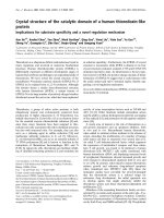
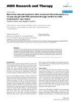
Biochemical assay of BAL fluid
Albumin content and LDH activity, measured in the acel-
lular BAL fluid, were used as indices of lung injury. Chal-
lenge with S. pneumoniae caused increases in BAL albumin
concentration and LDH activity in both groups, but these
parameters were significantly lower in L. casei treated mice
(Figure 1).
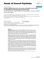
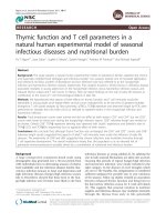
Fibrin(ogen) deposition in pulmonary tissue
Infected control animals showed fibrin(ogen) deposits in
the pleura. These deposits reached their highest intensity
at 10d post-infection (Figure 2). In the parenchyma, the
deposits were slightly positive with a focal pattern.
The animals treated with L casei showed fibrin deposits of
only in the pleura, with a focal pattern and lower intensity
than in the C group.
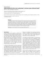
Local activation of coagulation
TATc levels were increased in BAL from both experimental
groups, showing highest values on d 1 post-infection (Fig-
ure 3A). Then, TATc concentration decreased gradually
until it reached initial values at 5 d post-infection. How-
ever, the levels of these complexes were lower in animals
supplemented with L. casei, which remained within the
normal range since d 2 post-infection.
Journal of Inflammation 2009, 6:28 />Page 4 of 10
(page number not for citation purposes)
Systemic activation of coagulation
The increase in TATc levels in BAL was accompanied by
increased systemic TATc levels since 12 h post-infection in
both groups. Mice treated with L. casei, returned to normal
values on d 5 after challenge, whereas the control group
continued with higher values (Figure 3B).
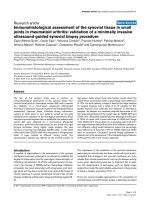
The percentage of prothrombin activity decreased on d 1
post-challenge in both experimental groups. However,
values were significantly lower in the control mice (Figure
4A). The L casei treated mice showed normal PT values
since d 5 post-infection, whereas the control group did
not reach normal values at any of the assessed periods.
After infection, APTT values were prolonged in both
experimental groups (Figure 4B). The mice supplemented
with L. casei normalized this parameter on d 5 post-infec-
tion, whereas the control group did so only on d 10 post-
infection.
Coagulation factors
FVII concentrations decreased in both groups after the
challenge, reaching minimum levels on d 1 post-infec-
tion. Only mice treated with L. casei normalized the FVII
Albumin and LDH in BALFigure 1
Albumin and LDH in BAL. Lactobacillus casei was orally
administrated at a dose of 10
9
cells for 2 d before challenge
with the pathogen; C group mice were infected without pre-
vious treatment. (A) Albumin content and (B) LDH activity in
BAL were evaluated. Results are expressed as means ± SD (n
= 5 or 6). *Significantly different from the C group and basal
values (p < 0.05).
Fibrin(ogen) deposition in pulmonary tissueFigure 2
Fibrin(ogen) deposition in pulmonary tissue. Lactobacil-
lus casei was orally administrated at a dose of 10
9
cells for 2 d
before challenge with the pathogen; C group mice were
infected without previous treatment. Panel A, C mice on d 0;
Panel B, C mice on d 5 post infection; Panel C, L casei mice
on d 5 post-infection.
Journal of Inflammation 2009, 6:28 />Page 5 of 10
(page number not for citation purposes)
values since d 5 post-infection (Figure 5A). These results
showed a similar behaviour to the prothrombin activity
described above.
FX values decreased in both groups after the infection,
although the animals supplemented with L. casei could
normalize this parameter on d 15 post-infection (Figure
5B). The C group showed lower values until the end of the
experiment.
No differences between groups were found in the levels of
FII during the studied period (Figure 5C).
The FV levels showed normal values during the whole
assayed period in both experimental groups (Figure 5D).
These results would indicate that liver functionality was
preserved.
The infection caused an increase in fibrinogen concentra-
tion in both groups on d 1 post-infection. After that, the
animals supplemented with L casei showed lower concen-
trations (p < 0.05) than control mice until the end of the
experiment (Figure 6A). The L. casei group returned to
nomal values on d 5 post-infection, while the C group did
so on d 10 post-infection.
Both groups showed increased FVIII levels after challenge
(Figure 6B). The peak was reached on d 1 post-infection,
and then levels dropped and returned to baseline within
d 4 post-infection with no differences between control
and treated groups.
Coagulation regulators in blood and lungs
The PC system provides important coagulation control.
We studied the levels of PCa in BAL and in plasma to eval-
utate the anticoagulant activity during the infection. After
challenge with S. pneumoniae, PCa increased in BAL in
both groups, reaching a peak on d 1 post-infection (Figure
7A). After that, the values dropped and remained
decreased until d 15 post-infection. No significantly dif-
ferences between control and treated groups were found
throughout the studied period. The PCa values in plasma
showed a different kinetic to the one described in BAL
(Figure 7B). The infection induced a significant decrease
in the plasma levels of PCa on d 1 post-infection in both
groups, returning to baseline on d 5 post-infection.
Levels of PAI-1 in BAL increased after infection in both
experimental groups, reaching a maximum on d 1 post
challenge (Figure 8A). However, the mice treated with L.
casei had significantly lower values than the control
group. The PAI-1 values returned to baseline in the treated
group sooner (d 5) than in the control (d 10).
Systemic PAI-1 levels showed a similar decrease in both
groups, reaching normal values on d 10 post-infection
(Figure 8B).
Thrombin-antithrombin complexes (TATc)Figure 3
Thrombin-antithrombin complexes (TATc). Lactobacil-
lus casei was orally administrated at a dose of 10
9
cells for 2 d
before challenge with the pathogen; C group mice were
infected without previous treatment. (A) TATc in BAL and
(B) TATc in plasma were studied. Results are expressed as
means ± SD (n = 5 or 6). *Significantly different from the C
group at the same time point (p < 0.05).
Prothrombin time and activated partial thromboplastin timeFigure 4
Prothrombin time and activated partial thrombo-
plastin time. Lactobacillus casei was orally administrated at a
dose of 10
9
cells for 2 d before challenge with the pathogen;
C group were infected without previous treatment. (A) Pro-
thrombin time and (B) activated partial thromboplastin time
were studied. Results are expressed as means ± SD (n = 5 or
6). *Significantly different from the C group (p < 0.05).
Journal of Inflammation 2009, 6:28 />Page 6 of 10
(page number not for citation purposes)
Cytokines
The levels of TNF-α, IL-1β and IL-6 in BAL before infection
were similar in both groups. After challenge with the path-
ogen, these cytokines increased significantly, reaching a
peak between 8 h and 12 h post-infection with higher val-
ues of TNF-α and IL-6 in the L. casei group. Afterwards, the
values of TNF-α and IL-1β decreased gradually until they
returned to base levels on d 5, whereas IL-6 concentration
remained elevated with significantly higher values in the
control group (Figure 9).
The serum levels of TNF-α, IL-1β and IL-6 augmented after
challenge, reaching the maximum values between 24 h
and 48 h post-infection. However, mice supplemented
with L. casei showed lower levels of TNF-α and IL-1β than
the control group on d 5 post-infection. Treatment with L
casei induced a stronger increase in IL-6, with values
higher than those in the control group until 48 h post-
infection. After that both groups showed similar values.
Coagulation factorsFigure 5
Coagulation factors. Lactobacillus casei was orally adminis-
trated at a dose of 10
9
cells for 2 d before challenge with the
pathogen; C group were infected without previous treat-
ment. (A) Factor VII, (B) factor X, (C) factor II and (D) factor
V activities were studied. Results are expressed as means ±
SD (n = 5 or 6). *Significantly different from the C group (p <
0.05).
Fibrinogen levels and factor VIII activityFigure 6
Fibrinogen levels and factor VIII activity. Lactobacillus
casei was orally administrated at a dose of 10
9
cells for 2 d
before challenge with the pathogen; C group were infected
without previous treatment. (A) Fibrinogen levels and (B)
factor VIII activity were studied. Results are expressed as
means ± SD (n = 5 or 6). *Significantly different from the C
group (p < 0.05).
Protein C activated (PC)Figure 7
Protein C activated (PC). Lactobacillus casei was orally
administrated at a dose of 10
9
cells for 2 d before challenge
with the pathogen; C group were infected without previous
treatment. (A) PC in BAL and (B) PC in plasma levels were
studied. Results are expressed as means ± SD (n = 5 or 6).
*Significantly different from the C group (p < 0.05).
Plasminogen activator inhibitor-1 (PAI-1)Figure 8
Plasminogen activator inhibitor-1 (PAI-1). Lactobacillus
casei was orally administrated at a dose of 10
9
cells for 2 d
before challenge with the pathogen; C group were infected
without previous treatment. (A) PAI-1 in BAL and (B) PAI-1
in plasma activity were studied. Results are expressed as
means ± SD (n = 5 or 6). *Significantly different from the C
group (p < 0.05).
Journal of Inflammation 2009, 6:28 />Page 7 of 10
(page number not for citation purposes)
The infection induced a progressive increase in the levels
of IL-4 in BAL and in serum in both experimental groups;
however, IL-4 values in the L. casei mice were significantly
higher than those in the control group (Figure 10).
Treatment with L. casei enhanced the levels of IL-10 in
BAL and in serum prior to infection (Figure 10). After 8 h
post-challenge, both groups showed a progressive
increase in IL-10 in BAL, which remained high up to d 5
post-infection. In the L. casei group, IL-10 in BAL was sig-
nificantly higher than in the control group since d 2 post-
infection. The values of serum IL-10 in the L. casei group
were higher than in the control group on d 3 and 5 post-
infection.
Discussion
Even though the inflammatory response and coagulation
activation exert an obvious protective function, the unco-
trolled functioning of these processes would be harmful
for the host.
Bearing in mind the previous experiences of our work
team concerning the ability of L casei to modulate the
immune response and protect mice against infection by S.
pneumoniae [9], we decided to investigate whether this
probiotic lactic acid bacteria could also regulate the hae-
mostatic processes during pneumonia and prevent exces-
sive fibrin formation [1], which increases the
inflammatory response even more [2].
In order to find out the intensity of the damage induced
by the pathogen at the lung level, we determined albumin
concentration and LDH activity in BAL [13]. We observed
that the S. pneumoniae induced increase in albumin con-
centration and in LDH activity in both groups, however
these alterations were significantly smaller in L. casei
treated mice. These results would indicate lower tissue
damage and improvement in the permeability of the alve-
olo capilar membrane. In adition, the supplemented ani-
mals showed lower deposits of fibrin in lung. This result
would be evidence for the inflammatory response modu-
lation [2].
To known the procoagulante state in lung, it was deter-
mined the levels of TATc in BAL. This marker was
increased in both groups on 1 d post-infection, but the
levels of these complexes were lower in animals supple-
mented with L. casei and remained within the normal
range since d 2 post-infection.
In order to study the procoagulante state at systemic level
we also determined TATc in plasma. The results evidenced
activation of the coagulation system in both groups since
12 h post-infection. Only the L. casei group reached de
normal values on d 5 after challenge.
On the basis of the fact that L. casei was able to regulate
fibrin deposition in lung during infection, we continued
IL-1β in BAL (A) and in serum (D); TNF-α in BAL(B) and in serum (E); IL-6 in BAL (C) and in serum (F) of mice fed L. casei for 2 d before (d0) and after challenge (d 1, 5, 10 y 15) with S. pneumoniaeFigure 9
IL-1β in BAL (A) and in serum (D); TNF-α in BAL(B)
and in serum (E); IL-6 in BAL (C) and in serum (F) of
mice fed L. casei for 2 d before (d0) and after chal-
lenge (d 1, 5, 10 y 15) with S. pneumoniae. Control mice
were challenged with the pathogen without previous treat-
ment. Results are expressed as means ± SD (n = 5 or 6).
Asterisks represent significant differences from the C group
at the same time point (*p < 0.05, **p < 0.01).
IL-4 in BAL (A) and in serum (C); IL-10 in BAL(B) and in serum (D) of mice fed L. casei for 2 d before (d0) and after challenge (d 1, 5, 10 y 15) with S. pneumoniaeFigure 10
IL-4 in BAL (A) and in serum (C); IL-10 in BAL(B)
and in serum (D) of mice fed L. casei for 2 d before
(d0) and after challenge (d 1, 5, 10 y 15) with S. pneu-
moniae. Control mice were challenged with the pathogen
without previous treatment. Results are expressed as means
± SD (n = 5 or 6). Asterisks represent significant differences
from the C group at the same time point (*p < 0.05, **p <
0.005).
Journal of Inflammation 2009, 6:28 />Page 8 of 10
(page number not for citation purposes)
to study its effects on different hemostatic plasmatic
parameters using our experimental model.
Considering that coagulation activation in lung is pre-
dominantly mediated by the extrinsic pathway, we inves-
tigated the possible alteration in prothrombin activity. We
observed that the pathogen induced a decrease in pro-
thrombin activity since d 1 post-infection in both experi-
mental groups. Similar findings were reported by Reitsma
et al. [14]. This behaviour could be attributed to the con-
sumption of coagulation factors of the extrinsic pathway
by its activation at the pulmonary level. This activation is
probable due the greater expression of FT induced by TNF-
α and IL-6 [10,15] whose level in serum and BAL were
singnificantly increased between 8 and 48 h post-infec-
tion. The early increase of TNF-α is required to an ade-
quate antibacterial response at an infection site [16].
Consequently, regulation of the inflammatory response
by anti-inflammatory cytokines is essential to prevents
damage to the host.
The animals that received L. casei recovered and finally
normalized the prothrombin activity in plasma on d 5
post-infection, while the control animals recovered par-
tially this parameter. This different behavior could be a
consequence of the effect of L. casei on cytokines release
[17]. Mice treated with L. casei showed lower serum levels
of TNF-α and IL-1 between d 3 and 5 after challenge. At
the same time the treated animals showed higher levels of
IL-10 and IL-4. This increase could help to reduce the pro-
duction of pro-inflammatory cytokines and prevent exces-
sive expression of FT [18-21].
The study of plasmatic levels of the coagulation factors
showed that FVII and FX followed a similar kinetic than
PT. Reitsma et al. [14] also observed a decrease in FVII and
FX in an endotoxemia model. We found that L. casei was
effective to normalize the activity of these coagulations
proteins. The beneficial effect of the lactic acid bacteria
could also be due to the balance between pro and anti-
inflammatory cytokines.
The levels of FII and FV were not significantly altered by
the infection, probably due to their longer half-life and to
the characteristics of the experimental model used.
In the present study we observed that infection induced
prolongation of the APTT test, probably because of the
thrombin generated by the extrinsic pathway. However,
the animals treated with L. casei reached normal values
earlier than the C.
On the basis of the hypothesis suggested by Reisman et al.
about the fact that high plasma levels of coagulation pro-
teins might reflect an inflammatory reaction, in this study
we performed determinations of FVIII and fibrinogen. The
infection induced an increase in FVIII during the first few
hours after its induction, reaching a maximum value at 24
h. Reitsma et al. also reported an increase in FVIII activity
in an model of endotoxemia [14]. In the present work, we
could not see any effect of L casei on FVIII plasma activity,
possibly because of that the changes are produced in few
hours after infection.
Fibrinogen is another coagulation factor commonly used
as an acute phase protein. We found that the infection
induced increase in fibrinogen since d 1 post-infection, an
effect that was regulated when L. casei was administered.
Similar result was reported with a functional food product
containing L. plantarum 299 v [22,23].
The activation of the coagulation mechanism during a
severe inflammatory process leads to a consumption of its
inhibitors in an attempt to control such activation. In this
process, the protein C system is altered, decreased plasma
levels being detected [24] as a consequence of its con-
sumption and decreased liver synthesis [1]. Besides,
thrombomodulin, the main PC co-factor, has been
proved to decrease its expression on endotelial cells due to
the action of cytokines such as TNF-α e IL-1β, leading to a
dysfunction in this system [10]. In our infection model,
PCa remained decreased in plasma and BAL during most
of the period studied, which would indicate that the
inflammatory response effectively damages this coagula-
tion control system. In the present study no recovery in
PCa levels by L. casei administration was observed.
Hemostasis is further controlled by the fibrinolytic sys-
tem, which degrade fibrin clots. The main inhibitor of the
plaminogen activators is PAI-1, which is produced by the
endothelium and the liver and increase in PAI-1 levels are
induced by TNF-α and IL-1β [25]. Thus, inhibition of the
fibrinolytic system is another event that facilitates fibrin
deposition. This inhibition might result from the increase
in pro-inflammatory cytokines [26]. Challenge with S.
pneumoniae increased significantly the values of PAI-1 en
BAL, leading to the local inhibition of fibrinolysis in the
lungs during the infection. However, L. casei treated mice
showed a less pronounced increase in PAI-1 in lung. This
lower inhibition of local fibrinolysis could account for the
fewer fibrinogen deposits observed in lung in this group.
The antiinflamatory effect of certain probiotic strains is
achieved though the induction of immunoregulatory
cytokines such as TGF-β, IL-10 and IL-4. The L. casei
group
showed levels of IL-10 and IL-4 in BAL and serum signifi-
cantly higher that those in the control group during the
late stage of the infection. This difference could be respon-
sible for the protective effect of the lactic acid bacterium
since IL-10 inhibits the synthesis of pro-inflammatory
Journal of Inflammation 2009, 6:28 />Page 9 of 10
(page number not for citation purposes)
cytokines such as TNF-α and IL-1 in vitro [27,28] and
attenuate the increase in PAI-1 concentrations during
human endotoxemia [29]. IL-4 had no significant effect
on PAI-1 production but can regulate the pro-coagulant
activity [19].
In conclusion
we showed that the preventive administration of L. casei
was effective to regulate coagulation activation and fibri-
nolysis inhibition during the infection, which led to a
decrease in fibrin deposits in lung. This protective effect of
L. casei would be mediated by the induction of higher lev-
els of anti-inflammatory interleukins such as in IL-4 and
IL-10, which were observed in our experimental model.
These interleukins would contribute to regulate the proin-
flammatory, procoagulant and antifibrinolytic effects of
TNF-α, IL-1β and IL-6.
This new line of research opens novel posibilities for the
application of probiotics in the prevention of pathologies
in which the inflammation-coagulation interaction plays
a major role. Diseases associated with high levels of PAI-1
such as cardiovascular disease or acute lung injury and
acute respiratory distress syndrome could be an appropri-
ate target. It is hoped that the knowledge gained in
unraveling the pathophysiology of coagulation and
inflammation will result in further refinements and
improved therapies for patients with severe systemic inju-
ries and septic shock.
Abbreviations
APTT: activated partial thromboplastin time; BAL: bron-
choalveolar lavage; IL: interleukin; L. case, Lactobacillus
casei CRL 431; NFM: non-fat milk; PAI-1: plasminogen
activator inhibitor -1; PBS: phosphate buffer saline; PC:
protein C; PCa: activated protein C; PT: prothrombin
time; S. pneumonie, Streptococcus pneumonie; TATc:
thrombin-antithrombin complexes; TNF-α: tumor necro-
sis factor alpha.
Competing interests
There are non-financial competing interests (political,
personal, religious, ideological, academic, intellectual,
commercial, or any other) to declare in relation to this
manuscript.
Authors' contributions
CH did the experimental work, the data analysis and pre-
pared the manuscript; JV contributed to the drafting of the
paper; HZ contributed with the experimental work; SA
contributed with the designs of study; GA revised the
manuscript for the intellectual content and gave final
approval. All authours have read and approved the final
version of the manuscript.
Acknowledgements
This work was supported by grants from CIUNT 26 D/202 and CIUNT 26
D/303. We wish to thank Mirta Hepner, Juan Pablo Frontrop and Graciela
Pieroni for their kind assistance with the PC assay.
References
1. Levi M, Poll T Van der, Buller HR: Bidirectional relation between
inflammation and coagulation. Circulation 2004, 109:2698-2704.
2. Abraham E: Tissue factor inhibition and clinical trial results of
tissue factor pathway inhibitor in sepsis. Crit Care Med 2000,
28(9 Suppl):S31-3.
3. Schultz MJ, Haitsma JJ, Zhang H, Slutsky AS: Pulmonary coagulop-
athy as a new target in therapeutic studies of acute lung
injury or pneumonia a review. Crit Care Med 2006, 34(3):871-7.
4. Ware LB, Bastarache JA, Wang L: Coagulation and fibrinolysis in
human acute lung injury new therapeutic targets? J Med
2005, 54(3):142-9.
5. Cross ML: Microbes versus microbes: immune signals gener-
ated by probiotic lactobacilli and their role in protection
against microbial pathogens. FEMS Immunol Med Microbiol 2002,
34(4):245-53.
6. Gill HS, Rutherfurd KJ, Prasad J, Gopal PK: Enhancement of natu-
ral and acquired immunity by Lactobacillus rhamnosus
(HN001), Lactobacillus acidophilus (HN017) and Bifidobacte-
rium lactis (HN019). Br J Nutr 2000, 83(2):167-76.
7. Ménard S, Candalh C, Bambou JC, Terpend K, Cerf-Bensussan N,
Heyman M: Lactic acid bacteria secrete metabolites retaining
anti-inflammatory properties after intestinal transport. Gut
2004, 53(6):821-8.
8. Perdigón G, Maldonado Galdeano C, Valdez JC, Medici M: Interac-
tion of lactic acid bacteria with the gut immune system. Eur
J Clin Nutr 2002, 56(Suppl 4):21-6.
9. Racedo S, Villena J, Medina M, Agüero G, Rodríguez V, Alvarez S:
Lactobacillus casei administration reduces lung injuries in a
Streptococcus pneumoniae infection in mice. Microbes Infect
2006, 8(9-10):2359-66.
10. Choi G, Hofstra JJ, Roelofs JJ, Rijneveld AW, Bresser P, Zee JS van
der, Florquin S, Poll T van der, Levi M, Schultz MJ: Antithrombin
inhibits bronchoalveolar activation of coagulation and limits
lung injury during Streptococcus pneumoniae pneumonia in
rats. Crit Care Med
2008, 1:204-10.
11. Agüero G, Villena J, Racedo S, Haro C, Alvarez S: Beneficial immu-
nomodulatory activity of Lactobacillus casei in malnourished
mice pneumonia: effect on inflammation and coagulation.
Nutrition 2006, 22(7-8):810-9.
12. Villena J, Racedo S, Agüero G, Bru E, Medina M, Alvarez S: Lactoba-
cillus casei improves resistance to pneumococcal respiratory
infection in malnourished mice. J Nutr 2005, 135(6):1462-9.
13. Wang E, Ouellet N, Simard M, Fillion I, Bergeron Y, Beauchamp D,
Bergeron M: Pulmonary and systemic host response to Strep-
tococcus pneumoniae and Klebsiella pneumoniae bacteremia
in normal and immunosuppressed mice. Infect Immun 2001,
69(9):5294-304.
14. Reitsma P, Branger J, Blink B Van Den, Weijer S, Poll T Van Der, Mei-
jers J: Procoagulant protein levels are differentially increased
during human endotoxemia. J Thromb Haemost 2003,
1(5):1019-23.
15. Kambas K, Markiewski MM, Pneumatikos IA, Rafail SS, Theodorou V,
Konstantonis D, Kourtzelis I, Doumas MN, Magotti P, Deangelis RA,
Lambris JD, Ritis KD: C5a and TNF-alpha up-regulate the
expression of tissue factor in intra-alveolar neutrophils of
patients with the acute respiratory distress syndrome. J
Immunol 2008, 180(11):7368-75.
16. Takashima K, Tateda K, Matsumoto T, Lizawa Y, Nakao M, Yamaguchi
K: Role of tumor necrosis factor alpha in pathogenesis of
pneumococcal pneumonia in mice. Infect Immun 1997,
65(1):257-260.
17. Galdeano CM, de Moreno de LeBlanc A, Vinderola G, Bonet ME, Per-
digón G: Proposed model: mechanisms of immunomodula-
tion induced by probiotic bacteria. Clin Vaccine Immunol 2007,
14(5):485-92.
18. Ramani M, Ollivier V, Khechai F, Vu T, Ternisien C, Bridey F, de Prost
D: Interleukin-10 inhibits endotoxin-induced tissue factor
mRNA production by human monocytes. FEBS Lett 1993,
334(1):114-6.
Publish with Bio Med Central and every
scientist can read your work free of charge
"BioMed Central will be the most significant development for
disseminating the results of biomedical research in our lifetime."
Sir Paul Nurse, Cancer Research UK
Your research papers will be:
available free of charge to the entire biomedical community
peer reviewed and published immediately upon acceptance
cited in PubMed and archived on PubMed Central
yours — you keep the copyright
Submit your manuscript here:
/>BioMedcentral
Journal of Inflammation 2009, 6:28 />Page 10 of 10
(page number not for citation purposes)
19. Ramani M, Ollivier V, Ternisien C, Vu T, Elbim C, Hakim J, de Prost
D: Interleukin 4 prevents the induction of tissue factor
mRNA in human monocytes in response to LPS or PMA
stimulation. Br J Haematol 1993, 85(3):462-8.
20. Pradier O, Gérard C, Delvaux A, Lybin M, Abramowicz D, Capel P,
Velu T, Goldman M: Interleukin-10 inhibits the induction of
monocyte procoagulant activity by bacterial lipopolysaccha-
ride. Eur J Immunol 1993, 23(10):2700-3.
21. Martin NB, Jamieson A, Tuffin DP: The effect of interleukin-4 on
tumour necrosis factor-alpha induced expression of tissue
factor and plasminogen activator inhibitor-1 in human
umbilical vein endothelial cells. Thromb Haemost 1993,
70(6):1037-42.
22. Naruszewicz M, Johansson M-L, Zapolska-Downar D, Bukowska H:
Effect of Lactobacillus plantarum 299 v on cardiovascular dis-
ease risk factors in smokers. Am J Clin Nutr 2002, 76:1249-55.
23. Molin G: Probiotics in foods not containing milk or milkcon-
stituents, with special reference to Lactobacillus plantarum
299 v. Am J Clin Nutr 2001, 73(suppl):380-5.
24. Esmon CT: Role of coagulation inhibitors in inflammation.
Thromb Haemost 2001, 86(1):51-6.
25. Schouten M, Joost Wiersinga W, Levi M, Poll T van der: Inflamma-
tion, endothelium, and coagulation in sepsis. J Leukoc Biol 2008,
83(3):536-45.
26. Poll T van der, Levi M, Büller HR, van Deventer SJ, de Boer JP, Hack
CE, et al.: Fibrinolytic response to tumor necrosis factor in
healthy subjects. J Exp Med 1991, 174:729-32.
27. Moore KW, O'Garra A, de Waal Malefyt R, Vieira P, Mosmann TR:
Interleukin 10. Annu Rev Immunol 1993, 11:165-90.
28. Isolauri E, Sütas Y, Kankaanpää P, Arvilommi H, Salminen S: Probiot-
ics: effects on immunity. Am J Clin Nutr 2001, 73(2
Suppl):444-450.
29. Pajkrt D, Poll T van der, Levi M, Cutler DL, Affrime MB, Ende A van
den, ten Cate JW, van Deventer SJ: Interleukin-10 inhibits activa-
tion of coagulation and fibrinolysis during human endotox-
emia. Blood 1997, 89(8):2701-5.