Báo cáo y học: " Isolated radial head dislocation, a rare and easily missed injury in the presence of major distracting injuries: a case report" ppsx
Bạn đang xem bản rút gọn của tài liệu. Xem và tải ngay bản đầy đủ của tài liệu tại đây (461.85 KB, 3 trang )
BioMed Central
Page 1 of 3
(page number not for citation purposes)
Journal of Medical Case Reports
Open Access
Case report
Isolated radial head dislocation, a rare and easily missed injury in
the presence of major distracting injuries: a case report
Ulfin Rethnam*, Rajam SU Yesupalan and Salah S Bastawrous
Address: Glan Clwyd Hospital, Rhyl, UK
Email: Ulfin Rethnam* - ; Rajam SU Yesupalan - ; Salah S Bastawrous - Salah.bastawrous@cd-
tr.wales.nhs.uk
* Corresponding author
Abstract
High velocity accidents can lead to major injuries – long bone fractures, abdominal trauma, pelvic
fractures and chest injuries. These injuries can act as distracting factors during the initial assessment
of a polytrauma patient and innocuous but significant smaller injuries can be missed. We present a
rare case of isolated anterolateral radial head dislocation in a polytrauma patient.
Background
Isolated dislocation of the radial head in adults is rare. If
neglected, these can cause restriction of forearm supina-
tion and pronation, secondary degenerative arthritis of
the elbow and distal radioulnar joints. This important
injury can easily be missed in the presence of major dis-
tracting injuries.
Case presentation
A 44-year old man presented to us with a high velocity
motorbike accident after a head-on collision with a truck.
On arrival to the A&E, he was alert and conscious but was
hypotensive and tachycardic. He complained of pain in
the groin and both knees. There was no significant past
history. Examination revealed extensive bruising of the
pelvic region, scrotal swelling and bilateral knee effusions.
Initial radiographs showed an open book type pelvic frac-
ture but no other bony injuries were identified. Stress
views of the knees in theatre revealed ligamentous laxity
bilaterally. The pelvis was stabilised with an external fixa-
tor after initial resuscitation and splints applied to both
knees.
12 hours later, the patient complained of pain in the right
elbow. There was no previous history of elbow injury or
arthritis. On examination, there was minimal swelling
over the elbow and tenderness over the radial head. There
was a flexion attitude of the right elbow. Although he had
good flexion and extension of the elbow, forearm prona-
tion and supination were restricted and painful. There was
no evidence of posterior interosseus nerve palsy. Radio-
graphs showed an anterolateral dislocation of the radial
head with no associated fractures of the radius, ulna or
disruption of the distal radioulnar joint. (Figure 1 &2)
Closed reduction was achieved by supinating the forearm
and applying pressure on the radial head following which
immobilisation was done in an above elbow plaster with
the forearm in supination and elbow in 90 degrees of flex-
ion. (Figure 3 &4) The elbow was tested for stability post
reduction and was found to be stable. On screening there
was no evidence of a coronoid or radial head fracture.
Immobilisation was continued for 3 weeks with serial
radiographs done at week 1 and 2 to make sure the radial
head was in reduced position. Elbow mobilisation was
started after removal of the plaster under supervision of
the physiotherapist. The patient was followed up at 3 and
6 months. At 6 months he had no residual pain at the
Published: 29 June 2007
Journal of Medical Case Reports 2007, 1:38 doi:10.1186/1752-1947-1-38
Received: 18 April 2007
Accepted: 29 June 2007
This article is available from: />© 2007 Rethnam et al; licensee BioMed Central Ltd.
This is an Open Access article distributed under the terms of the Creative Commons Attribution License ( />),
which permits unrestricted use, distribution, and reproduction in any medium, provided the original work is properly cited.
Journal of Medical Case Reports 2007, 1:38 />Page 2 of 3
(page number not for citation purposes)
elbow and movements were full elbow flexion & exten-
sion, full supination with restriction of last 10 degrees of
pronation. There was no evidence of instability of the
elbow.
Discussion
Isolated dislocation of the radial head without concomi-
tant ulnar fracture or humeroulnar subluxation in adults
is a rare injury. Most cases appear to be in children. Only
20 cases have been reported in adults in the last 30 years.
Most were treated conservatively with no recurrence. [1]
Anterior dislocation of the radial head is even rarer with
only 4 cases reported in the literature. [2] The mechanism
leading to an isolated radial dislocation has been vari-
ously described. Although most authors describe an indi-
rect mechanism, Takami et al described a direct trauma to
a semiflexed elbow leading to an anterior dislocation of
the radial head. [2] The postulated mechanism of injury
have been described as pronation of an extended elbow
[1] or traction injury to the right elbow and crush injury
to the forearm [3] although Bonatus et al speculated that
the injury occurred in a position of hyperextension and
supination. [4] Typical clinical presentation is a mainte-
nance of flexion and extension of the elbow following the
injury but loss of supination and pronation. [1] Reduc-
tion was achieved by a pronation maneuver. [4] Most
authors propose immobilization of the elbow in flexion
and supination in a plaster cast [5] while Bonatus et al [4]
& Negi et al [6] immobilised their cases in flexion & pro-
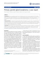
Post reduction lateral radiograph of the elbow showing the radial head in reduced positionFigure 3
Post reduction lateral radiograph of the elbow showing the
radial head in reduced position.
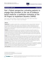
Radiograph of the elbow showing a dislocated radial headFigure 1
Radiograph of the elbow showing a dislocated radial head.
Radiograph of the elbow showing a dislocated radial headFigure 2
Radiograph of the elbow showing a dislocated radial head.
Publish with BioMed Central and every
scientist can read your work free of charge
"BioMed Central will be the most significant development for
disseminating the results of biomedical research in our lifetime."
Sir Paul Nurse, Cancer Research UK
Your research papers will be:
available free of charge to the entire biomedical community
peer reviewed and published immediately upon acceptance
cited in PubMed and archived on PubMed Central
yours — you keep the copyright
Submit your manuscript here:
/>BioMedcentral
Journal of Medical Case Reports 2007, 1:38 />Page 3 of 3
(page number not for citation purposes)
nation. The period of immobilisation varied from 10
days. [1] to 4 weeks. [5] Most acute cases can be reduced
closed and the functional outcome seems to be good post
reduction. If missed or neglected, an open reduction has
to be done with either an annular ligament reconstruction
[7] or a radial head excision deemed as the procedure of
choice [8].
We speculate the mechanism in our patient to be a hyper-
extension of the elbow with forearm in midprone posi-
tion leading to an anterolateral dislocation of the radial
head. The reduction was achieved in supination and
immobilisation of the elbow in flexion and supination
gave a favourable final outcome.
In the presence of major distracting injuries like long bone
fractures, pelvic fractures, chest and abdominal injuries,
an isolated radial head dislocation can be easily missed as
pain is masked by the presence of major distracting inju-
ries and flexion and extension of the elbow is normal. If
supination and pronation of the forearm is not assessed,
this injury can be missed resulting in degenerative arthritis
of the elbow and the distal radioulnar joints.
Conclusion
This case report has been prepared to stress the impor-
tance of a thorough secondary survey in patients with pol-
ytrauma after high impact motor vehicle accidents. A
proper secondary survey in patients with major distracting
injuries can prevent important injuries being missed.
Competing interests
The author(s) declare that they have no competing inter-
ests.
Authors' contributions
UR was involved in collecting patient details, reviewing
the literature and drafted the manuscript as the main
author.
RSUY was involved in reviewing the literature and proof
reading of the manuscript. RSUY has approved the final
manuscript.
SSB is the senior author and was responsible for final
proof reading of the article.
Acknowledgements
Written consent was obtained from the patient for publication of study.
Funding was neither sought nor obtained.
References
1. Obert L, Huot D, Lepage D, et al.: Isolated traumatic luxation of
the radial head in adults: report of a case and review of liter-
ature. Chir Main 2003, 22(4):216-9.
2. Takami H, Takahashi S, Ando M: Irreducible isolated dislocation
of the radial head. Clin Orthop Relat Res 1997:168-70.
3. Dhawan A, Hospodar PP: Isolated posttraumatic posterior dis-
location of the radial head in an adult. Am J Orthop 2002,
31(2):83-6.
4. Bonatus T, Chapman MW, Felix N: Traumatic anterior disloca-
tion of the radial head in an adult. J Orthop Trauma 1995,
9(5):441-4.
5. Yasuwaki Y, Itagane H, Nagata Y, Nishimoto S, Nakano A, Tanaka S:
Isolated lateral traumatic dislocation of the radial head in a
boy: case report. J Trauma 1993, 35(2):312-3.
6. Negi AK, Pestonji MD, Iyer S: Isolated posterior dislocation of
the radial head in an adult. J Postgrad Med 1992, 38(3):143.
7. Noyez JF: Isolated traumatic posterior dislocation of the
radial head: a report of two cases. Acta Orthop Belg 1996,
62(3):148-50.
8. Heidt RS Jr, Stern PJ: Isolated posterior dislocation of the radial
head. A case report. Clin Orthop Relat Res 1982:136-8.
Post reduction anteroposterior radiograph of the elbow showing the radial head in reduced positionFigure 4
Post reduction anteroposterior radiograph of the elbow
showing the radial head in reduced position.