Báo cáo khoa hoc:" Seizures as the first manifestation of chromosome 22q11.2 deletion syndrome in a 40-year old man: a case report" doc
Bạn đang xem bản rút gọn của tài liệu. Xem và tải ngay bản đầy đủ của tài liệu tại đây (471.97 KB, 5 trang )
BioMed Central
Page 1 of 5
(page number not for citation purposes)
Journal of Medical Case Reports
Open Access
Case report
Seizures as the first manifestation of chromosome 22q11.2 deletion
syndrome in a 40-year old man: a case report
Adriano R Tonelli*
1
, Kalyan Kosuri
1
, Sainan Wei
2
and Davoren Chick
1
Address:
1
Department of Internal Medicine, Michigan State University, East Lansing, Michigan, USA and
2
DNA Diagnostic and Cytogenetics
Laboratories, Department of Pediatrics and Human Development. Michigan State University, East Lansing, Michigan, USA
Email: Adriano R Tonelli* - ; Kalyan Kosuri - ; Sainan Wei - ;
Davoren Chick -
* Corresponding author
Abstract
Background: The microdeletion of chromosome 22q11.2 is the most common human deletion
syndrome. It typically presents early in life and is rarely considered in adult patients. As part of the
manifestations of this condition, patients can have parathyroid glandular involvement ranging from
hypocalcemic hypoparathyroidism to normocalcemia with normal parathryroid hormone levels.
The first manifestation of the syndrome might be seizures due to profound hypocalcemia.
Case presentation: A 40-year-old man without significant past medical history presented with a
new-onset generalized tonic-clonic seizure. He had no personal history of hypocalcemia or
seizures. Physical examination was remarkable for short stature, hypertelorism, prominent
forehead and nasal voice. His initial laboratory examination showed hypocalcemia (Calcium 5.2 mg/
dl and Calcium ionized 0.69 mmol/l) with hypoparathyroidism (Parathyroid hormone intact < 2.5
pg/ml. NV: 14–72 pg/ml). Urine Calcium was 3 mg/dl on a spot and 88 mg in a 24-hour urine
collection (NV: 100–300 mg/24 hs). The electrocardiogram showed a prolonged corrected QT
interval. Echocardiogram, abdominal ultrasound and electroencephalogram were normal. A
computer tomography of the brain showed basal ganglia calcification. The subtle physical findings
and the presence of idiopathic hypoparathyroidism motivated the performance of fluorescent in
situ hybridization which demonstrated a microdeletion on one of the homologs 22q11.2. The
patient was treated with calcium citrate and calcitriol with good response.
Conclusion: Microdeletion of chromosome 22q11.2 is among the most clinically variable
syndromes, with more than 180 features associated with the deletion. It has a variable phenotypical
expression, requiring a high level of awareness for its early diagnosis. Seizures, related to marked
hypocalcemia due to idiopathic hypoparathyroidism, might be the presenting feature in an adult
patient with this syndrome.
Background
The microdeletion of chromosome 22q11.2 is the most
common human deletion syndrome (1 in 4000 live
births) [1,2]. Most deletions occur de novo, with auto-
somal dominant inheritance observed in 15% of cases [3].
The deletion has been identified in most patients with
DiGeorge syndrome, velocardiofacial syndrome and
conotruncal anomaly face syndrome. The chromosome
Published: 3 December 2007
Journal of Medical Case Reports 2007, 1:167 doi:10.1186/1752-1947-1-167
Received: 14 April 2007
Accepted: 3 December 2007
This article is available from: />© 2007 Tonelli et al; licensee BioMed Central Ltd.
This is an Open Access article distributed under the terms of the Creative Commons Attribution License ( />),
which permits unrestricted use, distribution, and reproduction in any medium, provided the original work is properly cited.
Journal of Medical Case Reports 2007, 1:167 />Page 2 of 5
(page number not for citation purposes)
22q11.2 deletion syndrome is characterized by a congen-
ital failure in the development of the derivatives of various
pharyngeal arches and pouches with absent or small par-
athyroid glands [2]. It typically presents early in life and is
rarely considered in adult patients. The classical features
are conotruncal cardiac defects, dysmorphic facies, velo-
pharyngeal insufficiency, immunodeficiency, learning
disabilities and parathyroid dysfunction.
Parathyroid dysfunction in adults is generally due to
acquired conditions such as autoimmune processes or
injury of the parathyroid glands during surgery. Less com-
monly a genetic disorder like chromosome 22q11.2 dele-
tion syndrome can be the culprit condition. In this
syndrome the parathyroid glandular dysfunction ranges
from hypocalcemia with hypoparathyroidism to normo-
calcemia with normal parathryroid hormone levels [1-3].
Various forms of hypoparathryoidism have been reported
including late onset, transient, latent and recurrent [2].
Case presentation
A 40-year-old man presented with a new-onset general-
ized tonic-clonic seizure. He did not have any history of
hypocalcemia, seizures, psychiatric disorders or obvious
cognitive impairments. Physical examination was remark-
able for short stature, hypertelorism, hooded upper eye-
lids, prominent forehead, hypernasal speech and fullness
of the nose over the bridge. The etiology of the seizures
was attributed to the marked hypocalcemia noted on his
initial laboratory evaluation (Calcium 5.2 mg/dl and Cal-
cium ionized 0.69 mmol/l). Further work-up revealed
hypoparathyroidism (Parathyroid hormone intact < 2.5
pg/ml. NV: 14–72 pg/ml) and a normal total 25 hydroxy-
vitamin D (30 ng/ml. NV: 25–80 ng/ml). Thyroid stimu-
lation hormone was normal. Urine Calcium was 3 mg/dl
on a spot and 88 mg in a 24-hour urine collection (NV:
100–300 mg/24 hs).
The 12-lead electrocardiogram showed a prolonged cor-
rected QT interval. Echocardiogram, abdominal ultra-
sound and electroencephalogram were normal. A
computer tomography of the brain showed basal ganglia
calcification (figure 1). The presence of hypoparathy-
roidism and subtle dysmorphic facial features made chro-
mosome 22q11.2 deletion syndrome a possible
diagnosis. For this reason, fluorescent in situ hybridiza-
tion was performed using DNA fluorescent probes for the
DiGeorge/velocardiofacial syndrome critical region
(TUPLE1) at 22q11.2 and a control probe, arylsulfatase-A
(ARSA), at 22q13.3, from a commercially available source
(Vysis). This is a direct-labeled dual-color probe mixture
with TUPLE1 (HIRA) probe labeled in orange and ARSA
probe green (figure 2). At least 50 metaphase cells and
100 interphase cells were scored for both TUPLE1 and
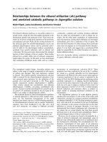
Brain computer tomography cuts of the patient, demonstrating basal ganglia and periventricular calcificationFigure 1
Brain computer tomography cuts of the patient, demonstrating basal ganglia and periventricular calcification.
Journal of Medical Case Reports 2007, 1:167 />Page 3 of 5
(page number not for citation purposes)
ARSA signals. All metaphase and interphase cells analyzed
show a deletion of TUPLE1 at 22q11.2 locus. The patient
was treated with calcium citrate and calcitriol. He
remained asymptomatic with normalization of his serum
calcium level.
Discussion
Microdeletion of chromosome 22q11.2 is among the
most clinically variable syndromes, with more than 180
features associated with the deletion [1,4]. The phenotype
develops over time; therefore the clinical presentation
may change [4]. It has a variable phenotypic expression,
with no pathognomonic or obligatory clinical features,
and requires a high level of awareness for its early diagno-
sis [1,5]. One of the clinical features of this syndrome is
seizures, which can be related to hypocalcemia, stroke,
cerebellar or cerebral cortical atrophy, or transient or
chronic ischemia [1,4,6-8]. A large cohort study showed
that 21% of the patients with chromosome 22q11.2 dele-
tion syndrome (62/290) had seizures, and at least 68%
(42/62) of the seizures were hypocalcemic in origin [8].
There are only a few reports in the medical literature
which describe new onset seizures caused by hypocal-
cemia in adolescence or adulthood (one report [7]), in
patients not previously diagnosed with chromosome
22q11.2 deletion syndrome [1,3,7,9,10].
Some patients present with asymptomatic hypocalcemia
and inappropriately low parathyroid hormone levels that
leads to fluorescence in situ hybridization and the diagno-
sis of chromosome 22q11.2 deletion syndrome [5,7,8].
The majority of patients are diagnosed soon after birth,
when the transport of calcium from mother to fetus is
abruptly interrupted. The low serum calcium level gener-
ally improves over the first year of life as the parathyroid
gland hypertrophies and the dietary calcium intake
Result of FISH analysis using LSI probe (TUPLE 1) from DiGeorge/velocardiofacial syndrome critical regionFigure 2
Result of FISH analysis using LSI probe (TUPLE 1) from DiGeorge/velocardiofacial syndrome critical region. TUPLE 1 (HIRA)
probe was labeled in Spectrum Orange and Arylsulfatase A (ARSA) in SpectrumGreen as control. Absence of the orange signal
indicates deletion of the TUPLE 1 locus at 22q11.2.
22q11.2 Del
22q11.2 Del
● Tuple1
● ARSA
NORMAL
NORMAL
● Tuple1
● ARSA
M
E
T
A
P
H
A
S
E
I
N
T
E
R
P
H
A
S
E
NORMAL
NORMAL
● Tuple1
● ARSA
22q11.2 Del
22q11.2 Del
● Tuple1
● ARSA
Journal of Medical Case Reports 2007, 1:167 />Page 4 of 5
(page number not for citation purposes)
increase. For this reason few older patients require ongo-
ing calcium supplementation [7,8,11].
Of note is that even when hypocalcemia is considered one
of the cardinal features of the syndrome, the prevalence of
this electrolyte abnormality depends on the selection cri-
teria used, ranging from 13 to 72%. Patients with pheno-
typic characteristics of the DiGeorge anomaly are more
likely to have clinical evidence of hypocalcemia or to have
calcium levels measured. A lower prevalence of hypocal-
cemia is seen in clinical presentations more consistent
with the velocardiofacial syndrome in which calcium lev-
els may not be routinely checked [11]. In a large European
cohort study of 558 patients known to have 22q11.2 dele-
tions, hypocalcemia was noted in 60% of the subjects of
whom 39% had presented with seizures [8].
Hypoparathyroidism can result in transient, persistent,
late-onset or latent hypocalcemia. Patients who are diag-
nosed later in life may have a latent form of hypoparath-
yroidism [2,10]. There are insufficient data to describe the
natural course of asymptomatic hypocalcemia diagnosed
in older patients. In spite of this it is clear that in some
patients, seizures may be the first manifestation of their
hypocalcemia, either spontaneously or precipitated dur-
ing periods of increased metabolic demand [3].
Patients with chromosome 22q11.2 deletion syndrome
can have a variety of brain abnormalities when assessed
by neuroimaging. Basal ganglia calcification, similar to
that seen in the patient presented in this case report, have
been rarely described in the literature. Possible explana-
tions include a low incidence of this finding or a lack of
computer tomography studies being performed on such
patients or being performed in patients who were too
young to have developed this abnormality. Most of the
patients with chromosome 22q11.2 syndrome were stud-
ied with magnetic resonance imaging, possibly missing
any calcification. The significance of this finding is unclear
[1,10-13].
Treatment of severe symptomatic hypocalcemia requires
prompt administration of parental calcium. In contrast,
asymptomatic hypocalcemia may be treated with oral cal-
cium and vitamin D supplements. Serum calcium levels
should be maintained in the low-normal range to mini-
mize hypercalciuria and the risk of development of renal
calculi [11].
In summary, the diagnosis of chromosome 22q11.2 dele-
tion syndrome during adulthood is uncommon. The con-
dition is probably under diagnosed. It should be
considered in adult patients presenting with idiopathic
hypoparathyroidism. Patients with this syndrome should
be informed of the symptoms that might occur with
hypocalcemia, have their calcium-parathyroid hormone
axis periodically checked and receive genetic counseled as
there is a 50% risk of having an affected offspring [1,5].
Conclusion
Chromosome 22q11.2 deletion syndrome has a variable
phenotypic expression. This diagnosis should be consid-
ered in adult patients presenting with idiopathic hypopar-
athyroidism. The absence of the classical features of this
condition should not exclude this pathology. Seizures,
related to marked hypocalcemia, might be the initial pres-
entation of this deletion. Treatment is relatively simple
and can bring about profound clinical improvement.
Competing interests
The author(s) declare that they have no competing inter-
ests.
Authors' contributions
AT: Involved in drafting of the manuscript, contributions
to conception and design, analysis of data;
KK: Involved in acquisition of data, conception and
design. Revised the manuscript;
SW: Carried out the molecular genetic studies. Revised the
manuscript;
DC: Contributed to conception and design. Revised the
manuscript.
Consent
The patient provided written informed consent for publi-
cation.
Acknowledgements
This manuscript received no funding.
References
1. Hiéronimus S, Bec-Roche M, Pedeutour F, Lambert JC, Wagner-Mal-
her K, Mas JC, Sadoul JL, Fénichel P: The spectrum of parathyroid
gland dysfunction associated with the microdeletion 22q11.
European Journal of Endocrinology 2006, 155:47-52.
2. Al-Jenaidi F, Makitie O, Grunebaum E, Sochett E: Parathyroid gland
dysfunction in 22q11.2 deletion syndrome. Horm Res 2007,
67:117-122.
3. Maalouf N, Sakhaee K, Odvina C: A case of chromosome 22q11
deletion syndrome diagnosed in a 32-year-old man with
hypoparathyroidism. J Clin Endocrinol Metab 2004, 89:4817-4820.
4. Robin N, Shprintzen R: Defining the clinical spectrum of dele-
tion 22q11.2. J Pediatr 2005, 147:90-96.
5. Taylor SC, Morris G, Wilson D, Davies SJ, Gregory JW: Hypopar-
athyroidism and 22q11 deletion syndrome. Arch Dis Child 2003,
88:520-522.
6. Shprintzen FJ: Velo-cardio-facial syndrome. In Management of
genetic syndromes 2nd edition. Edited by: Cassidy SB, Allanson J. New
York: Wiley; 2004:615-632.
7. Kar PS, Ogoe B, Meeking D: Di-George syndrome presenting
with hypocalcaemia in adulthood: two case reports and a
review. J Clin Pathol 2005, 58:655-657.
8. Ryan AK, Goodship JA, Wilson DI, Philip N, Levy A, Seidel H,
Schuffenhauer S, Oechsler H, Belohradsky B, Prieur M, Aurias A, Ray-
Publish with BioMed Central and every
scientist can read your work free of charge
"BioMed Central will be the most significant development for
disseminating the results of biomedical research in our lifetime."
Sir Paul Nurse, Cancer Research UK
Your research papers will be:
available free of charge to the entire biomedical community
peer reviewed and published immediately upon acceptance
cited in PubMed and archived on PubMed Central
yours — you keep the copyright
Submit your manuscript here:
/>BioMedcentral
Journal of Medical Case Reports 2007, 1:167 />Page 5 of 5
(page number not for citation purposes)
mond FL, Clayton-Smith J, Hatchwell E, McKeown C, Beemer FA,
Dallapiccola B, Novelli G, Hurst JA, Ignatius J, Green AJ, Winter RM,
Brueton L, Brøndum-Nielsen K, Scambler PJ, et al.: Spectrum of
clinical features associated with interstitial chromosome
22q11 deletions: a European collaborative study. J Med Genet
1997, 34:798-804.
9. Adachi M, Tachibana K, Masuno M, Makita Y, Maesaka H, Okada T,
Hizukuri K, Imaizumi K, Kuroki Y, Kurahashi H, Suwa S: Clinical
characteristics of children with hypoparathyroidism due to
22q11.2 microdeletion. Eur J Pediatr 1998, 157:34-38.
10. Scirè G, Dallapiccola B, Iannetti P, Bonaiuto F, Galasso C, Mingarelli
R, Boscherini B: Hypoparathyroidism as the major manifesta-
tion in two patients with 22q11 deletions. Am J Med Genet 1994,
52:478-482.
11. Weinzimer SA: Endocrine aspects of the 22q11.2 deletion syn-
drome. Genetics in Medicine 2001, 3:19-22.
12. Sieberer M, Haltenhof H, Haubitz B, Pabst B, Miller K, Garlipp P:
Basal ganglia calcification and psychosis in 22q11.2 deletion
syndrome. Eur Psychiatry 2005, 20:567-569.
13. Chow EW, Mikulis DJ, Zipursky RB, Scutt LE, Weksberg R, Bassett
AS: Qualitative MRI findings in adults with 22q11 deletion
syndrome and schizophrenia. Biol Psychiatry 1999, 46:1436-1442.